Best Seller
Human ICAM-1/CD54 Antibody (BBIG-I1 (11C81))
R&D Systems | Catalog # BBA3

Key Product Details
Species Reactivity
Validated:
Human
Cited:
Human, Mouse, Rat, Insect - Aedes albopictus (Asian tiger mosquito)
Applications
Validated:
Western Blot, Adhesion Blockade, Flow Cytometry, Immunocytochemistry, Immunoprecipitation
Cited:
Immunohistochemistry, Immunohistochemistry-Frozen, Western Blot, Neutralization, Flow Cytometry, Immunofluorescence, Immunocytochemistry, Immunoprecipitation, Bead-based Bioassay, Bioassay, Cell-based ELISA, ELISA Capture, ELISA Development, ELISA Development (Capture), Functional Assay, Inhibition
Label
Unconjugated
Antibody Source
Monoclonal Mouse IgG1 Clone # BBIG-I1 (11C81)
Loading...
Product Specifications
Immunogen
Activated HUVEC human umbilical vein endothelial cells
Specificity
This antibody was selected for its ability to stain human ICAM-1/CD54 transfected COS cells. It does not stain COS cells transfected with human E-Selectin, VCAM-1, or PECAM-1.
Clonality
Monoclonal
Host
Mouse
Isotype
IgG1
Endotoxin Level
<0.10 EU per 1 μg of the antibody by the LAL method.
Scientific Data Images for Human ICAM-1/CD54 Antibody (BBIG-I1 (11C81))
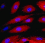
ICAM‑1/CD54 in C32 Human Cell Line.
ICAM‑1/CD54 was detected in live unfixed C32 human melanoma cell line using Mouse Anti-Human ICAM‑1/CD54 Monoclonal Antibody (Catalog # BBA3) at 5 µg/mL for 1 hour at 2° - 8° C. Cells were then fixed with 4% formaldehyde followed by incubation with the NorthernLights™ 557-conjugated Anti-Mouse IgG Secondary Antibody (red; NL007) and counterstained with DAPI (blue). Specific staining was localized to cell surface and cytoplasm.
Detection of Human ICAM‑1/CD54 by Western Blot.
Western blot shows lysates of C32. PVDF membrane was probed with 2 µg/mL of Mouse Anti-Human ICAM‑1/CD54 Monoclonal Antibody (Catalog # BBA3) followed by HRP-conjugated Anti-Mouse IgG Secondary Antibody (Catalog # HAF018). A specific band was detected for ICAM‑1/CD54 at approximately 85 kDa (as indicated). This experiment was conducted under non-reducing conditions and using Western Blot Buffer Group 1.
Detection of ICAM-1/CD54 by Immunohistochemistry
SERS-BFNP molecular imaging of atherosclerotic coronary arteries. A single human coronary artery was isolated from the heart of a patient undergoing heart transplantation surgery. The lumen of the artery segment was then injected with a mixture of anti-ICAM-1, anti-VCAM-1, anti-P-selectin, and isotype control BFNP, sutured closed, and incubated at 37 °C/5% CO2 for 12 h. Sutures were then removed and the artery segment was thoroughly washed prior to SERS spectroscopy and subsequent analysis of morphology, expression of adhesion molecules and SERS mapping. (B) Immunofluorescence staining for CD31, and expression of (C) ICAM-1, VCAM-1 and P-selectin are shown in red. Nuclei were counterstained using Hoechst 33342 (blue). Image collected and cropped by CiteAb from the following open publication (https://pubmed.ncbi.nlm.nih.gov/30613292), licensed under a CC-BY license. Not internally tested by R&D Systems.
Detection of ICAM‑1/CD54 in Human PBMCs by Flow Cytometry.
Human peripheral blood mononuclear cells (PBMCs) were stained with (A) Mouse Anti-Human ICAM-1/CD54 Monoclonal Antibody (Catalog # BBA3) or (B) Mouse IgG1 Isotype Control (MAB002) followed by anti-Mouse IgG PE-conjugated Secondary Antibody (F0102B) and Mouse Anti-Human CD14 APC-conjugated Monoclonal Antibody (FAB3832A). Staining was performed using our Staining Membrane-associated Proteins protocol.
Detection of ICAM-1/CD54 by Immunohistochemistry
In vivo SERS-BFNP molecular imaging of adhesion molecules. Following engraftment of human adipose, HANSG mice were allowed to recover for 3 weeks. Mice were then injected intravenously with 5 μg of human recombinant TNF-alpha 4 h prior to receiving an intravenous injection of BFNP. (A) Following SERS-BFNP molecular imaging, adipose grafts were excised and immunofluorescently stained for human (red) and murine (green) CD31. (B) Isotype control, ICAM-1, VCAM-1, and P-selectin staining are also shown in white counterstained with human CD31 (red). Nuclei were counterstained using Hoechst 33342 (blue). (C) To conduct SERS-BFNP molecular imaging, HANSG mice were anaesthetized and their adipose grafts non-invasively analyzed in vivo using SERS spectroscopy. (D) SERS spectra were acquired from mice that received a mixture of isotype-PPY, -BPE and -PYOT (blue spectra), or anti-P-selectin-PPY, anti-ICAM-1-BPE, anti-VCAM-1-PYOT BFNP (red spectra). The spectra shown are from 5 isotype vs. 5 targeted mice, with each spectrum acquired from a different mouse. (E) In addition to immunofluorescence microscopy, excised adipose grafts were analyzed using SERS microscopy. Detection of BFNP from sections of adipose tissue isolated from HANSG mice that received anti-ICAM-1 (purple), anti-VCAM-1 (red), and anti-P-selectin (blue) (upper panels) or Isotype-BPE (purple), Isotype-PYOT (red), and Isotype-PPY (blue) (lower panels) are shown superimposed on darkfield tissue images alongside a magnified image of Raman maps from the scanned areas (black boxes). The colored circles in the Raman map ((E) upper panel) correlate to the acquired spectra shown in (F) above their respective reference spectra. The optical image in (E) is a darkfield image. Scale bars: (A) = 1000 μm; (B) = 100 μm; (E) = 20 μm. Image collected and cropped by CiteAb from the following open publication (https://pubmed.ncbi.nlm.nih.gov/30613292), licensed under a CC-BY license. Not internally tested by R&D Systems.
Detection of ICAM-1/CD54 by Immunocytochemistry/ Immunofluorescence
Following stimulation, coronary artery endothelial cells (CAEC) express adhesion molecules detectable via immuno-SERS imaging in single and multiplex formats. (A) Fluorescence images of immunohistochemical staining of ICAM-1, VCAM-1 and P-selectin on CAEC in unstimulated and 10 ng/mL TNF-alpha -stimulated conditions. Isotype control, ICAM-1, VCAM-1 and P-selectin staining shown in green; nuclei were counterstained using Hoechst 33342 (blue). (B) CAEC were stimulated with 10 ng/mL TNF-alpha for 24 h, fixed in acetone, and incubated with isotype control, anti-ICAM-1, anti-VCAM-1 or anti-P-selectin BFNP or (C) with all BFNP simultaneously before being subjected to SERS mapping. (D) Representative spectra from anti-ICAM-1 (purple), anti-VCAM-1 (red) and anti-P-selectin (blue) BFNP acquired from the color-matched circles in (C) are shown above their respective reference spectra. Optical images in (B-C) are darkfield images. Scale bars = 20 μm. Results are representative of 3 independent experiments. Image collected and cropped by CiteAb from the following open publication (https://pubmed.ncbi.nlm.nih.gov/30613292), licensed under a CC-BY license. Not internally tested by R&D Systems.
Detection of ICAM-1/CD54 by Western Blot
Methotrexate (MTX) and Ad repress TNF-alpha -induced pro-inflammatory genes without affecting primary MIR181A-1 and MIR181B-1 expression.(A) Western blot analysis of VCAM-1, ICAM-1, and E-Selectin in HUVECs treated with or without MTX (10 µM) or Ad (50 µM), after stimulation of TNF-alpha (10 ng/ml) for 8 hr. Quantification of n = 3 independent experiments. (B) Real-time qPCR analysis of VCAM-1, ICAM-1, and E-Selectin in HUVECs treated with or without MTX (10 µM) or Ad (50 µM), after treatment with TNF-alpha (10 ng/ml) for 4 hr. Real-time qPCR analysis of (C) primary transcript of MIR181A1 or (D) primary transcript of MIR181B-1 in HUVECs treated with or without MTX (10 µM) or Ad (50 µM), after treatment with TNF-alpha (10 ng/ml) for 4 hr. (A–D), n = 3–6. *p<0.05; **p<0.01; ***p<0.001; ***p<0.0001. All values represent mean ± SEM. Image collected and cropped by CiteAb from the following open publication (https://pubmed.ncbi.nlm.nih.gov/33416495), licensed under a CC-BY license. Not internally tested by R&D Systems.
Detection of ICAM-1/CD54 by Western Blot
Induction of MIR181B expression by methotrexate (MTX) or adenosine is adenosine receptor A3 (ADORA3) dependent.(A) Knockdown for adenosine receptors A1, A2A, A2B, and A3 in HUVECs was performed to analyze MIR181B expression. three biological replicates. One-way ANOVA. (B) MIR181B expression in HUVECs transfected with Ctl-siRNA or ADORA3 siRNA after treatment with MTX (10 µM) or Ad (50 µM) or (C) treatment with TNF-alpha (10 ng/ml) alone or in combination MTX (10 µM) or Ad (50 µM). Three biological replicates. One-way ANOVA and Unpaired two-tailed Student t test. (D) Western blot analyses of VCAM-1, ICAM-1, and E-Selectin expression in HUVECs transfected with Ctl-siRNA or ADORA3 siRNA in the presence of TNF-alpha (10 ng/ml) in combination with either MTX (10 µM) or Ad (50 µM). Three biological replicates. Unpaired two-tailed Student t test. (E) in the presence of miRNA negative control (NS-m) or MIR181B mimics (181b-m) stimulated with TNF-alpha (10 ng/ml) or in combination with MTX (10 µM) or Ad (50 µM). Please see Figure 2—source data 1. Three biological replicates. Unpaired two-tailed Student t test. *p<0.05; **p<0.01; ***p<0.001; ****p<0.0001. n.s. indicated non significance. All values represent mean ± SEM.Figure 2—source data 1.Knockdown efficiency for adenosine receptor siRNAs.mRNA, and protein expression analysis for (A, B) ADORA3 siRNA, (C, D) ADORA1 siRNA, (E, F) ADORA2A siRNA and (G, H) ADORA2B siRNA compared to control siRNA in HUVECs transfected for 36 hr. (A–H), n = 3. ***p<0.001. All values represent mean ± SEM. Image collected and cropped by CiteAb from the following open publication (https://pubmed.ncbi.nlm.nih.gov/33416495), licensed under a CC-BY license. Not internally tested by R&D Systems.Applications for Human ICAM-1/CD54 Antibody (BBIG-I1 (11C81))
Application
Recommended Usage
Adhesion Blockade
The adhesion of HSB2 human peripheral blood acute lymphoblastic leukemia cells (5 x 10⁴ cells/well) to immobilized Recombinant Human ICAM-1/CD54 Fc Chimera (Catalog # 720-IC,12.5 µg/mL, 100 µL/well) was maximally inhibited (80-100%) by 10 µg/mL of the antibody.
Flow Cytometry
0.25 µg/106 cells
Sample: Human PBMC
Sample: Human PBMC
Immunocytochemistry
5-25 µg/mL
Sample: Live, unfixed C32 human melanoma cell line
Sample: Live, unfixed C32 human melanoma cell line
Immunoprecipitation
Davies, M.E., et al. (1991) Connect. Tissue Res. 26:207.
Western Blot
2 µg/mL
Sample: C32 human melanoma cell line
Sample: C32 human melanoma cell line
Reviewed Applications
Read 7 reviews rated 4.9 using BBA3 in the following applications:
Flow Cytometry Panel Builder
Bio-Techne Knows Flow Cytometry
Save time and reduce costly mistakes by quickly finding compatible reagents using the Panel Builder Tool.
Advanced Features
- Spectra Viewer - Custom analysis of spectra from multiple fluorochromes
- Spillover Popups - Visualize the spectra of individual fluorochromes
- Antigen Density Selector - Match fluorochrome brightness with antigen density

Formulation, Preparation, and Storage
Purification
Protein A or G purified from hybridoma culture supernatant
Reconstitution
Sterile PBS to a final concentration of 0.5 mg/mL.
Loading...
Formulation
Lyophilized from a 0.2 μm filtered solution in PBS with Trehalose.
Shipping
The product is shipped at ambient temperature. Upon receipt, store it immediately at the temperature recommended below.
Stability & Storage
Use a manual defrost freezer and avoid repeated freeze-thaw cycles.
- 12 months from date of receipt, -20 to -70 °C as supplied.
- 1 month, 2 to 8 °C under sterile conditions after reconstitution.
- 6 months, -20 to -70 °C under sterile conditions after reconstitution.
Calculators
Background: ICAM-1/CD54
Long Name
Intercellular Adhesion Molecule 1
Alternate Names
CD54, ICAM1
Gene Symbol
ICAM1
Additional ICAM-1/CD54 Products
Product Documents for Human ICAM-1/CD54 Antibody (BBIG-I1 (11C81))
Certificate of Analysis
To download a Certificate of Analysis, please enter a lot or batch number in the search box below.
Note: Certificate of Analysis not available for kit components.
Product Specific Notices for Human ICAM-1/CD54 Antibody (BBIG-I1 (11C81))
For research use only
Related Research Areas
Citations for Human ICAM-1/CD54 Antibody (BBIG-I1 (11C81))
 Powered by Bioz
Customer Reviews for Human ICAM-1/CD54 Antibody (BBIG-I1 (11C81)) (7)
4.9 out of 5
7 Customer Ratings
Have you used Human ICAM-1/CD54 Antibody (BBIG-I1 (11C81))?
Submit a review and receive an Amazon gift card!
$25/€18/£15/$25CAN/¥2500 Yen for a review with an image
$10/€7/£6/$10CAN/¥1110 Yen for a review without an image
Submit a review
Customer Images
Showing
1
-
5 of
7 reviews
Showing All
Filter By:
-
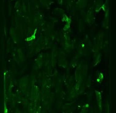
Application: Immunocytochemistry/ImmunofluorescenceSample Tested: HUVEC human umbilical vein endothelial cellsSpecies: HumanVerified Customer | Posted 07/10/2025

-
Application: Immunocytochemistry/ImmunofluorescenceSample Tested: Endothelial cellsSpecies: HumanVerified Customer | Posted 08/12/2021
-
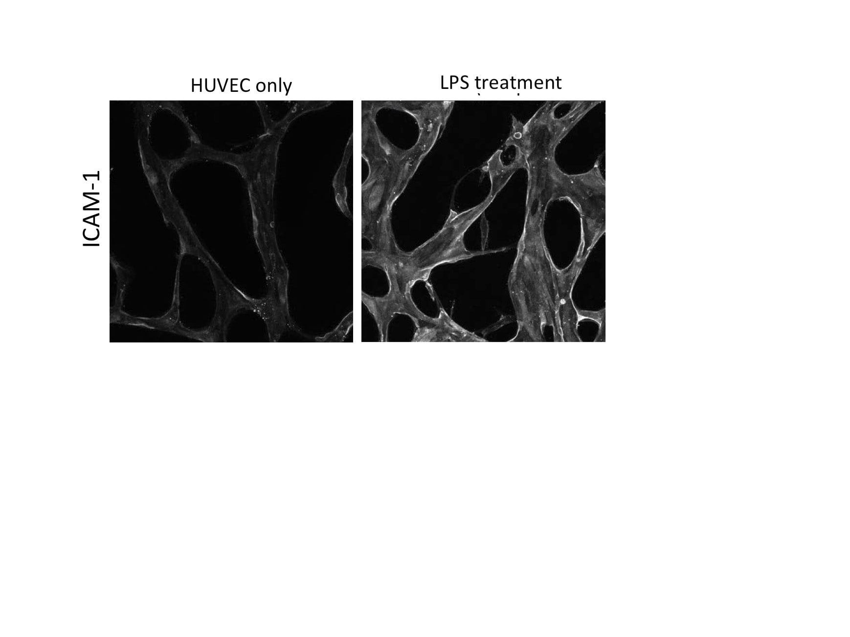
Application: Immunocytochemistry/ImmunofluorescenceSample Tested: HUVEC human umbilical vein endothelial cellsSpecies: HumanVerified Customer | Posted 04/26/2017HUVECs were stimulated with LPS (10 ng/mL)
-
Application: ImmunofluorescenceSample Tested: See PMID: 21277303Species: HumanVerified Customer | Posted 01/20/2015
-
Application: ImmunocytochemistrySample Tested: See PMID: 23104645Species: HumanVerified Customer | Posted 01/20/2015
-
Application: ImmunofluorescenceSample Tested: See PMID: 21164106Species: HumanVerified Customer | Posted 01/20/2015
-
Application: ImmunofluorescenceSample Tested: See PMID: 22560286Species: HumanVerified Customer | Posted 01/20/2015
There are no reviews that match your criteria.
Protocols
Find general support by application which include: protocols, troubleshooting, illustrated assays, videos and webinars.
- 7-Amino Actinomycin D (7-AAD) Cell Viability Flow Cytometry Protocol
- Appropriate Fixation of IHC/ICC Samples
- Cellular Response to Hypoxia Protocols
- ClariTSA™ Fluorophore Kits
- Detection & Visualization of Antibody Binding
- Extracellular Membrane Flow Cytometry Protocol
- Flow Cytometry Protocol for Cell Surface Markers
- Flow Cytometry Protocol for Staining Membrane Associated Proteins
- Flow Cytometry Staining Protocols
- Flow Cytometry Troubleshooting Guide
- ICC Cell Smear Protocol for Suspension Cells
- ICC Immunocytochemistry Protocol Videos
- ICC for Adherent Cells
- Immunocytochemistry (ICC) Protocol
- Immunocytochemistry Troubleshooting
- Immunofluorescence of Organoids Embedded in Cultrex Basement Membrane Extract
- Immunohistochemistry (IHC) and Immunocytochemistry (ICC) Protocols
- Immunoprecipitation Protocol
- Intracellular Flow Cytometry Protocol Using Alcohol (Methanol)
- Intracellular Flow Cytometry Protocol Using Detergents
- Intracellular Nuclear Staining Flow Cytometry Protocol Using Detergents
- Intracellular Staining Flow Cytometry Protocol Using Alcohol Permeabilization
- Intracellular Staining Flow Cytometry Protocol Using Detergents to Permeabilize Cells
- Preparing Samples for IHC/ICC Experiments
- Preventing Non-Specific Staining (Non-Specific Binding)
- Primary Antibody Selection & Optimization
- Propidium Iodide Cell Viability Flow Cytometry Protocol
- Protocol for Liperfluo
- Protocol for VisUCyte™ HRP Polymer Detection Reagent
- Protocol for the Characterization of Human Th22 Cells
- Protocol for the Characterization of Human Th9 Cells
- Protocol for the Fluorescent ICC Staining of Cell Smears - Graphic
- Protocol for the Fluorescent ICC Staining of Cultured Cells on Coverslips - Graphic
- Protocol for the Preparation and Fluorescent ICC Staining of Cells on Coverslips
- Protocol for the Preparation and Fluorescent ICC Staining of Non-adherent Cells
- Protocol for the Preparation and Fluorescent ICC Staining of Stem Cells on Coverslips
- Protocol for the Preparation of a Cell Smear for Non-adherent Cell ICC - Graphic
- Protocol: Annexin V and PI Staining by Flow Cytometry
- Protocol: Annexin V and PI Staining for Apoptosis by Flow Cytometry
- R&D Systems Quality Control Western Blot Protocol
- TUNEL and Active Caspase-3 Detection by IHC/ICC Protocol
- The Importance of IHC/ICC Controls
- Troubleshooting Guide: Fluorokine Flow Cytometry Kits
- Troubleshooting Guide: Western Blot Figures
- Western Blot Conditions
- Western Blot Protocol
- Western Blot Protocol for Cell Lysates
- Western Blot Troubleshooting
- Western Blot Troubleshooting Guide
- View all Protocols, Troubleshooting, Illustrated assays and Webinars

