IL-10 R beta, also known as IL-10 R2, mediates its biological activities via binding to a heteromeric receptor complex consisting of two distinct type II cytokine receptor subunits, the ligand binding IL-10 R alpha and the IL-10 R beta which does not bind IL-10 by itself but is required for signal transduction. The cDNA for human IL-10 R beta encodes a 325 amino acid (aa) type I transmembrane precursor protein with a 20 aa signal sequence, a 200 aa extracellular region, a 29 aa transmembrane segment and a 76 aa cytoplasmic domain. Within the extracellular region, there are two 100 aa subdomains that resemble the constant region of immunoglobulins. This structural motif is responsible for the alternative designation of IL-10 R beta as CRF2-4 (the 4th member of the cytokine receptor family class II/2). Human and mouse IL-10 R beta share approximately 69% aa sequence identity. Binding of the non-covalent IL-10 dimer to two IL-10 R alpha chains recruits two IL‑10 R beta chains resulting in the activation and phosphorylation of the signaling cascade involving JAK1, TYK2, and STAT3. IL-10 R beta is expressed ubiquitously. IL‑10 R beta is also a component of the IL-22 receptor complex consisting of the IL-10 R beta chain and IL-22 R, another type II cytokine receptor family member.
Human IL-10 R beta Antibody (90220)
R&D Systems | Catalog # MAB874

Key Product Details
Species Reactivity
Validated:
Human
Cited:
Human
Applications
Validated:
Western Blot, Neutralization, Flow Cytometry, CyTOF-ready
Cited:
Immunohistochemistry, Western Blot, Neutralization, Flow Cytometry
Label
Unconjugated
Antibody Source
Monoclonal Mouse IgG1 Clone # 90220
Loading...
Product Specifications
Immunogen
Mouse myeloma cell line NS0-derived recombinant human IL-10 R beta
Met20-Ser220
Accession # Q08334
Met20-Ser220
Accession # Q08334
Specificity
Detects human IL-10 R beta in direct ELISAs and Western blots. In direct ELISAs, no cross-reactivity with recombinant human (rh) IL‑10 R alpha, rhIFN-gamma RI, and rhIFN-gamma R2 is observed.
Clonality
Monoclonal
Host
Mouse
Isotype
IgG1
Endotoxin Level
<0.10 EU per 1 μg of the antibody by the LAL method.
Scientific Data Images for Human IL-10 R beta Antibody (90220)
IL‑10 Inhibition of IL‑1 beta secre-tion and Neutralization by Human IL‑10 R beta Antibody.
Recombinant Human IL-10 (Catalog # 217-IL) inhibits IL-1 beta secretion in LPS-activated human peripheral blood mononuclear cells (PBMC) in a dose-dependent manner (orange line), as measured by the Human IL-1 beta /IL-1F2 Quantikine ELISA Kit (Catalog # DLB50). IL-1 beta secre-tion inhibited by Recombinant Human IL-10 (0.25 ng/mL) is neutralized (green line) by increasing concentrations of Mouse Anti-Human IL-10 R beta Monoclonal Antibody (Catalog # MAB874). The ND50 is typically 0.5-2 µg/mL in the presence of LPS (0.25 ng/mL).
Detection of IL‑10 R beta in Human PBMC Monocytes by Flow Cytometry
Human PBMC monocytes were stained with Mouse Anti-Human IL-10 R beta Monoclonal Antibody (Catalog # MAB874, filled histogram) or isotype control antibody (Catalog # MAB002, open histogram) followed by PE-conjugated Anti-Mouse IgG Secondary Antibody (Catalog # F0102B). View our protocol for Staining Membrane-associated Proteins.Applications for Human IL-10 R beta Antibody (90220)
Application
Recommended Usage
CyTOF-ready
Ready to be labeled using established conjugation methods. No BSA or other carrier proteins that could interfere with conjugation.
Flow Cytometry
0.25 µg/106 cells
Sample: Human PBMC monocytes
Sample: Human PBMC monocytes
Western Blot
1 µg/mL
Sample: Recombinant Human IL‑10 R beta Fc Chimera (Catalog # 874-RB)
Sample: Recombinant Human IL‑10 R beta Fc Chimera (Catalog # 874-RB)
Neutralization
Measured by its ability to neutralize IL‑10 R beta -mediated IL‑10 response in LPS-activated human peripheral blood mononuclear cells (PBMC). Ralph, P. et al. (1991) J. Immunology 148:808. The Neutralization Dose (ND50) is typically 0.5-2 µg/mL in the presence of 0.25 ng/mL Recombinant Human IL‑10 and 0.25 ng/mL LPS.
Reviewed Applications
Read 1 review rated 3 using MAB874 in the following applications:
Flow Cytometry Panel Builder
Bio-Techne Knows Flow Cytometry
Save time and reduce costly mistakes by quickly finding compatible reagents using the Panel Builder Tool.
Advanced Features
- Spectra Viewer - Custom analysis of spectra from multiple fluorochromes
- Spillover Popups - Visualize the spectra of individual fluorochromes
- Antigen Density Selector - Match fluorochrome brightness with antigen density

Formulation, Preparation, and Storage
Purification
Protein A or G purified from hybridoma culture supernatant
Reconstitution
Reconstitute at 0.5 mg/mL in sterile PBS. For liquid material, refer to CoA for concentration.
Loading...
Formulation
Lyophilized from a 0.2 μm filtered solution in PBS with Trehalose. *Small pack size (SP) is supplied either lyophilized or as a 0.2 µm filtered solution in PBS.
Shipping
Lyophilized product is shipped at ambient temperature. Liquid small pack size (-SP) is shipped with polar packs. Upon receipt, store immediately at the temperature recommended below.
Stability & Storage
Use a manual defrost freezer and avoid repeated freeze-thaw cycles.
- 12 months from date of receipt, -20 to -70 °C as supplied.
- 1 month, 2 to 8 °C under sterile conditions after reconstitution.
- 6 months, -20 to -70 °C under sterile conditions after reconstitution.
Calculators
Background: IL-10 R beta
References
- Donnelly, R.P. et al. (2004) J. Leukoc. Biol. 76:314.
- Donnelly, R.P. et al. (1999) J. Interferon Cytokine Res. 19:563.
- Kotenko, S.V. et al. (2000) J. Biol. Chem. 276:2725.
- Liu, Y. et al. (1994) J. Immunol. 152:1821.
- Lutfalla, G. et al. (1993) Genomics 16:366.
- Kotenko, S.V. et al. (1997) EMBO J. 16:5894.
Long Name
Interleukin 10 Receptor beta
Alternate Names
CDw210b, CRFB4, D21S58, IL-10R2, IL-10Rb, IL10R beta, IL10RB
Gene Symbol
IL10RB
UniProt
Additional IL-10 R beta Products
Product Documents for Human IL-10 R beta Antibody (90220)
Certificate of Analysis
To download a Certificate of Analysis, please enter a lot or batch number in the search box below.
Note: Certificate of Analysis not available for kit components.
Product Specific Notices for Human IL-10 R beta Antibody (90220)
For research use only
Citations for Human IL-10 R beta Antibody (90220)
 Powered by Bioz
Customer Reviews for Human IL-10 R beta Antibody (90220) (1)
3 out of 5
1 Customer Rating
Have you used Human IL-10 R beta Antibody (90220)?
Submit a review and receive an Amazon gift card!
$25/€18/£15/$25CAN/¥2500 Yen for a review with an image
$10/€7/£6/$10CAN/¥1110 Yen for a review without an image
Submit a review
Customer Images
Showing
1
-
1 of
1 review
Showing All
Filter By:
-
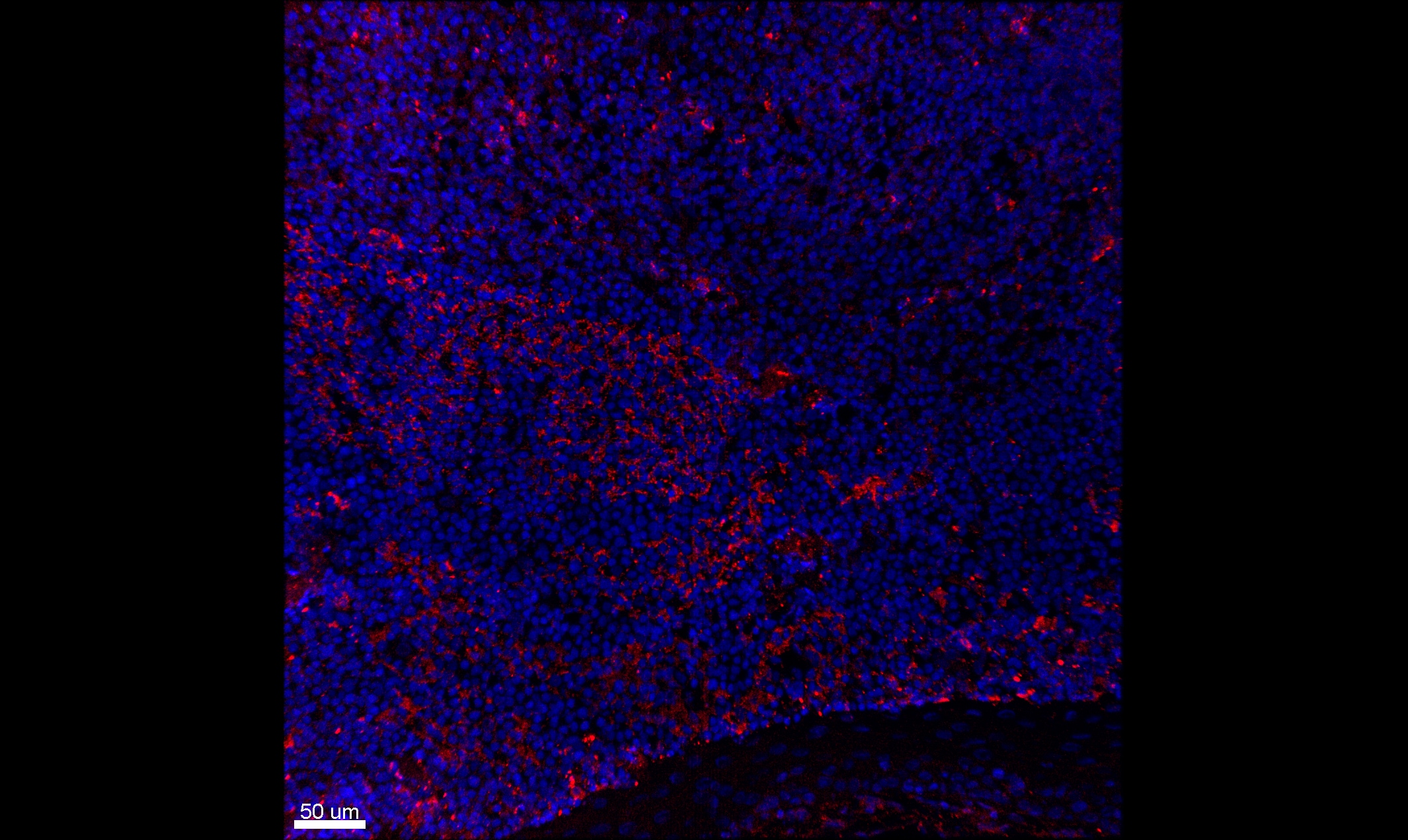
Application: Immunohistochemistry-FrozenSample Tested: Human Tonsil tissueSpecies: HumanVerified Customer | Posted 01/15/2021Human tonsil stained for IL10Rb (red) and counterstained with DAPI (blue)Acetone fixation

There are no reviews that match your criteria.
Protocols
Find general support by application which include: protocols, troubleshooting, illustrated assays, videos and webinars.
- 7-Amino Actinomycin D (7-AAD) Cell Viability Flow Cytometry Protocol
- Cellular Response to Hypoxia Protocols
- Extracellular Membrane Flow Cytometry Protocol
- Flow Cytometry Protocol for Cell Surface Markers
- Flow Cytometry Protocol for Staining Membrane Associated Proteins
- Flow Cytometry Staining Protocols
- Flow Cytometry Troubleshooting Guide
- Intracellular Flow Cytometry Protocol Using Alcohol (Methanol)
- Intracellular Flow Cytometry Protocol Using Detergents
- Intracellular Nuclear Staining Flow Cytometry Protocol Using Detergents
- Intracellular Staining Flow Cytometry Protocol Using Alcohol Permeabilization
- Intracellular Staining Flow Cytometry Protocol Using Detergents to Permeabilize Cells
- Propidium Iodide Cell Viability Flow Cytometry Protocol
- Protocol for Liperfluo
- Protocol for the Characterization of Human Th22 Cells
- Protocol for the Characterization of Human Th9 Cells
- Protocol: Annexin V and PI Staining by Flow Cytometry
- Protocol: Annexin V and PI Staining for Apoptosis by Flow Cytometry
- R&D Systems Quality Control Western Blot Protocol
- Troubleshooting Guide: Fluorokine Flow Cytometry Kits
- Troubleshooting Guide: Western Blot Figures
- Western Blot Conditions
- Western Blot Protocol
- Western Blot Protocol for Cell Lysates
- Western Blot Troubleshooting
- Western Blot Troubleshooting Guide
- View all Protocols, Troubleshooting, Illustrated assays and Webinars
Loading...
Associated Pathways
