MKI67 (also Ki67) is a 350-400 kDa nuclear protein that belongs to a molecular group comprised of mitotic chromosome-associated proteins. Ki67 was originally recognized as an antigen associated with the monoclonal Ki67 antibody raised against Hodgkin's lymphoma nuclear material. Ki67 is contextually expressed, being potentially found in all cells that are not in the Go phase of the cell cycle. Thus, MKI67 qualifies as a cell proliferation marker. Functionally, Ki67 is known to interact with 160 kDa Hklp2, a protein that promotes centrosome separation and spindle bipolarity. It also directly interacts with NIFK, and apparently binds to UBF, thus playing a role in rRNA synthesis. Human MKI67 is 3256 amino acids (aa) in length. It contains one FHA domain (aa 8-98), followed by at least 24 utilized Ser/Thr phosphorylation sites and sixteen 120 aa repeats (aa 1000-2928) that are interspersed with at least 90 additional utilized phosphorylation sites. There are two potential isoform variants. One isoform is 315-345 kDa in size and shows a deletion of aa 136-495, while a second isoform contains a 58 aa substitution for aa 1-513. Over aa 3120-3256, human Ki67 shares 46% aa sequence identity with the mouse ortholog to Ki67.

Key Product Details
Validated by
Knockout/Knockdown
Species Reactivity
Validated:
Human
Cited:
Human, Mouse, Transgenic Mouse
Applications
Validated:
Knockout Validated, Immunohistochemistry, Dual RNAscope ISH-IHC Compatible, Immunocytochemistry, Simple Western
Cited:
Immunohistochemistry, Immunohistochemistry-Paraffin, Western Blot, Immunocytochemistry
Label
Unconjugated
Antibody Source
Polyclonal Sheep IgG
Loading...
Product Specifications
Immunogen
E. coli-derived recombinant human Ki67/MKI67
Asn3120-Ile3256
Accession # P46013
Asn3120-Ile3256
Accession # P46013
Specificity
Detects human Ki67/MKI67 in direct ELISAs. In direct ELISAs, approximately 10% cross-reactivity with recombinant mouse Ki67/MKI67 is observed.
Clonality
Polyclonal
Host
Sheep
Isotype
IgG
Scientific Data Images for Human Ki67/MKI67 Antibody
Ki67/MKI67 in A549 Human Cell Line.
Ki67/MKI67 was detected in immersion fixed A549 human lung carcinoma cell line using Sheep Anti-Human Ki67/MKI67 Antigen Affinity-purified Polyclonal Antibody (Catalog # AF7617) at 5 µg/mL for 3 hours at room temperature. Cells were stained using the Northern-Lights™ 557-conjugated Anti-Sheep IgG Secondary Antibody (red; NL010) and counterstained with DAPI (blue). Specific staining was localized to nuclei and nucleoli. View our protocol for Fluorescent ICC Staining of Cells on Coverslips.
Ki67/MKI67 in Human Breast Cancer Tissue.
Ki67/MKI67 was detected in immersion fixed paraffin-embedded sections of human breast cancer tissue using Sheep Anti-Human Ki67/MKI67 Antigen Affinity-purified Polyclonal Antibody (Catalog # AF7617) at 1 µg/mL overnight at 4 °C. Before incubation with the primary antibody, tissue was subjected to heat-induced epitope retrieval using Antigen Retrieval Reagent-Basic (CTS013). Tissue was stained using the Anti-Sheep HRP-DAB Cell & Tissue Staining Kit (brown; Catalog # CTS019) and counterstained with hematoxylin (blue). Specific staining was localized to the nuclei of epithelial cells. View our protocol for Chromogenic IHC Staining of Paraffin-embedded Tissue Sections.
Detection of Human Ki67/MKI67 by Simple WesternTM.
Simple Western lane view shows lysates of HeLa human cervical epithelial carcinoma cell line and MCF‑7 human breast cancer cell line, loaded at 0.2 mg/mL. A specific band was detected for Ki67/MKI67 at approximately 320 kDa (as indicated) using 20 µg/mL of Sheep Anti-Human Ki67/MKI67 Antigen Affinity-purified Polyclonal Antibody (Catalog # AF7617) followed by 1:50 dilution of HRP-conjugated Anti-Sheep IgG Secondary Antibody (HAF016). This experiment was conducted under reducing conditions and using the 66-440 kDa separation system.
Detection of Human Ki67/MKI67 by Simple WesternTM.
Simple Western lane view shows lysates of HeLa human cervical epithelial carcinoma cell line and Ki67 knockout HeLa cell line (KO), loaded at 0.2 mg/mL. A specific band was detected for Ki67/MKI67 at approximately 325 kDa (as indicated) using 20 µg/mL of Sheep Anti-Human Ki67/MKI67 Antigen Affinity-purified Polyclonal Antibody (Catalog # AF7617) followed by 1:50 dilution of HRP-conjugated Anti-Sheep IgG Secondary Antibody (HAF016). GAPDH (AF5718) is shown as a loading control. This experiment was conducted under reducing conditions and using the 66-440 kDa separation system.
Ki67/MKI67 Specificity is Shown by Immunocytochemistry in Knockout Cell Line.
Ki67/MKI67 was detected in immersion fixed HeLa human cervical epithelial carcinoma cell line but is not detected in Ki67/MKI67 knockout (KO) HeLa cell line using Sheep Anti-Human Ki67/MKI67 Antigen Affinity-purified Polyclonal Antibody (Catalog # AF7617) at 1 µg/mL for 3 hours at room temperature. Cells were stained using the NorthernLights™ 557-conjugated Anti-Sheep IgG Secondary Antibody (red; NL010) and counterstained with DAPI (blue). Specific staining was localized to nuclei. View our protocol for Fluorescent ICC Staining of Cells on Coverslips.
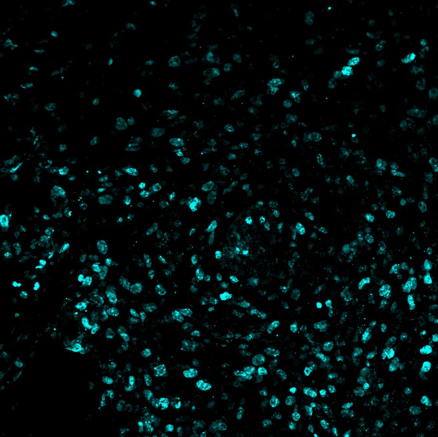
Ki-67/MKI67 in human breast cancer using Dual RNAscope®ISH and IHC.
MKi67 mRNA (red) and MKi67 protein (green) were detected in formalin-fixed paraffin-embedded tissue sections of human breast cancer. ACD’s Integrated Co-Detection Workflow was performed using ACD RNAScope Probe Hs-MKI67 (Catalog # 591771) and sheep anti-human Ki67/MKI67 polyclonal antibody (AF7617) at 5 μg/mL. Tissue was stained using RNAscope® 2.5 HD Detection Kit-RED (Catalog # 322360) and RNAscope® 2.5 LS Green Accessory Pack (Catalog # 322550). Tissue was counterstained with 50% hematoxylin (blue).Applications for Human Ki67/MKI67 Antibody
Application
Recommended Usage
Dual RNAscope ISH-IHC Compatible
5-25 µg/mL
Sample: Formalin-fixed paraffin-embedded tissue sections of human breast cancer.
Sample: Formalin-fixed paraffin-embedded tissue sections of human breast cancer.
Immunocytochemistry
5-15 µg/mL
Sample: Immersion fixed A549 human lung carcinoma cell line
Sample: Immersion fixed A549 human lung carcinoma cell line
Immunohistochemistry
5-15 µg/mL
Sample: Immersion fixed paraffin-embedded sections of human breast cancer tissue subjected to heat-induced epitope retrieval using Antigen Retrieval Reagent-Basic (Catalog # CTS013)
Sample: Immersion fixed paraffin-embedded sections of human breast cancer tissue subjected to heat-induced epitope retrieval using Antigen Retrieval Reagent-Basic (Catalog # CTS013)
Knockout Validated
Ki67/MKI67 is specifically detected in HeLa human cervical epithelial carcinoma parental cell line but is not detectable in Ki67/MKI67 knockout HeLa cell line.
Simple Western
20 µg/mL
Sample: HeLa human cervical epithelial carcinoma cell line and MCF‑7 human breast cancer cell line.
Sample: HeLa human cervical epithelial carcinoma cell line and MCF‑7 human breast cancer cell line.
Reviewed Applications
Read 2 reviews rated 5 using AF7617 in the following applications:
Formulation, Preparation, and Storage
Purification
Antigen Affinity-purified
Reconstitution
Sterile PBS to a final concentration of 0.2 mg/mL. For liquid material, refer to CoA for concentration.
Loading...
Formulation
Lyophilized from a 0.2 μm filtered solution in PBS with Trehalose. *Small pack size (SP) is supplied either lyophilized or as a 0.2 µm filtered solution in PBS.
Shipping
Lyophilized product is shipped at ambient temperature. Liquid small pack size (-SP) is shipped with polar packs. Upon receipt, store immediately at the temperature recommended below.
Stability & Storage
Use a manual defrost freezer and avoid repeated freeze-thaw cycles.
- 12 months from date of receipt, -20 to -70 °C as supplied.
- 1 month, 2 to 8 °C under sterile conditions after reconstitution.
- 6 months, -20 to -70 °C under sterile conditions after reconstitution.
Calculators
Background: Ki67/MKI67
Long Name
Antigen Identified by Monoclonal Antibody Ki67
Alternate Names
Ki-67, KIA, MIB-1, MKI67, PPP1R105, TSG126
Gene Symbol
MKI67
UniProt
Additional Ki67/MKI67 Products
Product Documents for Human Ki67/MKI67 Antibody
Certificate of Analysis
To download a Certificate of Analysis, please enter a lot or batch number in the search box below.
Note: Certificate of Analysis not available for kit components.
Product Specific Notices for Human Ki67/MKI67 Antibody
For research use only
Related Research Areas
Citations for Human Ki67/MKI67 Antibody
 Powered by Bioz
Customer Reviews for Human Ki67/MKI67 Antibody (2)
5 out of 5
2 Customer Ratings
Have you used Human Ki67/MKI67 Antibody?
Submit a review and receive an Amazon gift card!
$25/€18/£15/$25CAN/¥2500 Yen for a review with an image
$10/€7/£6/$10CAN/¥1110 Yen for a review without an image
Submit a review
Customer Images
Showing
1
-
2 of
2 reviews
Showing All
Filter By:
-
Application: ImmunohistochemistrySample Tested: Breast cancer tissueSpecies: HumanVerified Customer | Posted 11/28/2024Human, 1:200

-
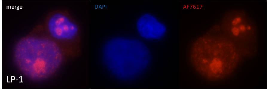
Application: ImmunocytochemistrySample Tested: Multiple myeloma cells line LP-1 cytospinsSpecies: HumanVerified Customer | Posted 08/24/2015IFF/ICC staining of LP-1 cells with AF7617
There are no reviews that match your criteria.
Protocols
Find general support by application which include: protocols, troubleshooting, illustrated assays, videos and webinars.
- Antigen Retrieval Protocol (PIER)
- Antigen Retrieval for Frozen Sections Protocol
- Appropriate Fixation of IHC/ICC Samples
- Cellular Response to Hypoxia Protocols
- Chromogenic IHC Staining of Formalin-Fixed Paraffin-Embedded (FFPE) Tissue Protocol
- Chromogenic Immunohistochemistry Staining of Frozen Tissue
- ClariTSA™ Fluorophore Kits
- Detection & Visualization of Antibody Binding
- Fluorescent IHC Staining of Frozen Tissue Protocol
- Graphic Protocol for Heat-induced Epitope Retrieval
- Graphic Protocol for the Preparation and Fluorescent IHC Staining of Frozen Tissue Sections
- Graphic Protocol for the Preparation and Fluorescent IHC Staining of Paraffin-embedded Tissue Sections
- Graphic Protocol for the Preparation of Gelatin-coated Slides for Histological Tissue Sections
- ICC Cell Smear Protocol for Suspension Cells
- ICC Immunocytochemistry Protocol Videos
- ICC for Adherent Cells
- IHC Sample Preparation (Frozen sections vs Paraffin)
- ISH-IHC Protocol for Chromogenic Detection on Formalin Fixed Paraffin Embedded (FFPE) Tissue
- Immunocytochemistry (ICC) Protocol
- Immunocytochemistry Troubleshooting
- Immunofluorescence of Organoids Embedded in Cultrex Basement Membrane Extract
- Immunofluorescent IHC Staining of Formalin-Fixed Paraffin-Embedded (FFPE) Tissue Protocol
- Immunohistochemistry (IHC) and Immunocytochemistry (ICC) Protocols
- Immunohistochemistry Frozen Troubleshooting
- Immunohistochemistry Paraffin Troubleshooting
- Preparing Samples for IHC/ICC Experiments
- Preventing Non-Specific Staining (Non-Specific Binding)
- Primary Antibody Selection & Optimization
- Protocol for Heat-Induced Epitope Retrieval (HIER)
- Protocol for Making a 4% Formaldehyde Solution in PBS
- Protocol for VisUCyte™ HRP Polymer Detection Reagent
- Protocol for the Fluorescent ICC Staining of Cell Smears - Graphic
- Protocol for the Fluorescent ICC Staining of Cultured Cells on Coverslips - Graphic
- Protocol for the Preparation & Fixation of Cells on Coverslips
- Protocol for the Preparation and Chromogenic IHC Staining of Frozen Tissue Sections
- Protocol for the Preparation and Chromogenic IHC Staining of Frozen Tissue Sections - Graphic
- Protocol for the Preparation and Chromogenic IHC Staining of Paraffin-embedded Tissue Sections
- Protocol for the Preparation and Chromogenic IHC Staining of Paraffin-embedded Tissue Sections - Graphic
- Protocol for the Preparation and Fluorescent ICC Staining of Cells on Coverslips
- Protocol for the Preparation and Fluorescent ICC Staining of Non-adherent Cells
- Protocol for the Preparation and Fluorescent ICC Staining of Stem Cells on Coverslips
- Protocol for the Preparation and Fluorescent IHC Staining of Frozen Tissue Sections
- Protocol for the Preparation and Fluorescent IHC Staining of Paraffin-embedded Tissue Sections
- Protocol for the Preparation of Gelatin-coated Slides for Histological Tissue Sections
- Protocol for the Preparation of a Cell Smear for Non-adherent Cell ICC - Graphic
- TUNEL and Active Caspase-3 Detection by IHC/ICC Protocol
- The Importance of IHC/ICC Controls
- Troubleshooting Guide: Immunohistochemistry
- View all Protocols, Troubleshooting, Illustrated assays and Webinars
Loading...