

Scientific images may be resized, cropped, or adjusted for brightness or contrast for presentation purposes. These updates are limited to presentation and do not affect the underlying scientific interpretation of the data. Where available, additional source data may be provided upon request.
Key Product Details
Species Reactivity
Validated:
Cited:
Applications
Validated:
Cited:
Label
Antibody Source
Product Specifications
Immunogen
Pro17-His257
Accession # ABM86208
Specificity
Clonality
Host
Isotype
Scientific Data Images for Human LILRB4/CD85k/ILT3 Antibody
Detection of LILRB4/CD85k/ILT3 by Western Blot
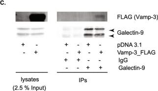
Galectin-9 interacts with Vamp-3 in DCs. A moDCs were lysed and whole-cell extracts incubated with either anti-galectin-9 specific antibody or an isotype control (goat IgG). All interacting proteins were isolated and identified by mass-spectrometry. Volcano plot depicts differentially enriched galectin-9 (right) and isotype (left) interacting proteins. B moDCs were prepped as per (A) but immunoprecipitated (IP) complexes were resolved and probed with galectin-9 and Vamp-3 specific antibodies. Graph shows quantification of Vamp-3 content in each sample using ImageJ. Data shown is representative of 5 individual experiments. C THP-1 cells were transfected with empty vector or plasmid containing 3 × FLAG_Vamp-3, lysed and whole-cell extracts incubated with either anti-galectin-9 specific antibody or an isotype control (goat IgG). IP complexes were resolved and probed with galectin-9 and Vamp-3 specific antibodies. Immunoblot shown is representative of four independent experiments. D NT or gal9 siRNA transfected moDCs were treated with LPS or DMSO as negative control for 6 h prior to being lysed and whole lysates probed against the specified proteins. Tubulin was used as loading control. Immunoblot shown is representative of three independent experiments. Graph shows quantification of Vamp-3 content in each sample using ImageJ. E. Quantification of Vamp-3 and SNAP23 levels shown in (D). Graphs depict Vamp-3 and SNAP23 levels in galectin-9-depleted moDCs normalised to tubulin and made relative to the corresponding untreated NT siRNA sample. Graph shows mean ± SD of three independent donors. **p < 0.005 Image collected and cropped by CiteAb from the following open publication (https://pubmed.ncbi.nlm.nih.gov/37755527), licensed under a CC-BY license. Not internally tested by R&D Systems.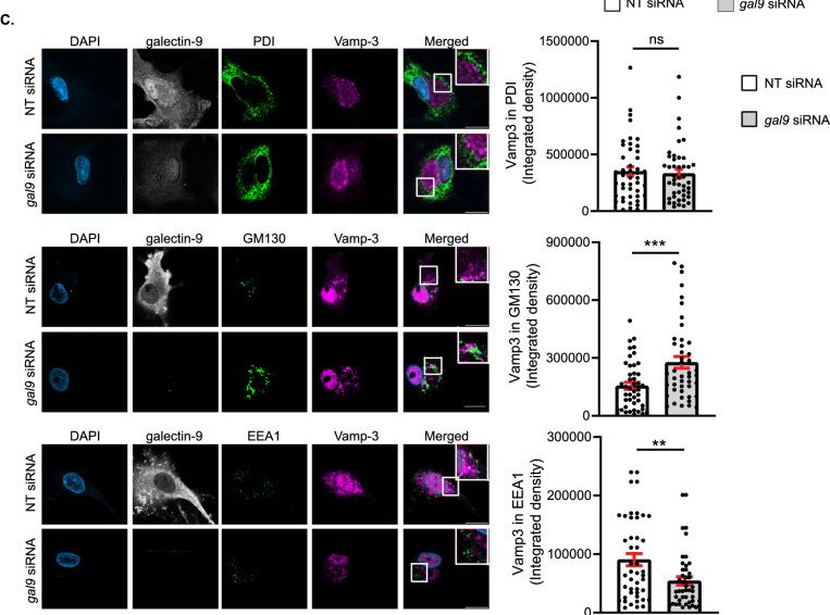
Detection of LILRB4/CD85k/ILT3 by Immunocytochemistry/ Immunofluorescence
Galectin-9 regulates Vamp-3 intracellular localisation. A NT and gal9 siRNA transfected moDCs were incubated with LPS for 4 h prior to being fixed in methanol and stained for Vamp-3, galectin-9 and DAPI for nuclear localisation. Representative images for each treatment are shown. Graphs: fluorescent cross-sections as indicated depicting Vamp-3 intensity across the cell. Scale bar: 10 µm. B Line scan quantification of cells shown in (A). Data represent mean Vamp-3 intensity for the specified distance from the nuclei ± SEM of one representative donor out of three. Fifteen cells were analysed per condition. Two-way ANOVA was performed for statistical analysis. C Representative images of DCs treated as in (A), fixed in 4% PFA and stained against galectin-9, Vamp-3, PDI, GM130, EEA1 and DAPI for nuclear localisation. Graph depicts Vamp-3 integrated density values for three independent donors ± SEM. Fifteen cells were analysed per donor. Unpaired t-test was conducted between NT and gal9 siRNA samples. Please note that PFA-fixed cells also show Vamp-3 nuclear staining as a result of the fixation process. n.s. p > 0.05; **p < 0.01; ***p < 0.001; ****p < 0.0001 Image collected and cropped by CiteAb from the following open publication (https://pubmed.ncbi.nlm.nih.gov/37755527), licensed under a CC-BY license. Not internally tested by R&D Systems.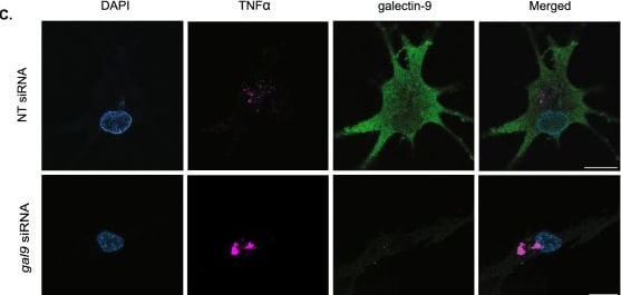
Detection of LILRB4/CD85k/ILT3 by Immunocytochemistry/ Immunofluorescence
Galectin-9-depleted moDCs eliminate intracellular cytokines via lysosomes. A, B Day 6 NT and gal9 siRNA-transfected moDCs were treated with LPS (1 µg/ml) alone or with Bafilomycin (200 µM) for 6 h after which cells were harvested and intracellular levels of TNF alpha determined by flow cytometry. Graph represents the % of TNF alpha positive cells (A) or the amount of TNF alpha present intracellularly represented by the mean fluorescence TNF alpha intensity (B). Graphs show mean value ± SEM of three independent donors. Paired students t-test was conducted between NT and gal9 siRNA-transfected cells. C Representative images from NT and gal9 siRNA moDCs treated as per (A). Scale bar: 10 µm. D Confocal microscopy quantification of the number of TNF alpha -positive cells (%) (left) and the integrated TNF alpha intensity of such cells (right) in NT and gal9 siRNA DCs treated with LPS and Bafilomycin. Data represent mean ± SEM of two independent donors. Unpaired Student's t-test was conducted between NT and gal9 siRNA-transfected cells. *p < 0.05; **p < 0.005 Image collected and cropped by CiteAb from the following open publication (https://pubmed.ncbi.nlm.nih.gov/37755527), licensed under a CC-BY license. Not internally tested by R&D Systems.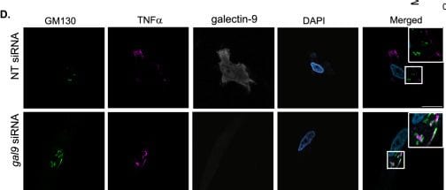
Detection of LILRB4/CD85k/ILT3 by Immunocytochemistry/ Immunofluorescence
Post-Golgi cytokine trafficking is impaired upon galectin-9 depletion. A Scheme depicting the experimental design based on the RUSH system. In the absence of biotin (left panel), the TNF alpha _GFP is trapped at the ER by the lumenal Str-KDEL hook and no cytokine trafficking post-ER can occur. When biotin is added (right panel), biotin outcompetes the interaction with streptavidin, allowing TNF alpha _GFP to freely traffic to the Golgi and the endosomal compartments. B NT and gal9 siRNA transfected moDCs were transfected with a GFP-labelled TNF alpha RUSH construct and treated with biotin to induce GFP-TNF alpha trafficking. Images represent snapshots of live-cell imaging GFP-TNF alpha at the indicated time points. Scale bar: 10 µm. C Quantification of images shown in (B). Data represent mean intensity ± SEM from two independent donors. D moDCs treated as per (B) were incubated with biotin for 30 min prior to being fixed and stained for GM130 as Golgi marker, galectin-9 and DAPI for nuclear localisation. Representative images are shown. Scale bar: 10 µm. E Representative line scans of cells shown in (D) depicting GM130 (yellow) and TNF alpha (in red) intensity across the cell. F. Mander's correlation coefficient was calculated for images shown in (D). Paired Student’s t-test was conducted between NT and gal9 siRNA transfected moDCs. Data represent mean correlation coefficient ± SEM of two independent donors. Fifteen cells were analysed for each condition per donor. ***p < 0.001 Image collected and cropped by CiteAb from the following open publication (https://pubmed.ncbi.nlm.nih.gov/37755527), licensed under a CC-BY license. Not internally tested by R&D Systems.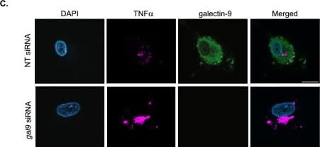
Detection of LILRB4/CD85k/ILT3 by Immunocytochemistry/ Immunofluorescence
Cytokines accumulate intracellularly upon galectin-9 depletion. A Day 5 NT and gal9 siRNA transfected moDCs were treated for 6 h with 1 µg/ml LPS alone or in combination with the Golgi inhibitors Brefeldin A (BFA, 10 µg/ml) and Monensin (Mon, 2 µM) prior to being harvested and the intracellular levels of TNF alpha analysed by flow cytometry. Dotted line shows TNF alpha levels in untreated cells. Dark grey depicts NT siRNA DCs treated with LPS (1 µg/ml) and light grey NT siRNA cells treated with LPS + the Golgi inhibitors. Green depicts gal9 siRNA DCs treated with LPS and magenta shows gal9 siRNA treated with LPS + Golgi inhibitors. B Quantification and statistical analysis of experiments depicted in (A). Results show the mean value ± SEM for four independent donors. Unpaired students t-test was conducted between NT and gal9 siRNA-transfected cells. C Representative images from results shown in (A) of cells treated with LPS only and stained against TNF alpha, galectin-9 and DAPI for nuclear localisation. Scale bar: 10 µm. D, E Confocal microscopy quantification of the number of TNF alpha positive cells (%) (D) and the integrated TNF alpha intensity of such cells (E) in NT and gal9 siRNA DCs treated with LPS. Data are representative of four independent donors. Unpaired Student's t-test was conducted between NT and gal9 siRNA-transfected cells. **p < 0.005, *p < 0.05 Image collected and cropped by CiteAb from the following open publication (https://pubmed.ncbi.nlm.nih.gov/37755527), licensed under a CC-BY license. Not internally tested by R&D Systems.Applications for Human LILRB4/CD85k/ILT3 Antibody
CyTOF-ready
Flow Cytometry
Sample: Human whole blood monocytes
Western Blot
Sample: Recombinant Human LILRB4/CD85k/ILT3
Reviewed Applications
Read 2 reviews rated 4 using AF2425 in the following applications:
Flow Cytometry Panel Builder
Bio-Techne Knows Flow Cytometry
Save time and reduce costly mistakes by quickly finding compatible reagents using the Panel Builder Tool.
Advanced Features
- Spectra Viewer - Custom analysis of spectra from multiple fluorochromes
- Spillover Popups - Visualize the spectra of individual fluorochromes
- Antigen Density Selector - Match fluorochrome brightness with antigen density

Formulation, Preparation, and Storage
Purification
Reconstitution
Reconstitute at 0.2 mg/mL in sterile PBS. For liquid material, refer to CoA for concentration.
Formulation
Shipping
Stability & Storage
- 12 months from date of receipt, -20 to -70 °C as supplied.
- 1 month, 2 to 8 °C under sterile conditions after reconstitution.
- 6 months, -20 to -70 °C under sterile conditions after reconstitution.
Calculators
Background: LILRB4/CD85k/ILT3
Long Name
Alternate Names
Gene Symbol
UniProt
Additional LILRB4/CD85k/ILT3 Products
Product Documents for Human LILRB4/CD85k/ILT3 Antibody
Certificate of Analysis
To download a Certificate of Analysis, please enter a lot or batch number in the search box below.
Note: Certificate of Analysis not available for kit components.
Product Specific Notices for Human LILRB4/CD85k/ILT3 Antibody
For research use only
Citations for Human LILRB4/CD85k/ILT3 Antibody
 Powered by Bioz
Customer Reviews for Human LILRB4/CD85k/ILT3 Antibody (2)
Have you used Human LILRB4/CD85k/ILT3 Antibody?
Submit a review and receive an Amazon gift card!
$25/€18/£15/$25CAN/¥2500 for a review with an image
$10/€7/£6/$10CAN/¥1110 for a review without an image
Submit a review
Customer Images
-
Application: ELISASample Tested: SerumSpecies: HumanVerified Customer | Posted 11/29/2022

-
Application: ELISASample Tested: Serum and Plasma and AML-193 human acute monocytic leukemia cell lineSpecies: HumanVerified Customer | Posted 07/07/2020
There are no reviews that match your criteria.
Protocols
Find general support by application which include: protocols, troubleshooting, illustrated assays, videos and webinars.
- 7-Amino Actinomycin D (7-AAD) Cell Viability Flow Cytometry Protocol
- Cellular Response to Hypoxia Protocols
- Extracellular Membrane Flow Cytometry Protocol
- Flow Cytometry Protocol for Cell Surface Markers
- Flow Cytometry Protocol for Staining Membrane Associated Proteins
- Flow Cytometry Staining Protocols
- Flow Cytometry Troubleshooting Guide
- Intracellular Flow Cytometry Protocol Using Alcohol (Methanol)
- Intracellular Flow Cytometry Protocol Using Detergents
- Intracellular Nuclear Staining Flow Cytometry Protocol Using Detergents
- Intracellular Staining Flow Cytometry Protocol Using Alcohol Permeabilization
- Intracellular Staining Flow Cytometry Protocol Using Detergents to Permeabilize Cells
- Propidium Iodide Cell Viability Flow Cytometry Protocol
- Protocol for Liperfluo
- Protocol for the Characterization of Human Th22 Cells
- Protocol for the Characterization of Human Th9 Cells
- Protocol: Annexin V and PI Staining by Flow Cytometry
- Protocol: Annexin V and PI Staining for Apoptosis by Flow Cytometry
- R&D Systems Quality Control Western Blot Protocol
- Troubleshooting Guide: Fluorokine Flow Cytometry Kits
- Troubleshooting Guide: Western Blot Figures
- Western Blot Conditions
- Western Blot Protocol
- Western Blot Protocol for Cell Lysates
- Western Blot Troubleshooting
- Western Blot Troubleshooting Guide
- View all Protocols, Troubleshooting, Illustrated assays and Webinars