As the first member of membrane type (MT) MMPs, MMP-14, also known as MT1-MMP, plays an important role in extracellular matrix (ECM) remodeling by being able to degrade type I collagen, activate pro-MMP-2 and process cell adhesion molecules such as CD44 and Integrin alpha V (1). MMP-14 is therefore a key enzyme in many physiological and pathological processes such as angiogenesis and tumor invasion. Structurally, MMP-14 consists of the following domains: a pro domain containing the furin cleavage site, a catalytic domain containing the zinc-binding site, a hinge region, a hemopexin-like domain, a transmembrane domain, and a cytoplamasic tail (2).
Human MMP-14/MT1-MMP Antibody (5H2)
R&D Systems | Catalog # MAB918

Key Product Details
Species Reactivity
Validated:
Human
Cited:
Human
Applications
Validated:
Immunohistochemistry, Western Blot, Immunoprecipitation
Cited:
Western Blot, Immunocytochemistry
Label
Unconjugated
Antibody Source
Monoclonal Mouse IgG1 Clone # 5H2
Loading...
Product Specifications
Immunogen
Human MMP-14 synthetic peptide
AYIREGHEKQA
Accession # P50281
AYIREGHEKQA
Accession # P50281
Specificity
Detects recombinant human (rh) MMP-14 in Western blots. In Western blots, no cross-reactivity with recombinant human (rh) MMP-1, -2, -7, -8, -9, -10, -12, -13 or the catalytic domains of rhMMP-15 and -16.
Clonality
Monoclonal
Host
Mouse
Isotype
IgG1
Scientific Data Images for Human MMP-14/MT1-MMP Antibody (5H2)
Detection of Human MMP‑14/MT1‑MMP by Western Blot.
Western blot shows lysates of human placenta tissue. PVDF membrane was probed with 2 µg/mL of Mouse Anti-Human MMP-14/MT1-MMP Monoclonal Antibody (Catalog # MAB918) followed by HRP-conjugated Anti-Mouse IgG Secondary Antibody (Catalog # HAF018). Specific bands were detected for MMP-14/MT1-MMP at approximately 50 kDa and 62 kDa (as indicated). This experiment was conducted under reducing conditions and using Immunoblot Buffer Group 1.
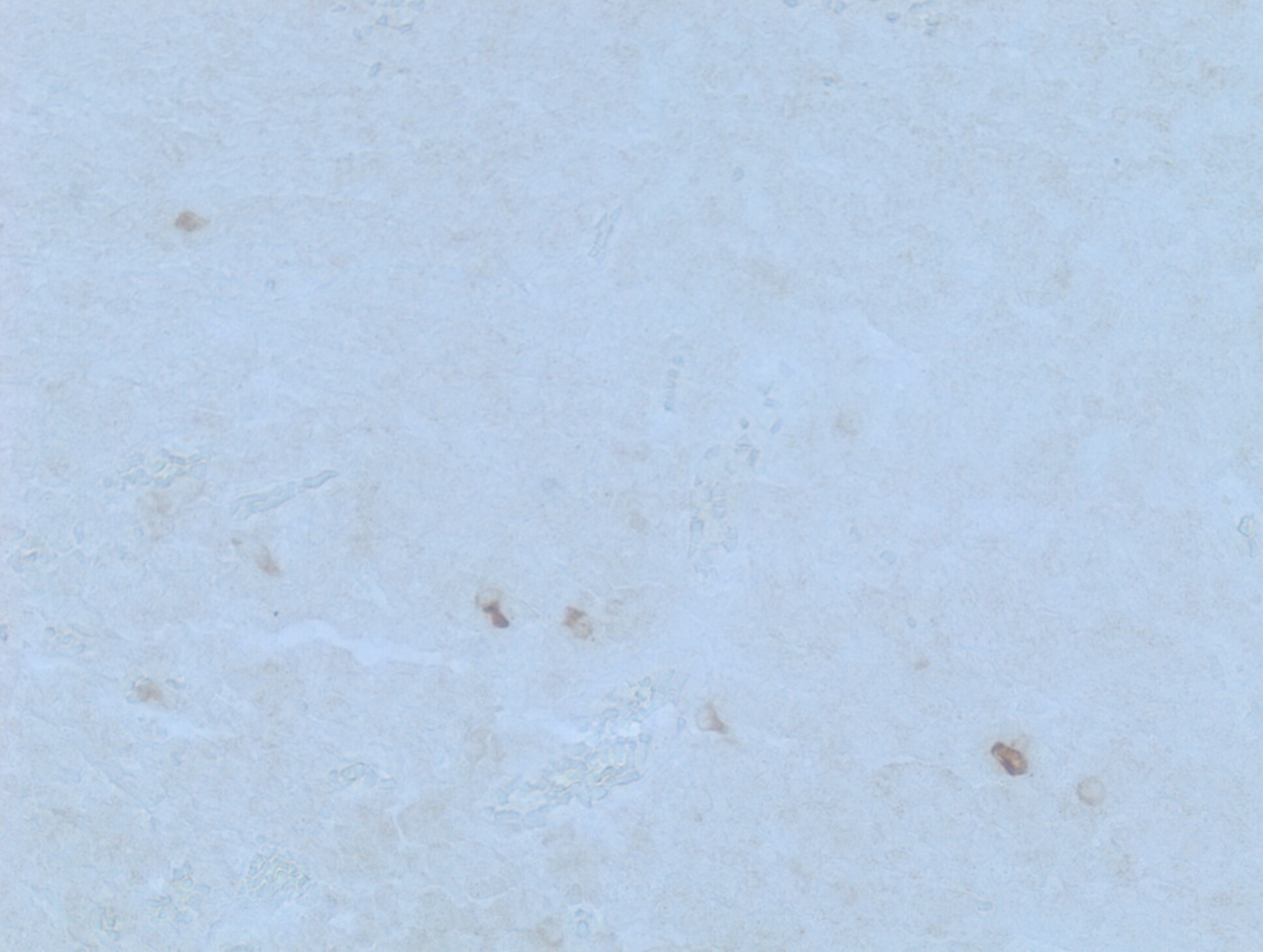
MMP‑14 in Human Benign Nodular Hyperplasia.
MMP-14 was detected in immersion fixed paraffin-embedded sections of human benign nodular hyperplasia using 25 µg/mL Mouse Anti-Human MMP-14 Monoclonal Antibody (Catalog # MAB918) overnight at 4 °C. Tissue was stained with the Anti-Mouse HRP-AEC Cell & Tissue Staining Kit (red; Catalog # CTS003) and counterstained with hematoxylin (blue). View our protocol for Chromogenic IHC Staining of Paraffin-embedded Tissue Sections.Applications for Human MMP-14/MT1-MMP Antibody (5H2)
Application
Recommended Usage
Immunohistochemistry
8-25 µg/mL
Sample: Immersion fixed paraffin-embedded sections of human benign nodular hyperplasia
Sample: Immersion fixed paraffin-embedded sections of human benign nodular hyperplasia
Immunoprecipitation
25 µg/mL
Sample: Conditioned cell culture medium spiked with Recombinant Human MMP‑14 (Catalog # 918‑MP), see our available Western blot detection antibodies
Sample: Conditioned cell culture medium spiked with Recombinant Human MMP‑14 (Catalog # 918‑MP), see our available Western blot detection antibodies
Western Blot
2 µg/mL
Sample: Human placenta tissue
Sample: Human placenta tissue
Reviewed Applications
Read 1 review rated 4 using MAB918 in the following applications:
Formulation, Preparation, and Storage
Purification
Protein A or G purified from hybridoma culture supernatant
Reconstitution
Reconstitute at 0.5 mg/mL in sterile PBS. For liquid material, refer to CoA for concentration.
Loading...
Formulation
Lyophilized from a 0.2 μm filtered solution in TBS with Trehalose. *Small pack size (SP) is supplied either lyophilized or as a 0.2 µm filtered solution in PBS.
Shipping
Lyophilized product is shipped at ambient temperature. Liquid small pack size (-SP) is shipped with polar packs. Upon receipt, store immediately at the temperature recommended below.
Stability & Storage
Use a manual defrost freezer and avoid repeated freeze-thaw cycles.
- 12 months from date of receipt, -20 to -70 °C as supplied.
- 1 month, 2 to 8 °C under sterile conditions after reconstitution.
- 6 months, -20 to -70 °C under sterile conditions after reconstitution.
Calculators
Background: MMP-14/MT1-MMP
References
- Seike, M. (2003) Cancer Lett. 194:1.
- Sato, H. et al. (1994) Nature 370:61.
Long Name
Matrix Metalloproteinase 14/Membrane Type 1 MMP
Alternate Names
MMP-X1, MMP14, MT-MMP1, MT1-MMP
Entrez Gene IDs
4323 (Human)
Gene Symbol
MMP14
UniProt
Additional MMP-14/MT1-MMP Products
Product Documents for Human MMP-14/MT1-MMP Antibody (5H2)
Certificate of Analysis
To download a Certificate of Analysis, please enter a lot or batch number in the search box below.
Note: Certificate of Analysis not available for kit components.
Product Specific Notices for Human MMP-14/MT1-MMP Antibody (5H2)
For research use only
Related Research Areas
Citations for Human MMP-14/MT1-MMP Antibody (5H2)
 Powered by Bioz
Customer Reviews for Human MMP-14/MT1-MMP Antibody (5H2) (1)
4 out of 5
1 Customer Rating
Have you used Human MMP-14/MT1-MMP Antibody (5H2)?
Submit a review and receive an Amazon gift card!
$25/€18/£15/$25CAN/¥2500 Yen for a review with an image
$10/€7/£6/$10CAN/¥1110 Yen for a review without an image
Submit a review
Customer Images
Showing
1
-
1 of
1 review
Showing All
Filter By:
-
Application: Immunohistochemistry-ParaffinSample Tested: TonsilSpecies: HumanVerified Customer | Posted 04/15/2019Human tonsil FFPE tissue stained with anti-MMP14 antibody after HIER (pH6)

There are no reviews that match your criteria.
Protocols
Find general support by application which include: protocols, troubleshooting, illustrated assays, videos and webinars.
- Antigen Retrieval Protocol (PIER)
- Antigen Retrieval for Frozen Sections Protocol
- Appropriate Fixation of IHC/ICC Samples
- Cellular Response to Hypoxia Protocols
- Chromogenic IHC Staining of Formalin-Fixed Paraffin-Embedded (FFPE) Tissue Protocol
- Chromogenic Immunohistochemistry Staining of Frozen Tissue
- ClariTSA™ Fluorophore Kits
- Detection & Visualization of Antibody Binding
- Fluorescent IHC Staining of Frozen Tissue Protocol
- Graphic Protocol for Heat-induced Epitope Retrieval
- Graphic Protocol for the Preparation and Fluorescent IHC Staining of Frozen Tissue Sections
- Graphic Protocol for the Preparation and Fluorescent IHC Staining of Paraffin-embedded Tissue Sections
- Graphic Protocol for the Preparation of Gelatin-coated Slides for Histological Tissue Sections
- IHC Sample Preparation (Frozen sections vs Paraffin)
- Immunofluorescent IHC Staining of Formalin-Fixed Paraffin-Embedded (FFPE) Tissue Protocol
- Immunohistochemistry (IHC) and Immunocytochemistry (ICC) Protocols
- Immunohistochemistry Frozen Troubleshooting
- Immunohistochemistry Paraffin Troubleshooting
- Immunoprecipitation Protocol
- Preparing Samples for IHC/ICC Experiments
- Preventing Non-Specific Staining (Non-Specific Binding)
- Primary Antibody Selection & Optimization
- Protocol for Heat-Induced Epitope Retrieval (HIER)
- Protocol for Making a 4% Formaldehyde Solution in PBS
- Protocol for VisUCyte™ HRP Polymer Detection Reagent
- Protocol for the Preparation & Fixation of Cells on Coverslips
- Protocol for the Preparation and Chromogenic IHC Staining of Frozen Tissue Sections
- Protocol for the Preparation and Chromogenic IHC Staining of Frozen Tissue Sections - Graphic
- Protocol for the Preparation and Chromogenic IHC Staining of Paraffin-embedded Tissue Sections
- Protocol for the Preparation and Chromogenic IHC Staining of Paraffin-embedded Tissue Sections - Graphic
- Protocol for the Preparation and Fluorescent IHC Staining of Frozen Tissue Sections
- Protocol for the Preparation and Fluorescent IHC Staining of Paraffin-embedded Tissue Sections
- Protocol for the Preparation of Gelatin-coated Slides for Histological Tissue Sections
- R&D Systems Quality Control Western Blot Protocol
- TUNEL and Active Caspase-3 Detection by IHC/ICC Protocol
- The Importance of IHC/ICC Controls
- Troubleshooting Guide: Immunohistochemistry
- Troubleshooting Guide: Western Blot Figures
- Western Blot Conditions
- Western Blot Protocol
- Western Blot Protocol for Cell Lysates
- Western Blot Troubleshooting
- Western Blot Troubleshooting Guide
- View all Protocols, Troubleshooting, Illustrated assays and Webinars
Loading...