
Insulin is a disulfide-linked heterodimeric protein secreted by the pancreatic Islets of Langerhans. Mature insulin is generated by the proteolytic removal of a peptide from proinsulin. It is involved in the regulation of glucose metabolism through interactions with the Insulin Receptor.
Insulin Antibody (182410)
R&D Systems | Catalog # MAB1417


Scientific images may be resized, cropped, or adjusted for brightness or contrast for presentation purposes. These updates are limited to presentation and do not affect the underlying scientific interpretation of the data. Where available, additional source data may be provided upon request.
Loading...
Key Product Details
Species Reactivity
Validated:
Human, Mouse, Bovine
Cited:
Human, Mouse, Transgenic Mouse
Applications
Validated:
Immunohistochemistry, Immunocytochemistry
Cited:
Immunohistochemistry, Immunohistochemistry-Paraffin, Immunohistochemistry-Frozen, Flow Cytometry, Immunocytochemistry
Label
Unconjugated
Antibody Source
Monoclonal Rat IgG2A Clone # 182410
Loading...
Product Specifications
Immunogen
E. coli-derived recombinant human Insulin
Specificity
Detects bovine, human, and mouse insulin.
Clonality
Monoclonal
Host
Rat
Isotype
IgG2A
Scientific Data Images for Insulin Antibody (182410)
Insulin in beta TC‑6 Mouse Cell Line.
Insulin was detected in immersion fixed beta TC-6 mouse beta cell insulinoma cell line using Human/Mouse/Bovine Insulin Monoclonal Antibody (Catalog # MAB1417) at 10 µg/mL for 3 hours at room temperature. Cells were stained using the NorthernLights™ 557-conjugated Anti-Rat IgG Secondary Antibody (red; NL013) and counterstained with DAPI (blue). View our protocol for Fluorescent ICC Staining of Cells on Coverslips.
Insulin in Human Pancreas.
Insulin was detected in immersion fixed paraffin-embedded sections of human pancreas using Rat Anti-Human/Mouse/Bovine Insulin Monoclonal Antibody (Catalog # MAB1417) at 0.5 µg/mL for 1 hour at room temperature followed by incubation with the Anti-Rat IgG VisUCyte™ HRP Polymer Antibody (VC005). Before incubation with the primary antibody, tissue was subjected to heat-induced epitope retrieval using Antigen Retrieval Reagent-Basic (CTS013). Tissue was stained using DAB (brown) and counterstained with hematoxylin (blue). Specific staining was localized to cytoplasm and plasma membrane in islet cells. Staining was performed using our protocol for IHC Staining with VisUCyte HRP Polymer Detection Reagents.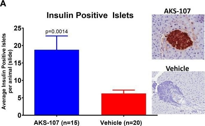
Detection of Insulin by Immunohistochemistry
Preservation of pancreatic islet insulin-producing beta cells from female NOD mice treated with AKS-107. Four to 6 week-old female NOD mice received i.p. injections of Vehicle or AKS-107 at 2 mg/kg twice weekly for 16 weeks (see Figure 8E for diabetes incidence) in which mice were sacrificed at 26 weeks of age for insulin- and C-peptide-specific cell staining via immunohistochemistry analyses of whole pancreas samples. Briefly, whole pancreata were harvested, formalin fixed, embedded, and sectioned such that longitudinal slices of pancreas were placed on slides. Sections were stained for insulin using a rat anti-insulin primary Ab (R & D systems, #MAB1417) and a donkey anti-rat IgG secondary Ab conjugated to Alexa488, and for leukocyte infiltration (insulitis) using Hematoxylin. Slides were scanned on an Aperio whole slide scanner (20x magnification), and then analyzed in a blinded fashion. One slide was analyzed per animal and for each animal the number of insulin islets per slide were counted. Mean insulin (A) and C-peptide (B) positive islets per slide per animal are shown (p, student’s t-test) beside representative images of positive (AKS-107-treated) and negative (Vehicle-treated) islet samples. Image collected and cropped by CiteAb from the following open publication (https://www.frontiersin.org/articles/10.3389/fimmu.2024.1367514/full), licensed under a CC-BY license. Not internally tested by R&D Systems.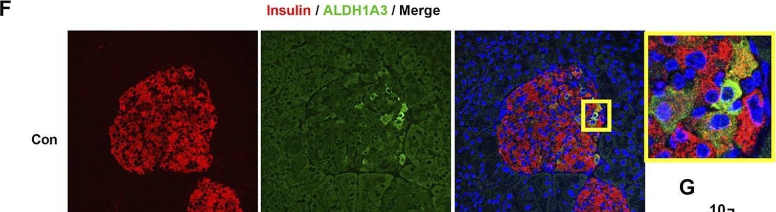
Detection of Insulin by Immunohistochemistry
SZ-A amended beta -cell dysfunction in type 2 diabetic KKAy mice. (A,B) An intraperitoneal glucose-stimulated insulin secretion test (IPGSIST) was executed in KKAy mice after treating with SZ-A (200 mg/kg) for 8 weeks (n = 5 mice per group). (A) Blood glucose and insulin levels at 0 min before injection of glucose. (B) Blood glucose and the percentage of increased blood insulin at 30 min after glucose (2.0 g/kg) load. (C–G) Immunohistochemistry of the pancreas of KKAy mice in the Con group and SZ-A (200 mg/kg) group (n = 5 mice per group). beta Cells were labeled with insulin antibody (red), alpha cells were labeled with glucagon antibody (green), dedifferentiated cells were labeled with ALDH1A3 antibody (green). (D) The percentage of beta -cell/islet area; (E) the percentage of alpha -cell/islet area; and (G) the ratio of ALDH1A3-positive beta cells/ beta cells. All data are expressed as the mean ± SEM. *p < 0.05, **p < 0.01, ***p < 0.001, vs. the Con group. Image collected and cropped by CiteAb from the following open publication (https://www.frontiersin.org/articles/10.3389/fphar.2022.841981/full), licensed under a CC-BY license. Not internally tested by R&D Systems.Applications for Insulin Antibody (182410)
Application
Recommended Usage
Immunocytochemistry
8-25 µg/mL
Sample: Immersion fixed beta TC-6 mouse beta cell insulinoma cell line
Sample: Immersion fixed beta TC-6 mouse beta cell insulinoma cell line
Immunohistochemistry
0.5-25 µg/mL
Sample: Immersion fixed paraffin-embedded sections of human pancreas
Sample: Immersion fixed paraffin-embedded sections of human pancreas
Formulation, Preparation, and Storage
Purification
Protein A or G purified from hybridoma culture supernatant
Reconstitution
Reconstitute at 0.5 mg/mL in sterile PBS. For liquid material, refer to CoA for concentration.
Loading...
Formulation
Lyophilized from a 0.2 μm filtered solution in PBS with Trehalose. See Certificate of Analysis for details.<br/>*Small pack size (-SP) is supplied either lyophilized or as a 0.2 µm filtered solution in PBS.
Shipping
Lyophilized product is shipped at ambient temperature. Liquid small pack size (-SP) is shipped with polar packs. Upon receipt, store immediately at the temperature recommended below.
Stability & Storage
Use a manual defrost freezer and avoid repeated freeze-thaw cycles.
- 12 months from date of receipt, -20 to -70 °C as supplied.
- 1 month, 2 to 8 °C under sterile conditions after reconstitution.
- 6 months, -20 to -70 °C under sterile conditions after reconstitution.
Calculators
Background: Insulin
Alternate Names
IDDM2, ILPR, INS, IRDN, MODY10
Gene Symbol
INS
Additional Insulin Products
Product Documents for Insulin Antibody (182410)
Certificate of Analysis
To download a Certificate of Analysis, please enter a lot or batch number in the search box below.
Note: Certificate of Analysis not available for kit components.
Product Specific Notices for Insulin Antibody (182410)
For research use only
Citations for Insulin Antibody (182410)
 Powered by Bioz
Customer Reviews for Insulin Antibody (182410)
There are currently no reviews for this product. Be the first to review Insulin Antibody (182410) and earn rewards!
Have you used Insulin Antibody (182410)?
Submit a review and receive an Amazon gift card!
$25/€18/£15/$25CAN/¥2500 for a review with an image
$10/€7/£6/$10CAN/¥1110 for a review without an image
Submit a review
Protocols
Find general support by application which include: protocols, troubleshooting, illustrated assays, videos and webinars.
- Antigen Retrieval Protocol (PIER)
- Antigen Retrieval for Frozen Sections Protocol
- Appropriate Fixation of IHC/ICC Samples
- Cellular Response to Hypoxia Protocols
- Chromogenic IHC Staining of Formalin-Fixed Paraffin-Embedded (FFPE) Tissue Protocol
- Chromogenic Immunohistochemistry Staining of Frozen Tissue
- ClariTSA™ Fluorophore Kits
- Detection & Visualization of Antibody Binding
- Fluorescent IHC Staining of Frozen Tissue Protocol
- Graphic Protocol for Heat-induced Epitope Retrieval
- Graphic Protocol for the Preparation and Fluorescent IHC Staining of Frozen Tissue Sections
- Graphic Protocol for the Preparation and Fluorescent IHC Staining of Paraffin-embedded Tissue Sections
- Graphic Protocol for the Preparation of Gelatin-coated Slides for Histological Tissue Sections
- ICC Cell Smear Protocol for Suspension Cells
- ICC Immunocytochemistry Protocol Videos
- ICC for Adherent Cells
- IHC Sample Preparation (Frozen sections vs Paraffin)
- Immunocytochemistry (ICC) Protocol
- Immunocytochemistry Troubleshooting
- Immunofluorescence of Organoids Embedded in Cultrex Basement Membrane Extract
- Immunofluorescent IHC Staining of Formalin-Fixed Paraffin-Embedded (FFPE) Tissue Protocol
- Immunohistochemistry (IHC) and Immunocytochemistry (ICC) Protocols
- Immunohistochemistry Frozen Troubleshooting
- Immunohistochemistry Paraffin Troubleshooting
- Preparing Samples for IHC/ICC Experiments
- Preventing Non-Specific Staining (Non-Specific Binding)
- Primary Antibody Selection & Optimization
- Protocol for Heat-Induced Epitope Retrieval (HIER)
- Protocol for Making a 4% Formaldehyde Solution in PBS
- Protocol for VisUCyte™ HRP Polymer Detection Reagent
- Protocol for the Fluorescent ICC Staining of Cell Smears - Graphic
- Protocol for the Fluorescent ICC Staining of Cultured Cells on Coverslips - Graphic
- Protocol for the Preparation & Fixation of Cells on Coverslips
- Protocol for the Preparation and Chromogenic IHC Staining of Frozen Tissue Sections
- Protocol for the Preparation and Chromogenic IHC Staining of Frozen Tissue Sections - Graphic
- Protocol for the Preparation and Chromogenic IHC Staining of Paraffin-embedded Tissue Sections
- Protocol for the Preparation and Chromogenic IHC Staining of Paraffin-embedded Tissue Sections - Graphic
- Protocol for the Preparation and Fluorescent ICC Staining of Cells on Coverslips
- Protocol for the Preparation and Fluorescent ICC Staining of Non-adherent Cells
- Protocol for the Preparation and Fluorescent ICC Staining of Stem Cells on Coverslips
- Protocol for the Preparation and Fluorescent IHC Staining of Frozen Tissue Sections
- Protocol for the Preparation and Fluorescent IHC Staining of Paraffin-embedded Tissue Sections
- Protocol for the Preparation of Gelatin-coated Slides for Histological Tissue Sections
- Protocol for the Preparation of a Cell Smear for Non-adherent Cell ICC - Graphic
- TUNEL and Active Caspase-3 Detection by IHC/ICC Protocol
- The Importance of IHC/ICC Controls
- Troubleshooting Guide: Immunohistochemistry
- View all Protocols, Troubleshooting, Illustrated assays and Webinars


