
Key Product Details
Species Reactivity
Validated:
Human, Mouse
Cited:
Human, Mouse, Rat, Porcine, Bovine, Transgenic Mouse
Applications
Validated:
Immunohistochemistry, Blockade of Receptor-ligand Interaction
Cited:
Immunohistochemistry, Immunohistochemistry-Paraffin, Western Blot, Neutralization, Immunocytochemistry, Functional Assay
Label
Unconjugated
Antibody Source
Polyclonal Goat IgG
Loading...
Product Specifications
Immunogen
Mouse myeloma cell line NS0-derived recombinant mouse Gremlin
Lys25-Asp184
Accession # O70326
Lys25-Asp184
Accession # O70326
Specificity
Detects human and mouse Gremlin in direct ELISAs.
Clonality
Polyclonal
Host
Goat
Isotype
IgG
Endotoxin Level
<0.10 EU per 1 μg of the antibody by the LAL method.
Scientific Data Images for Gremlin Antibody
Gremlin in Human Breast Cancer Tissue.
Gremlin was detected in immersion fixed paraffin-embedded sections of human breast cancer tissue using Goat Anti-Human/Mouse Gremlin Antigen Affinity-purified Polyclonal Antibody (Catalog # AF956) at 3 µg/mL for 1 hour at room temperature followed by incubation with the Anti-Goat IgG VisUCyte™ HRP Polymer Antibody (Catalog # VC004). Before incubation with the primary antibody, tissue was subjected to heat-induced epitope retrieval using Antigen Retrieval Reagent-Basic (Catalog # CTS013). Tissue was stained using DAB (brown) and counterstained with hematoxylin (blue). Specific staining was localized to cytoplasm. View our protocol for IHC Staining with VisUCyte HRP Polymer Detection Reagents.
Gremlin in Embryonic Mouse Ribs.
Gremlin was detected in immersion fixed frozen sections of embryonic mouse ribs (15 d.p.c.) using 15 µg/mL Goat Anti-Human/Mouse Gremlin Antigen Affinity-purified Polyclonal Antibody (Catalog # AF956) overnight at 4 °C. Tissue was stained with the Anti-Goat HRP-DAB Cell & Tissue Staining Kit (brown; Catalog # CTS008) and counterstained with hematoxylin (blue). View our protocol for Chromogenic IHC Staining of Frozen Tissue Sections.
Detection of Gremlin by Immunohistochemistry
Inverse relationship between expression of Grem1 protein and pSmad1/5 staining in mouse intestine. Sequential FFPE sections (5 μm) of mouse intestine were stained for Grem1 (A) or pSmad1/5 (B) as described in Methods. (C) Positively stained cells (empty bars) or villi tips (filled bars) were quantified in the indicated regions using Image J and data were plotted as mean pixel intensity –/+ SEM (n = 3 mice, 3 independent regions of intestine quantified per mouse). *p< 0.05, ***p< 0.001 using two-way ANOVA and Bonferroni post hoc test. Image collected and cropped by CiteAb from the following open publication (https://pubmed.ncbi.nlm.nih.gov/31384391), licensed under a CC-BY license. Not internally tested by R&D Systems.
Detection of Gremlin by Immunohistochemistry
Distinct pattern of endogenous Grem1 expression in mouse colon. Sections (5 μm) from FFPE colon samples (n = 4) from wild-type or Grem1-/- mice were processed for in situ hybridisation (A left) and immunohistochemistry (A right; B.) as described in Methods. Positive Grem1 mRNA and protein staining was imaged using DAB (brown) and sections were counterstained using haematoxylin and imaged using PathXL. (A) Grem1 mRNA is visible as brown, punctate staining in the muscularis mucosa layer. Scale bars 100 μm. (B) Grem1 protein staining is evident as brown staining in the muscularis layer and the base of the colonic crypts (left; scale bar 100 μm). Grem1 protein staining in the musclaris layer as well as cells of the colonic crypt. CBCC, crypt base columnar cells; P, Paneth cells; TA, transit-amplifying cells. Image collected and cropped by CiteAb from the following open publication (https://pubmed.ncbi.nlm.nih.gov/31384391), licensed under a CC-BY license. Not internally tested by R&D Systems.
Detection of Gremlin by Western Blot
VEGFR2-MEK-ERK axis mediated the enhancement of FAO from Gremlin 1. (A). Cell viability assay of human intestinal fibroblast cells CCD-18Co and CCD-112Co cells treated with 200 ng/ml Gremlin 1 and Rapamycin (10 nM), LY3214996 (10 nM) or stattic (10 μM) for 24 h. (B). Immunoblotting analysis of VEGFR2-MEK-ERK signaling in human intestinal fibroblast cells CCD-18Co and CCD-112Co cells treated with 200 ng/ml Gremlin 1 and LY3214996 (10 nM) for 24 h. Actin acted as loading control and re-used for illustrative purposes. (C). Immunofluorescence staining of a-SMA in human intestinal fibroblast cells CCD-18Co and CCD-112Co cells treated with 200 ng/ml Gremlin 1 and LY3214996 (10 nM). (D). The intensity statistical results of a-SMA immunofluorescence staining from five random fields of slides. E. OCR measurement of CCD-18Co and CCD-112Co cells treated with 200 ng/ml Gremlin 1 and LY3214996 (10 nM) for 24 h. p values are derived from one-way ANOVA analysis. Dunnett’s test was used to analysis the difference between control group to the other groups. **p ≤ 0.01; ***p ≤ 0.001. Image collected and cropped by CiteAb from the following open publication (https://pubmed.ncbi.nlm.nih.gov/33967807), licensed under a CC-BY license. Not internally tested by R&D Systems.
Detection of Gremlin by Western Blot
BMP2, TGF-beta 1 and BMP8A show different effects on the expressional induction of Nog, Grem1 and Grem2. C3H10T1/2 cells were treated with BMP2 (1 nM), TGF-beta 1 (0.3 nM), BMP2 plus with TGF-beta 1 or BMP8A (1 nM) for 24 h. The transcript levels (upper panel) and encoding protein amounts (lower panel) of Nog (A), Grem1 (B) and Grem2 (C) were then evaluated. beta -actin amounts served as loading controls in immunoblotting. D To explore how BMP8A induces Grem1 expression, cells were treated with BMP8A in the absence or presence of SB431542 (SB) or dorsomorphin (DM) as indicated. Quantification data are presented as means ± SEM. *P < 0.05, ***P < 0.001 compared to no-treatment control. # P < 0.001 compared between indicated groups Image collected and cropped by CiteAb from the following open publication (https://pubmed.ncbi.nlm.nih.gov/36443839), licensed under a CC-BY license. Not internally tested by R&D Systems.
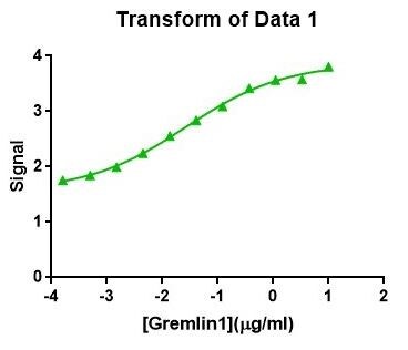
Mouse Gremlin ELISA Standard Curve
Recombinant Mouse Gremlin (Catalog # 956-GR) was serially diluted and captured by Rat Anti-Mouse Gremlin Monoclonal Antibody (Catalog # MAB956) coated on a Clear Polystyrene Microplate (Catalog # DY990). Goat Anti-Human/Mouse Gremlin Antigen Affinity-purified Polyclonal Antibody (Catalog # AF956) was biotinylated and incubated with the protein captured on the plate. Detection of the standard curve was achieved by incubating Streptavidin-HRP (Catalog # DY998)Applications for Gremlin Antibody
Application
Recommended Usage
Blockade of Receptor-ligand Interaction
Immunohistochemistry
3-15 µg/mL
Sample: Immersion fixed frozen sections of embryonic mouse ribs (15 d.p.c.), and immersion fixed paraffin-embedded sections of human breast cancer tissue
Sample: Immersion fixed frozen sections of embryonic mouse ribs (15 d.p.c.), and immersion fixed paraffin-embedded sections of human breast cancer tissue
Reviewed Applications
Read 2 reviews rated 4 using AF956 in the following applications:
Formulation, Preparation, and Storage
Purification
Antigen Affinity-purified
Reconstitution
Reconstitute at 0.2 mg/mL in sterile PBS. For liquid material, refer to CoA for concentration.
Loading...
Formulation
Lyophilized from a 0.2 μm filtered solution in PBS with Trehalose. *Small pack size (SP) is supplied either lyophilized or as a 0.2 µm filtered solution in PBS.
Shipping
Lyophilized product is shipped at ambient temperature. Liquid small pack size (-SP) is shipped with polar packs. Upon receipt, store immediately at the temperature recommended below.
Stability & Storage
Use a manual defrost freezer and avoid repeated freeze-thaw cycles.
- 12 months from date of receipt, -20 to -70 °C as supplied.
- 1 month, 2 to 8 °C under sterile conditions after reconstitution.
- 6 months, -20 to -70 °C under sterile conditions after reconstitution.
Calculators
Background: Gremlin
References
- Hsu, D.R. et al. (1998) Mol. Cell 1:673.
- Merino, R. et al. (1999) Development 126:5515.
- Shi, W. et al. (2001) Am. J. Physiol. Lung Cell Mol. Physiol. 280:L1030.
- Topol, L.Z. et al. (2000) J. Biol. Chem. 275:8785.
Alternate Names
DAND2, DRM, GREM1
Gene Symbol
GREM1
UniProt
Additional Gremlin Products
Product Documents for Gremlin Antibody
Certificate of Analysis
To download a Certificate of Analysis, please enter a lot or batch number in the search box below.
Note: Certificate of Analysis not available for kit components.
Product Specific Notices for Gremlin Antibody
For research use only
Related Research Areas
Citations for Gremlin Antibody
 Powered by Bioz
Customer Reviews for Gremlin Antibody (2)
4 out of 5
2 Customer Ratings
Have you used Gremlin Antibody?
Submit a review and receive an Amazon gift card!
$25/€18/£15/$25CAN/¥2500 Yen for a review with an image
$10/€7/£6/$10CAN/¥1110 Yen for a review without an image
Submit a review
Customer Images
Showing
1
-
2 of
2 reviews
Showing All
Filter By:
-
Application: ELISASample Tested: Recombinant proteinSpecies: HumanVerified Customer | Posted 07/09/2018

-
Application: Immunohistochemistry-ParaffinSample Tested: See PMID 23832098Species: RatVerified Customer | Posted 01/12/2015
There are no reviews that match your criteria.
Protocols
Find general support by application which include: protocols, troubleshooting, illustrated assays, videos and webinars.
- Antigen Retrieval Protocol (PIER)
- Antigen Retrieval for Frozen Sections Protocol
- Appropriate Fixation of IHC/ICC Samples
- Cellular Response to Hypoxia Protocols
- Chromogenic IHC Staining of Formalin-Fixed Paraffin-Embedded (FFPE) Tissue Protocol
- Chromogenic Immunohistochemistry Staining of Frozen Tissue
- ClariTSA™ Fluorophore Kits
- Detection & Visualization of Antibody Binding
- Fluorescent IHC Staining of Frozen Tissue Protocol
- Graphic Protocol for Heat-induced Epitope Retrieval
- Graphic Protocol for the Preparation and Fluorescent IHC Staining of Frozen Tissue Sections
- Graphic Protocol for the Preparation and Fluorescent IHC Staining of Paraffin-embedded Tissue Sections
- Graphic Protocol for the Preparation of Gelatin-coated Slides for Histological Tissue Sections
- IHC Sample Preparation (Frozen sections vs Paraffin)
- Immunofluorescent IHC Staining of Formalin-Fixed Paraffin-Embedded (FFPE) Tissue Protocol
- Immunohistochemistry (IHC) and Immunocytochemistry (ICC) Protocols
- Immunohistochemistry Frozen Troubleshooting
- Immunohistochemistry Paraffin Troubleshooting
- Preparing Samples for IHC/ICC Experiments
- Preventing Non-Specific Staining (Non-Specific Binding)
- Primary Antibody Selection & Optimization
- Protocol for Heat-Induced Epitope Retrieval (HIER)
- Protocol for Making a 4% Formaldehyde Solution in PBS
- Protocol for VisUCyte™ HRP Polymer Detection Reagent
- Protocol for the Preparation & Fixation of Cells on Coverslips
- Protocol for the Preparation and Chromogenic IHC Staining of Frozen Tissue Sections
- Protocol for the Preparation and Chromogenic IHC Staining of Frozen Tissue Sections - Graphic
- Protocol for the Preparation and Chromogenic IHC Staining of Paraffin-embedded Tissue Sections
- Protocol for the Preparation and Chromogenic IHC Staining of Paraffin-embedded Tissue Sections - Graphic
- Protocol for the Preparation and Fluorescent IHC Staining of Frozen Tissue Sections
- Protocol for the Preparation and Fluorescent IHC Staining of Paraffin-embedded Tissue Sections
- Protocol for the Preparation of Gelatin-coated Slides for Histological Tissue Sections
- TUNEL and Active Caspase-3 Detection by IHC/ICC Protocol
- The Importance of IHC/ICC Controls
- Troubleshooting Guide: Immunohistochemistry
- View all Protocols, Troubleshooting, Illustrated assays and Webinars
Loading...