PHOX2B (Paired mesoderm homeobox protein 2B; also NBPhox) is a 33-35 kDa member of the paired homeobox family of transcription factors. It is not actually a mesoderm-associated protein, but is instead apparently restricted to neuronal precursors and mature neurons. It is found in glutamatergic neurons of the NTS and in neurons that demarcate the respiratory chemoreception pathway. In addition, PHOX2B is essential to the development of noradrenergic adrenal chromaffin and sympathetic motor neurons. Human PHOX2B is 314 amino acids (aa) in length. It contains one DNA binding homeobox domain (aa 98-157), and two poly-Ala sequences, one between aa 159-167, and another between aa 241-260. Alanine extensions involving anywhere from 5 to 11 Ala residues may exist. These change the nature of the PHOX2B molecule. While wild-type PHOX2B is nuclear, Ala extensions promote its retention in the cytoplasm with loss of activity. PHOX2B may potentially form oligomers with itself, or it paralogue, PHOX2A. Human and mouse PHOX2B are identical in aa sequence.

Key Product Details
Species Reactivity
Validated:
Human, Mouse
Cited:
Human, Mouse, Transgenic Mouse
Applications
Validated:
Immunocytochemistry
Cited:
Immunohistochemistry, Flow Cytometry, Immunocytochemistry
Label
Unconjugated
Antibody Source
Polyclonal Goat IgG
Loading...
Product Specifications
Immunogen
E. coli-derived recombinant human PHOX2B
Met1-Glu94
Accession # Q99453
Met1-Glu94
Accession # Q99453
Specificity
Detects human PHOX2B in direct ELISAs.
Clonality
Polyclonal
Host
Goat
Isotype
IgG
Scientific Data Images for PHOX2B Antibody
PHOX2B in IMR-32 Human Cell Line.
PHOX2B was detected in immersion fixed IMR-32 human neuroblastoma cell line using Goat Anti-Human PHOX2B Antigen Affinity-purified Polyclonal Antibody (Catalog # AF4940) at 10 µg/mL for 3 hours at room temperature. Cells were stained using the NorthernLights™ 557-conjugated Anti-Goat IgG Secondary Antibody (red, upper panel; Catalog # NL001) and counterstained with DAPI (blue, lower panel). Specific staining was localized to nuclei and cytoplasm. View our protocol for Fluorescent ICC Staining of Cells on Coverslips.
Detection of PHOX2B by Immunohistochemistry
GPR4 transcript and protein expression in the retrotrapezoid nucleus (RTN). A, RNAscope multiplex in situ hybridization (ISH) labeling for Gpr4 and the RTN marker Nmb at bregma level −6.48 mm. Arrowheads indicate RTN neurons that coexpress Nmb and Gpr4; arrows indicate more dorsally located neurons with high levels of Nmb that do not express Gpr4 (Shi et al., 2017). B, C, HA immunostaining in the RTN of Gpr4HA/HA (B) and wild-type Gpr4+/+ (C) mice; RTN neurons are identified by expression of PHOX2B. D, Representative maps of PHOX2B+/HA+ cells and PHOX2B-only cells through the rostrocaudal extent of the RTN (top, bregma −5.8 to −7.08), and average distribution of HA+ cells through the RTN (bottom). Data were averaged (±SEM) from four mice; scale bar, 50 µm. Image collected and cropped by CiteAb from the following open publication (https://www.eneuro.org/lookup/doi/10.1523/ENEURO.0002-24.2024), licensed under a CC-BY license. Not internally tested by R&D Systems.Applications for PHOX2B Antibody
Application
Recommended Usage
Immunocytochemistry
5-15 µg/mL
Sample: Immersion fixed IMR-32 human neuroblastoma cell line
Sample: Immersion fixed IMR-32 human neuroblastoma cell line
Reviewed Applications
Read 1 review rated 5 using AF4940 in the following applications:
Formulation, Preparation, and Storage
Purification
Antigen Affinity-purified
Reconstitution
Reconstitute at 0.2 mg/mL in sterile PBS. For liquid material, refer to CoA for concentration.
Loading...
Formulation
Lyophilized from a 0.2 μm filtered solution in PBS with Trehalose. *Small pack size (SP) is supplied either lyophilized or as a 0.2 µm filtered solution in PBS.
Shipping
Lyophilized product is shipped at ambient temperature. Liquid small pack size (-SP) is shipped with polar packs. Upon receipt, store immediately at the temperature recommended below.
Stability & Storage
Use a manual defrost freezer and avoid repeated freeze-thaw cycles.
- 12 months from date of receipt, -20 to -70 °C as supplied.
- 1 month, 2 to 8 °C under sterile conditions after reconstitution.
- 6 months, -20 to -70 °C under sterile conditions after reconstitution.
Calculators
Background: PHOX2B
Long Name
Paired-like Homeobox 2b
Alternate Names
NBPhox, PMX2B
Gene Symbol
PHOX2B
UniProt
Additional PHOX2B Products
Product Documents for PHOX2B Antibody
Certificate of Analysis
To download a Certificate of Analysis, please enter a lot or batch number in the search box below.
Note: Certificate of Analysis not available for kit components.
Product Specific Notices for PHOX2B Antibody
For research use only
Related Research Areas
Citations for PHOX2B Antibody
 Powered by Bioz
Customer Reviews for PHOX2B Antibody (1)
5 out of 5
1 Customer Rating
Have you used PHOX2B Antibody?
Submit a review and receive an Amazon gift card!
$25/€18/£15/$25CAN/¥2500 Yen for a review with an image
$10/€7/£6/$10CAN/¥1110 Yen for a review without an image
Submit a review
Customer Images
Showing
1
-
1 of
1 review
Showing All
Filter By:
-
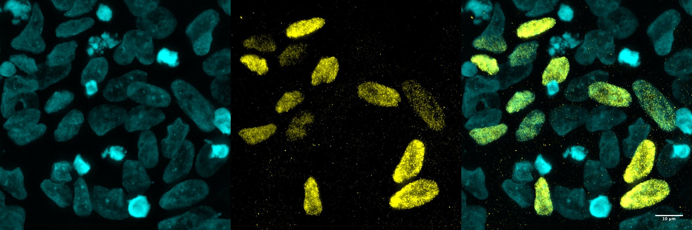
Application: Immunocytochemistry/ImmunofluorescenceSample Tested: hiPSC-derived sympathoblastsSpecies: HumanVerified Customer | Posted 07/28/2025PHOX2B was detected in 4%PFA-fixed hiPSC-derived sympathoblasts using Goat Anti-Human PHOX2B Antigen Affinity-purified Polyclonal Antibody (Catalog # AF4940) at 5 µg/mL for 1 hour at room temperature. Cells were then stained with a Cy3-conjugated donkey anti-goat IgG secondary antibody (yellow), and counterstained with DAPI (cyan). Specific staining was localized to the nuclei.

There are no reviews that match your criteria.
Protocols
Find general support by application which include: protocols, troubleshooting, illustrated assays, videos and webinars.
- Appropriate Fixation of IHC/ICC Samples
- Cellular Response to Hypoxia Protocols
- ClariTSA™ Fluorophore Kits
- Detection & Visualization of Antibody Binding
- ICC Cell Smear Protocol for Suspension Cells
- ICC Immunocytochemistry Protocol Videos
- ICC for Adherent Cells
- Immunocytochemistry (ICC) Protocol
- Immunocytochemistry Troubleshooting
- Immunofluorescence of Organoids Embedded in Cultrex Basement Membrane Extract
- Immunohistochemistry (IHC) and Immunocytochemistry (ICC) Protocols
- Preparing Samples for IHC/ICC Experiments
- Preventing Non-Specific Staining (Non-Specific Binding)
- Primary Antibody Selection & Optimization
- Protocol for VisUCyte™ HRP Polymer Detection Reagent
- Protocol for the Fluorescent ICC Staining of Cell Smears - Graphic
- Protocol for the Fluorescent ICC Staining of Cultured Cells on Coverslips - Graphic
- Protocol for the Preparation and Fluorescent ICC Staining of Cells on Coverslips
- Protocol for the Preparation and Fluorescent ICC Staining of Non-adherent Cells
- Protocol for the Preparation and Fluorescent ICC Staining of Stem Cells on Coverslips
- Protocol for the Preparation of a Cell Smear for Non-adherent Cell ICC - Graphic
- TUNEL and Active Caspase-3 Detection by IHC/ICC Protocol
- The Importance of IHC/ICC Controls
- View all Protocols, Troubleshooting, Illustrated assays and Webinars
Loading...