I kappa B kinase alpha (IKK alpha ) is also known as IKBKA, CHUK, and IKK1. The classical active IKK complex, composed of IKK alpha, IKK beta, and two forms of processed IKK gamma, phosphorylates and inactivates I kappa B, resulting in the release and nuclear translocation of active NF-kappa B. IKK alpha contains kinase, leucine zipper, and helix-loop-helix domains, and heterodimerizes with IKK beta. NF-kappa B-inducing kinase (NIK) phosphorylates and activates IKK alpha.

Key Product Details
Species Reactivity
Validated:
Human, Mouse, Rat
Cited:
Human
Applications
Validated:
Western Blot, Immunocytochemistry
Cited:
Western Blot
Label
Unconjugated
Antibody Source
Polyclonal Sheep IgG
Loading...
Product Specifications
Immunogen
E. coli-derived recombinant human IKK alpha
Gly375-Glu745
Accession # O15111
Gly375-Glu745
Accession # O15111
Specificity
Detects human, mouse, and rat IKK alpha. Does not cross-react with recombinant human (rh) IKK beta, rhIKK gamma, or rhIKK epsilon.
Clonality
Polyclonal
Host
Sheep
Isotype
IgG
Scientific Data Images for IKK alpha Antibody
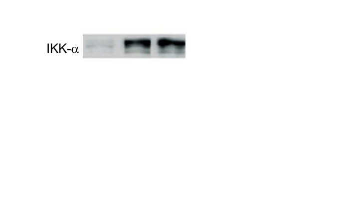
Detection of Human/Mouse/Rat IKK alpha by Western Blot.
Western blot shows lysates of C2C12 mouse myoblast cell line, Rat-2 rat embryonic fibroblast cell line, and U937 human histiocytic lymphoma cell lines. PVDF membrane was probed with 1 µg/mL of Sheep Anti-Human/Mouse/Rat IKKa Antigen Affinity-purified Polyclonal Antibody (Catalog # AF3768) followed by HRP-conjugated Anti-Sheep IgG Secondary Antibody (Catalog # HAF016). A specific band was detected for IKKa at approximately 85 kDa (as indicated). This experiment was conducted under reducing conditions and using Immunoblot Buffer Group 1.
IKK alpha in HCT‑116 Human Cell Line.
IKKa was detected in immersion fixed HCT-116 human colorectal carcinoma cell line using Sheep Anti-Human/Mouse/Rat IKKa Antigen Affinity-purified Polyclonal Antibody (Catalog # AF3768) at 15 µg/mL for 3 hours at room temperature. Cells were stained using the NorthernLights™ 557-conjugated Anti-Sheep IgG Secondary Antibody (red; Catalog # NL010) and counterstained with DAPI (blue). Specific staining was localized to cytoplasmic. View our protocol for Fluorescent ICC Staining of Cells on Coverslips.Applications for IKK alpha Antibody
Application
Recommended Usage
Immunocytochemistry
5-15 µg/mL
Sample: Immersion fixed HCT-116 human colorectal carcinoma cell line and human peripheral blood mononuclear cells treated with PMA and calcium ionomycin
Sample: Immersion fixed HCT-116 human colorectal carcinoma cell line and human peripheral blood mononuclear cells treated with PMA and calcium ionomycin
Western Blot
1 µg/mL
Sample: C2C12 mouse myoblast cell line, Rat-2 rat embryonic fibroblast cell line, and U937 human histiocytic lymphoma cell lines
Sample: C2C12 mouse myoblast cell line, Rat-2 rat embryonic fibroblast cell line, and U937 human histiocytic lymphoma cell lines
Reviewed Applications
Read 4 reviews rated 4.3 using AF3768 in the following applications:
Formulation, Preparation, and Storage
Purification
Antigen Affinity-purified
Reconstitution
Reconstitute at 0.2 mg/mL in sterile PBS. For liquid material, refer to CoA for concentration.
Loading...
Formulation
Lyophilized from a 0.2 μm filtered solution in PBS with Trehalose. *Small pack size (SP) is supplied either lyophilized or as a 0.2 µm filtered solution in PBS.
Shipping
Lyophilized product is shipped at ambient temperature. Liquid small pack size (-SP) is shipped with polar packs. Upon receipt, store immediately at the temperature recommended below.
Stability & Storage
Use a manual defrost freezer and avoid repeated freeze-thaw cycles.
- 12 months from date of receipt, -20 to -70 °C as supplied.
- 1 month, 2 to 8 °C under sterile conditions after reconstitution.
- 6 months, -20 to -70 °C under sterile conditions after reconstitution.
Calculators
Background: IKK alpha
Long Name
IkB Kinase alpha
Alternate Names
CHUK, IkBKalpha, IKK1, NFKBIKA, TCF16
Gene Symbol
CHUK
UniProt
Additional IKK alpha Products
Product Documents for IKK alpha Antibody
Certificate of Analysis
To download a Certificate of Analysis, please enter a lot or batch number in the search box below.
Note: Certificate of Analysis not available for kit components.
Product Specific Notices for IKK alpha Antibody
For research use only
Citations for IKK alpha Antibody
 Powered by Bioz
Customer Reviews for IKK alpha Antibody (4)
4.3 out of 5
4 Customer Ratings
Have you used IKK alpha Antibody?
Submit a review and receive an Amazon gift card!
$25/€18/£15/$25CAN/¥2500 Yen for a review with an image
$10/€7/£6/$10CAN/¥1110 Yen for a review without an image
Submit a review
Customer Images
Showing
1
-
4 of
4 reviews
Showing All
Filter By:
-
Application: ELISASample Tested: Multiple Myelome cell lineSpecies: HumanVerified Customer | Posted 01/16/2020

-
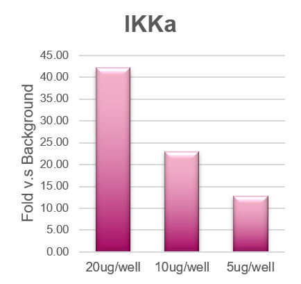
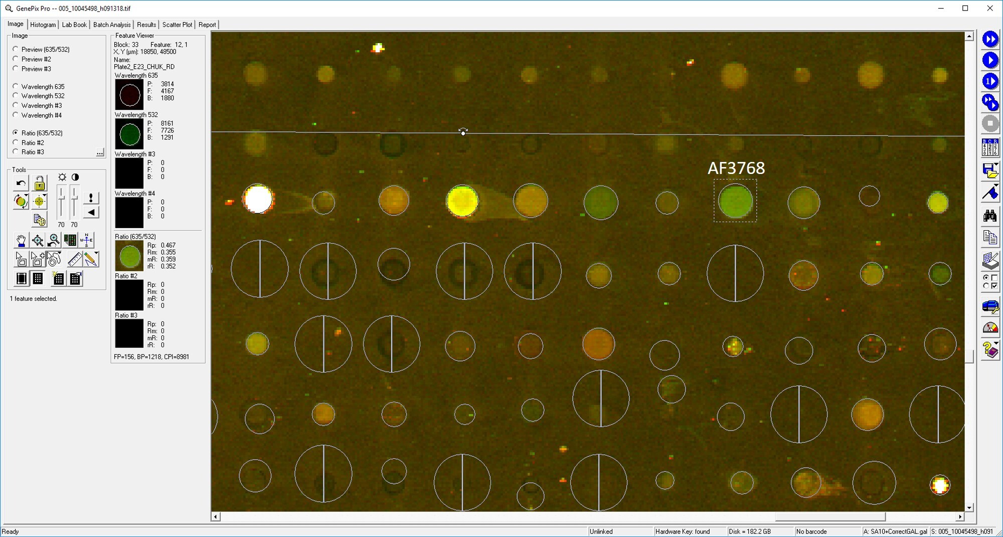
Application: MicroarraysSample Tested: EDTA PlasmaSpecies: HumanVerified Customer | Posted 01/03/2020
-
Application: MicroarraySample Tested: EDTA PlasmaSpecies: HumanVerified Customer | Posted 11/02/2018
-
Application: Western BlotSample Tested: HEK293 human embryonic kidney cell lineSpecies: HumanVerified Customer | Posted 07/17/2018
There are no reviews that match your criteria.
Protocols
Find general support by application which include: protocols, troubleshooting, illustrated assays, videos and webinars.
- Appropriate Fixation of IHC/ICC Samples
- Cellular Response to Hypoxia Protocols
- ClariTSA™ Fluorophore Kits
- Detection & Visualization of Antibody Binding
- ICC Cell Smear Protocol for Suspension Cells
- ICC Immunocytochemistry Protocol Videos
- ICC for Adherent Cells
- Immunocytochemistry (ICC) Protocol
- Immunocytochemistry Troubleshooting
- Immunofluorescence of Organoids Embedded in Cultrex Basement Membrane Extract
- Immunohistochemistry (IHC) and Immunocytochemistry (ICC) Protocols
- Preparing Samples for IHC/ICC Experiments
- Preventing Non-Specific Staining (Non-Specific Binding)
- Primary Antibody Selection & Optimization
- Protocol for VisUCyte™ HRP Polymer Detection Reagent
- Protocol for the Fluorescent ICC Staining of Cell Smears - Graphic
- Protocol for the Fluorescent ICC Staining of Cultured Cells on Coverslips - Graphic
- Protocol for the Preparation and Fluorescent ICC Staining of Cells on Coverslips
- Protocol for the Preparation and Fluorescent ICC Staining of Non-adherent Cells
- Protocol for the Preparation and Fluorescent ICC Staining of Stem Cells on Coverslips
- Protocol for the Preparation of a Cell Smear for Non-adherent Cell ICC - Graphic
- R&D Systems Quality Control Western Blot Protocol
- TUNEL and Active Caspase-3 Detection by IHC/ICC Protocol
- The Importance of IHC/ICC Controls
- Troubleshooting Guide: Western Blot Figures
- Western Blot Conditions
- Western Blot Protocol
- Western Blot Protocol for Cell Lysates
- Western Blot Troubleshooting
- Western Blot Troubleshooting Guide
- View all Protocols, Troubleshooting, Illustrated assays and Webinars


