
Key Product Details
Species Reactivity
Validated:
Cited:
Applications
Validated:
Cited:
Label
Antibody Source
Product Specifications
Immunogen
Pro2-Ala115
Accession # AAA36315
Specificity
Clonality
Host
Isotype
Scientific Data Images for MIF Antibody
Detection of Human MIF by Western Blot.
Western blot shows lysates of THP-1 human acute monocytic leukemia cell line and U937 human histiocytic lymphoma cell line. PVDF membrane was probed with 0.5 µg/mL of Goat Anti-Human/Mouse/Rat MIF Antigen Affinity-purified Polyclonal Antibody (Catalog # AF-289-PB) followed by HRP-conjugated Anti-Goat IgG Secondary Antibody (HAF017). A specific band was detected for MIF at approximately 12 kDa (as indicated). This experiment was conducted under reducing conditions and using Immunoblot Buffer Group 1.
Detection of Mouse and Rat MIF by Western Blot.
Western blot shows lysates of J774A.1 mouse reticulum cell sarcoma macrophage cell line and NR8383 rat alveolar macrophage cell line. PVDF membrane was probed with 0.5 µg/mL of Goat Anti-Human/Mouse/Rat MIF Antigen Affinity-purified Polyclonal Antibody (Catalog # AF-289-PB) followed by HRP-conjugated Anti-Goat IgG Secondary Antibody (HAF017). A specific band was detected for MIF at approximately 12-14 kDa (as indicated). This experiment was conducted under reducing conditions and using Immunoblot Buffer Group 1.
Detection of Human MIF by Simple WesternTM.
Simple Western lane view shows lysates of THP‑1 human acute monocytic leukemia cell line, loaded at 0.2 mg/mL. A specific band was detected for MIF at approximately 12 kDa (as indicated) using 10 µg/mL of Goat Anti-Human/Mouse/Rat MIF Antigen Affinity-purified Polyclonal Antibody (Catalog # AF-289-PB). This experiment was conducted under reducing conditions and using the 12-230 kDa separation system.
Detection of MIF in THP-1 cells by Flow Cytometry.
THP-1 cells were stained with Goat Anti-Human/Mouse/Rat MIF Antigen Affinity-purified Polyclonal Antibody (Catalog # AF-289-PB, filled histogram) or isotype control antibody (Catalog # AB-108-C, open histogram), followed by Phycoerythrin-conjugated Anti-Goat IgG Secondary Antibody (Catalog # F0107). To facilitate intracellular staining, cells were fixed and permeabilized with FlowX FoxP3 Fixation & Permeabilization Buffer Kit (Catalog # FC012). View our protocol for Staining Intracellular Molecules.Applications for MIF Antibody
CyTOF-ready
Intracellular Staining by Flow Cytometry
Sample: THP-1 cells fixed and permeabilized with FlowX FoxP3 Fixation & Permeabilization Buffer Kit (Catalog # FC012).
Simple Western
Sample: THP‑1 human acute monocytic leukemia cell line
Western Blot
Sample: THP‑1 human acute monocytic leukemia cell line, U937 human histiocytic lymphoma cell line, J774A.1 mouse reticulum cell sarcoma macrophage cell line, and NR8383 rat alveolar macrophage cell line
Reviewed Applications
Read 2 reviews rated 5 using AF-289-PB in the following applications:
Flow Cytometry Panel Builder
Bio-Techne Knows Flow Cytometry
Save time and reduce costly mistakes by quickly finding compatible reagents using the Panel Builder Tool.
Advanced Features
- Spectra Viewer - Custom analysis of spectra from multiple fluorochromes
- Spillover Popups - Visualize the spectra of individual fluorochromes
- Antigen Density Selector - Match fluorochrome brightness with antigen density

Formulation, Preparation, and Storage
Purification
Reconstitution
Reconstitute at 0.2 mg/mL in sterile PBS. For liquid material, refer to CoA for concentration.
Formulation
Shipping
Stability & Storage
- 12 months from date of receipt, -20 to -70 °C as supplied.
- 1 month, 2 to 8 °C under sterile conditions after reconstitution.
- 6 months, -20 to -70 °C under sterile conditions after reconstitution.
Calculators
Background: MIF
References
- Norand, E.F. and M. Leech (2005) Front. Biosci. 10:12.
- Donn, R.P. and D.W. Ray (2004) J. Endocrinol. 182:1.
- Calandra, T. and T. Roger (2003) Nat. Rev. Immunol. 3:791.
- Kozak, C.A. et al. (1995) Genomics 27:405.
- Weiser, W.Y. et al. (1989) Proc. Natl. Acad. Sci. USA 86:7522.
- Paralkar, V. and G. Wistow (1994) Genomics 19:48.
- Wistow, G.J. et al. (1993) Proc. Natl. Acad. Sci. USA 90:1272.
- Flieger, O. et al. (2003) FEBS Lett. 551:78.
- Philo, J.S. et al. (2004) Biophys. Chem. 108:77.
- Sun, H-W. et al. (1996) Protein Eng. 9:631.
- Stamps, S.L. et. al. (2000) Biochemistry 39:9671.
- Nguyen, M.T. et al. (2003) J. Biol. Chem. 278:33654.
- Sato, A. et al. (2003) Dev. Comp. Immunol. 27:401.
- Bernhagen, J. et al. (1994) Biochemistry 33:14144.
- Leng, L. et al. (2003) J. Exp. Med. 197:1467.
- Meyer-Siegler, K.L. and P.L. Vera (2005) J. Urol. 173:615.
Long Name
Alternate Names
Gene Symbol
UniProt
Additional MIF Products
Product Documents for MIF Antibody
Certificate of Analysis
To download a Certificate of Analysis, please enter a lot or batch number in the search box below.
Note: Certificate of Analysis not available for kit components.
Product Specific Notices for MIF Antibody
For research use only
Related Research Areas
Citations for MIF Antibody
 Powered by Bioz
Customer Reviews for MIF Antibody (2)
Have you used MIF Antibody?
Submit a review and receive an Amazon gift card!
$25/€18/£15/$25CAN/¥2500 Yen for a review with an image
$10/€7/£6/$10CAN/¥1110 Yen for a review without an image
Submit a review
Customer Images
-
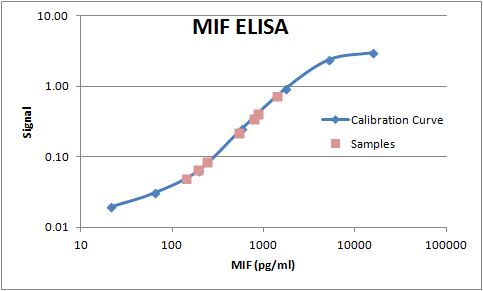
Application: ELISASample Tested: Serum and PlasmaSpecies: HumanVerified Customer | Posted 07/19/2019

-
Application: ELISASample Tested: Serum and PlasmaSpecies: HumanVerified Customer | Posted 11/07/2017This polyclonal was paired up with the monoclonal MAB289 to build a sandwich ELISA for the measurement of human MIF in serum and plasma samples.
There are no reviews that match your criteria.
Protocols
Find general support by application which include: protocols, troubleshooting, illustrated assays, videos and webinars.
- 7-Amino Actinomycin D (7-AAD) Cell Viability Flow Cytometry Protocol
- Cellular Response to Hypoxia Protocols
- Extracellular Membrane Flow Cytometry Protocol
- Flow Cytometry Protocol for Cell Surface Markers
- Flow Cytometry Protocol for Staining Membrane Associated Proteins
- Flow Cytometry Staining Protocols
- Flow Cytometry Troubleshooting Guide
- Intracellular Flow Cytometry Protocol Using Alcohol (Methanol)
- Intracellular Flow Cytometry Protocol Using Detergents
- Intracellular Nuclear Staining Flow Cytometry Protocol Using Detergents
- Intracellular Staining Flow Cytometry Protocol Using Alcohol Permeabilization
- Intracellular Staining Flow Cytometry Protocol Using Detergents to Permeabilize Cells
- Propidium Iodide Cell Viability Flow Cytometry Protocol
- Protocol for Liperfluo
- Protocol for the Characterization of Human Th22 Cells
- Protocol for the Characterization of Human Th9 Cells
- Protocol: Annexin V and PI Staining by Flow Cytometry
- Protocol: Annexin V and PI Staining for Apoptosis by Flow Cytometry
- R&D Systems Quality Control Western Blot Protocol
- Troubleshooting Guide: Fluorokine Flow Cytometry Kits
- Troubleshooting Guide: Western Blot Figures
- Western Blot Conditions
- Western Blot Protocol
- Western Blot Protocol for Cell Lysates
- Western Blot Troubleshooting
- Western Blot Troubleshooting Guide
- View all Protocols, Troubleshooting, Illustrated assays and Webinars
FAQs for MIF Antibody
-
Q: What does the PB in AF-289-PB mean?
A: The PB in AF-289-PB is a part of the catalog code for this antibody and it appears on the vial lable for both the retail(100 µg) and SP (25 µg) size of the antibody.