The c-Jun N-terminal Kinases (JNKs) are part of the MAPK (mitogen-activated protein kinase) system that transmits signals from the extracellular milieu to both the cytoplasm and nucleus of the cell. Following perturbation at the cell membrane, MEKKs/MAP3Ks are initially activated, followed by their activation of MKKs/MAP2Ks, and MKKs activation of MAPKs/MAP(1)Ks. There are three classes of MAPKs: ERKs, p38 Kinases and JNKs. JNKs are 45-55 kDa protein products of three genes which, through alternative splicing, generate up to 10 possible isoforms. The phosphorylation targets for MAPKs vary, but include p53, c-MYC, ATF2 and c-Jun, the latter molecule representing the namesake for the enzyme group. The three human JNKs share approximately 80% aa sequence identity. JNKs from human, mouse and rat all contain a conserved Met-Met-Thr(183)-Pro-Tyr(185)-Val-Val motif that undergoes dual phosphorylation by MMK4 and MMK7 to activate the different JNKs. Activated by environmental stresses and inflammatory cytokines, JNKs translocate to the nucleus where they regulate the activity of several transcription factors; including the c-Jun component of AP-1 and ATF-2.
phospho-JNK (T183/Y185) Antibody (1006A)
R&D Systems | Catalog # MAB1205
Recombinant Monoclonal Antibody.
 by Western Blot.")
Key Product Details
Validated by
Biological Validation
Species Reactivity
Validated:
Human, Mouse, Rat
Cited:
Human, Mouse, Capsaspora owczarzaki
Applications
Validated:
Western Blot, Immunocytochemistry, Simple Western
Cited:
Western Blot
Label
Unconjugated
Antibody Source
Recombinant Monoclonal Rabbit IgG Clone # 1006A
Loading...
Product Specifications
Immunogen
Phosphopeptide containing human, rat, and mouse JNK1 T183/Y185 site
Specificity
Detects human, mouse and rat p46 and p54 JNK when dually phosphorylated at sites homologous to T183/Y185 of JNK1 and JNK2, and T221/Y223 of JNK3 in Western blots.
Clonality
Monoclonal
Host
Rabbit
Isotype
IgG
Scientific Data Images for phospho-JNK (T183/Y185) Antibody (1006A)
Detection of Human and Mouse Phospho-JNK (T183/Y185) by Western Blot.
Western blot shows lysates of HeLa human cervical epithelial carcinoma cell line and 293T human embryonic kidney cell line untreated (-) or treated (+) with 20 mJ/cm2ultraviolet light (UV) followed by a 30 minute recovery. PVDF membrane was probed with 1 µg/ml of Rabbit Anti-Human/Mouse/Rat Phospho-JNK (T183/Y185) Monoclonal Antibody (Catalog # MAB1205), followed by HRP-conjugated Anti-Rabbit IgG Secondary Antibody (Catalog # HAF008). Specific bands were detected for Phospho-JNK (T183/Y185) at approximately 46 and 54 kDa (as indicated). This experiment was conducted under reducing conditions and using Immunoblot Buffer Group 1. in HEK293 Human Cell Line.")
Phospho-JNK (T183/Y185) in HEK293 Human Cell Line.
JNK phosphorylated at T183/Y185 was detected in immersion fixed HEK293 human embryonic kidney cell line untreated (lower panel) or treated with UV radiation (upper panel) using Rabbit Anti-Human/Mouse/Rat Phospho-JNK (T183/Y185) Monoclonal Antibody (Catalog # MAB1205) at 25 µg/ml for 3 hours at room temperature. Cells were stained using the NorthernLights™ 557-conjugated Anti-Rabbit IgG Secondary Antibody (red; Catalog # NL004) and counterstained with DAPI. Filamentous actin was stained with fluorescein-conjugated phalloidin (green). Specific staining was localized to nuclei. View our protocol for Fluorescent ICC Staining of Cells on Coverslips. by Simple Western<SUP>TM</SUP>.")
Detection of Human Phospho-JNK (T183/Y185) by Simple WesternTM.
Simple Western lane view shows lysates of HEK293T human embryonic kidney cell line untreated (-) or treated (+) with 20 J/m2ultraviolet light (UV) followed by a 30 minute recovery, loaded at 0.2 mg/mL. A specific band was detected for Phospho-JNK (T183/Y185) at approximately 46 and 56 kDa (as indicated) using 20 μg/ml of Rabbit Anti-Human/Mouse/Rat Phospho-JNK (T183/Y185) Monoclonal Antibody (Catalog # MAB1205). This experiment was conducted under reducing conditions and using the 12-230 kDa separation system. Non-specific interaction with the 230 kDa Simple Western standard may be seen with this antibody.
Detection of Human JNK1/2/3 by Simple Western
ACPA inhibits p-Akt, induces p-JNK and affects levels of specific metabolites in NSCLC lines.a Principal component analysis (PCA) score plot: Metabolomics profiling of control and ACPA-treated A549, H1299, H358, and H838 cells. b Changes in variable importance in projection (VIP) values for 19 metabolites in A549 cells. c, d, e Changes in VIP values for 20 metabolites in H1299, H358, and H838 cells. Significantly changed metabolites (*p < 0.05, indicated by arrows) were matched to apoptotic pathways. f, g, h, i Increase and decrease in several metabolites of ACPA-treated A549, H1299, H358, and H838 cells (*p < 0.05). j Simple Western showing total Akt, p-Akt (S473), total JNK46 and JNK54 and p-JNK46 and p-JNK54 (T183/Y185) in A549 cells at 24 hours after treatment with IC50 dose of ACPA. k Relative expression levels of Akt and p-Akt for control and ACPA-treated A549 cells after normalization by total vinculin protein. l Relative expression levels of JNK (46 and 54 kDa) and p-JNK for control and ACPA-treated A549 cells after normalization by total vinculin protein. *p < 0.05, Student’s t-test. All tests were done in quadruplicates. Image collected and cropped by CiteAb from the following publication (https://pubmed.ncbi.nlm.nih.gov/33431819), licensed under a CC-BY license. Not internally tested by R&D Systems.
Detection of Human JNK1/2/3 by Western Blot
Effect of LSW treatment on TNF alpha -mediated NF-kappa B and p38/JNK signaling in EA.hy926 cells. The cells were treated with LSW (50 μM) for 18 h before TNF alpha (10 ng/mL) stimulation for 15 min, followed by the detecting the protein expression of I kappa B alpha (A), p65 (B), p38 (C), and JNK (D) by Western blotting. Protein bands of I kappa B alpha were normalized to GAPDH; bands of the phosphorylated p65, p38, and JNK were normalized to their total forms. Data were normalized to the untreated group (Untr). *, p < 0.05; **, p < 0.01; ****, p < 0.0001, ns, not significant. Image collected and cropped by CiteAb from the following open publication (https://pubmed.ncbi.nlm.nih.gov/36359987), licensed under a CC-BY license. Not internally tested by R&D Systems.Applications for phospho-JNK (T183/Y185) Antibody (1006A)
Application
Recommended Usage
Immunocytochemistry
10-35 µg/mL
Sample: Immersion fixed HEK293 human embryonic kidney cell line treated with UV radiation
Sample: Immersion fixed HEK293 human embryonic kidney cell line treated with UV radiation
Simple Western
10-25 µg/mL
Sample: HEK293T human embryonic kidney cell line treated with ultraviolet light (UV)
Sample: HEK293T human embryonic kidney cell line treated with ultraviolet light (UV)
Western Blot
1 µg/mL
Sample: HeLa human cervical epithelial carcinoma cell line and 293T human embryonic kidney cell line treated with ultraviolet light (UV)
Sample: HeLa human cervical epithelial carcinoma cell line and 293T human embryonic kidney cell line treated with ultraviolet light (UV)
Reviewed Applications
Read 3 reviews rated 4.7 using MAB1205 in the following applications:
Formulation, Preparation, and Storage
Purification
Protein A or G purified from cell culture supernatant
Reconstitution
Reconstitute at 0.5 mg/mL in sterile PBS. For liquid material, refer to CoA for concentration.
Loading...
Formulation
Lyophilized from a 0.2 μm filtered solution in PBS with Trehalose. *Small pack size (SP) is supplied either lyophilized or as a 0.2 µm filtered solution in PBS.
Shipping
Lyophilized product is shipped at ambient temperature. Liquid small pack size (-SP) is shipped with polar packs. Upon receipt, store immediately at the temperature recommended below.
Stability & Storage
Use a manual defrost freezer and avoid repeated freeze-thaw cycles.
- 12 months from date of receipt, -20 to -70 °C as supplied.
- 1 month, 2 to 8 °C under sterile conditions after reconstitution.
- 6 months, -20 to -70 °C under sterile conditions after reconstitution.
Calculators
Background: JNK
Long Name
C-Jun N-terminal Kinase
Alternate Names
c-Jun N-terminal kinase 1, EC 2.7.11, EC 2.7.11.24, JNK1 alpha protein kinase, JNK1 beta protein kinase, JNK1JNK1A2, JNK-46, JUN N-terminal kinase, MAP kinase 8, MAPK 8, mitogen-activated protein kinase 8, mitogen-activated protein kinase 8 isoform JNK1 alpha1, mitogen-activated protein kinase 8 isoform JNK1 beta2, PRKM8JNK, protein kinase JNK1, SAPK1JNK21B1/2, Stress-activated protein kinase 1, Stress-activated protein kinase JNK1
Additional JNK Products
Product Documents for phospho-JNK (T183/Y185) Antibody (1006A)
Certificate of Analysis
To download a Certificate of Analysis, please enter a lot or batch number in the search box below.
Note: Certificate of Analysis not available for kit components.
Product Specific Notices for phospho-JNK (T183/Y185) Antibody (1006A)
For research use only
Citations for phospho-JNK (T183/Y185) Antibody (1006A)
 Powered by Bioz
Customer Reviews for phospho-JNK (T183/Y185) Antibody (1006A) (3)
4.7 out of 5
3 Customer Ratings
Have you used phospho-JNK (T183/Y185) Antibody (1006A)?
Submit a review and receive an Amazon gift card!
$25/€18/£15/$25CAN/¥2500 Yen for a review with an image
$10/€7/£6/$10CAN/¥1110 Yen for a review without an image
Submit a review
Customer Images

Showing
1
-
3 of
3 reviews
Showing All
Filter By:
-
Application: Western BlotSample Tested: HeLa human cervical epithelial carcinoma cell lineSpecies: HumanVerified Customer | Posted 11/03/2021

-
Application: Chromatin ImmunoprecipitationSample Tested: 3T3-L1 mouse embryonic fibroblast adipose-like cell lineSpecies: Cynomolgus MonkeyVerified Customer | Posted 08/01/2017
-
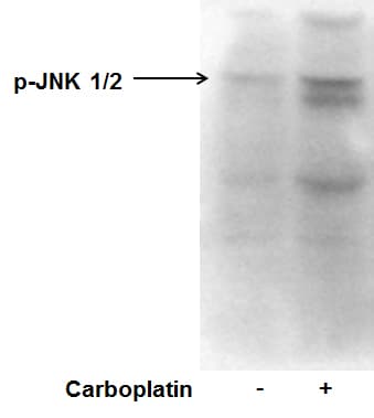
Application: Western BlotSample Tested: Human cancer cell whole cell lysateSpecies: HumanVerified Customer | Posted 10/04/2015p-JNK expression in MDA-MB-231 in response to carboplatin treatment.
There are no reviews that match your criteria.
Protocols
Find general support by application which include: protocols, troubleshooting, illustrated assays, videos and webinars.
- Appropriate Fixation of IHC/ICC Samples
- Cellular Response to Hypoxia Protocols
- ClariTSA™ Fluorophore Kits
- Detection & Visualization of Antibody Binding
- ICC Cell Smear Protocol for Suspension Cells
- ICC Immunocytochemistry Protocol Videos
- ICC for Adherent Cells
- Immunocytochemistry (ICC) Protocol
- Immunocytochemistry Troubleshooting
- Immunofluorescence of Organoids Embedded in Cultrex Basement Membrane Extract
- Immunohistochemistry (IHC) and Immunocytochemistry (ICC) Protocols
- Preparing Samples for IHC/ICC Experiments
- Preventing Non-Specific Staining (Non-Specific Binding)
- Primary Antibody Selection & Optimization
- Protocol for VisUCyte™ HRP Polymer Detection Reagent
- Protocol for the Fluorescent ICC Staining of Cell Smears - Graphic
- Protocol for the Fluorescent ICC Staining of Cultured Cells on Coverslips - Graphic
- Protocol for the Preparation and Fluorescent ICC Staining of Cells on Coverslips
- Protocol for the Preparation and Fluorescent ICC Staining of Non-adherent Cells
- Protocol for the Preparation and Fluorescent ICC Staining of Stem Cells on Coverslips
- Protocol for the Preparation of a Cell Smear for Non-adherent Cell ICC - Graphic
- R&D Systems Quality Control Western Blot Protocol
- TUNEL and Active Caspase-3 Detection by IHC/ICC Protocol
- The Importance of IHC/ICC Controls
- Troubleshooting Guide: Western Blot Figures
- Western Blot Conditions
- Western Blot Protocol
- Western Blot Protocol for Cell Lysates
- Western Blot Troubleshooting
- Western Blot Troubleshooting Guide
- View all Protocols, Troubleshooting, Illustrated assays and Webinars
Loading...