Human/Mouse/Rat SOD2/Mn‑SOD Antibody
R&D Systems | Catalog # MAB3419

Key Product Details
Species Reactivity
Validated:
Human, Mouse, Rat
Cited:
Human, Mouse, Porcine
Applications
Validated:
Western Blot, Immunocytochemistry
Cited:
Western Blot, Immunocytochemistry
Label
Unconjugated
Antibody Source
Monoclonal Mouse IgG1 Clone # 349810
Loading...
Product Specifications
Immunogen
E. coli-derived recombinant human SOD2/Mn-SOD
Lys25-Lys222
Accession # P04179
Lys25-Lys222
Accession # P04179
Specificity
Detects human, mouse, and rat SOD2 in Western blots. In Western blots, no cross-reactivity with recombinant human SOD1 or SOD3 is observed.
Clonality
Monoclonal
Host
Mouse
Isotype
IgG1
Scientific Data Images for Human/Mouse/Rat SOD2/Mn‑SOD Antibody
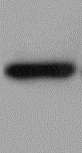
Detection of Human/Mouse/Rat SOD2/Mn‑SOD by Western Blot.
Western blot shows lysates of MCF-7 human breast cancer cell line, NIH-3T3 mouse embryonic fibroblast cell line, and L6 rat myoblast cell line. PVDF membrane was probed with 0.5 µg/mL Mouse Anti-Human/Mouse/Rat SOD2/Mn-SOD Monoclonal Antibody (Catalog # MAB3419) followed by HRP-conjugated Anti-Mouse IgG Secondary Antibody (Catalog # HAF007). For additional reference, recombinant human SOD1, SOD2, and SOD3 (1 ng/lane) were included. A specific band for SOD2/Mn-SOD was detected at approximately 22 kDa (as indicated). This experiment was conducted under reducing conditions and using Immunoblot Buffer Group 2.
SOD2/Mn-SOD in MCF‑7 Human Cell Line.
SOD2/Mn-SOD was detected in immersion fixed MCF-7 human breast cancer cell line using Mouse Anti-Human/Mouse/Rat SOD2/Mn-SOD Monoclonal Antibody (Catalog # MAB3419) at 25 µg/mL for 3 hours at room temperature. Cells were stained using the NorthernLights™ 557-conjugated Anti-Mouse IgG Secondary Antibody (red; Catalog # NL007) and counterstained with DAPI (blue). Specific staining was localized to cytoplasm. View our protocol for Fluorescent ICC Staining of Cells on Coverslips.
SOD2/Mn‑SOD in HL‑60 Human Cell Line.
SOD2/Mn-SOD was detected in immersion fixed HL-60 human acute promyelocytic leukemia cell line using Mouse Anti-Human/Mouse/Rat SOD2/Mn-SOD Monoclonal Antibody (Catalog # MAB3419) at 3 µg/mL for 3 hours at room temperature. Cells were stained using the NorthernLights™ 557-conjugated Anti-Mouse IgG Secondary Antibody (red; Catalog # NL007) and counterstained with DAPI (blue). Specific staining was localized to cytoplasm. View our protocol for Fluorescent ICC Staining of Non-adherent Cells.
Detection of Human SOD2/Mn-SOD by Western Blot
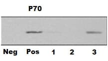
FAM210B is transported into and is localized in the mitochondria. (a) Schematic representation of FAM210B domains based on the primary structure of FAM210B. (b) Confocal microscopy of HeLa cells transfected with GFP-tagged human FAM210B and RFP-tagged mitochondria. FAM210B-GFP, GFP sequence introduced at the C terminus of FAM210B; FAM210B (MTS)-GFP, the MTS (aa 1–47) of FAM210B was added to the GFP N terminus; FAM210B ( delta MTS)-GFP, GFP was added to the C terminus of MTS (1–47)-deleted CRIF1. Scale bar, 20 mm. (c) Confocal microscopy of HeLa cells transfected with GFP-tagged human FAM210B and RFP-tagged endoplasmic reticulum. (d) Western blotting analysis following subcellular fractionation of GFP-tagged human FAM210B HeLa cells. (e) Western blotting analysis following subcellular fractionation of endogenous in HeLa cells. (f) The mitochondria of HeLa cells were swollen and sonicated to disrupt membranes, washed with alkali buffer (pH 11.5) to detach loosely associated proteins from membranes, and then re-isolated by ultracentrifugation. The supernatant (Supe) and membrane fractions (Pellet) were subjected to western blotting for FAM210B, TOM20, or MnSOD. (g) Mitochondria isolated from HeLa cells were subjected to proteinase K (PK) proteolysis to digest exposed proteins, and detergent (SDS) was used to disrupt both IMMs (inner membrane of mitochondria) and OMMs (outer membrane of mitochondria). The lysates were resolved and subjected to immunoblot analyses. The submitochondrial markers used are Tom20 (OMM), Cyt C (Cytochrome c, intermembrane space), NDUFS1 (IMM), and MnSOD (mitochondrial matrix) Image collected and cropped by CiteAb from the following publication (https://pubmed.ncbi.nlm.nih.gov/28594398), licensed under a CC-BY license. Not internally tested by R&D Systems.
Detection of Human SOD2/Mn-SOD by Western Blot
FAM210B is transported into and is localized in the mitochondria. (a) Schematic representation of FAM210B domains based on the primary structure of FAM210B. (b) Confocal microscopy of HeLa cells transfected with GFP-tagged human FAM210B and RFP-tagged mitochondria. FAM210B-GFP, GFP sequence introduced at the C terminus of FAM210B; FAM210B (MTS)-GFP, the MTS (aa 1–47) of FAM210B was added to the GFP N terminus; FAM210B ( delta MTS)-GFP, GFP was added to the C terminus of MTS (1–47)-deleted CRIF1. Scale bar, 20 mm. (c) Confocal microscopy of HeLa cells transfected with GFP-tagged human FAM210B and RFP-tagged endoplasmic reticulum. (d) Western blotting analysis following subcellular fractionation of GFP-tagged human FAM210B HeLa cells. (e) Western blotting analysis following subcellular fractionation of endogenous in HeLa cells. (f) The mitochondria of HeLa cells were swollen and sonicated to disrupt membranes, washed with alkali buffer (pH 11.5) to detach loosely associated proteins from membranes, and then re-isolated by ultracentrifugation. The supernatant (Supe) and membrane fractions (Pellet) were subjected to western blotting for FAM210B, TOM20, or MnSOD. (g) Mitochondria isolated from HeLa cells were subjected to proteinase K (PK) proteolysis to digest exposed proteins, and detergent (SDS) was used to disrupt both IMMs (inner membrane of mitochondria) and OMMs (outer membrane of mitochondria). The lysates were resolved and subjected to immunoblot analyses. The submitochondrial markers used are Tom20 (OMM), Cyt C (Cytochrome c, intermembrane space), NDUFS1 (IMM), and MnSOD (mitochondrial matrix) Image collected and cropped by CiteAb from the following publication (https://pubmed.ncbi.nlm.nih.gov/28594398), licensed under a CC-BY license. Not internally tested by R&D Systems.Applications for Human/Mouse/Rat SOD2/Mn‑SOD Antibody
Application
Recommended Usage
Immunocytochemistry
1-25 µg/mL
Sample: Immersion fixed MCF-7 human breast cancer cells and HL‑60 human acute promyelocytic leukemia cell line
Sample: Immersion fixed MCF-7 human breast cancer cells and HL‑60 human acute promyelocytic leukemia cell line
Western Blot
0.5 µg/mL
Sample: MCF-7 human breast cancer cell line, NIH-3T3 mouse embryonic fibroblast cell line, and L6 rat myoblast cell line
Sample: MCF-7 human breast cancer cell line, NIH-3T3 mouse embryonic fibroblast cell line, and L6 rat myoblast cell line
Reviewed Applications
Read 2 reviews rated 5 using MAB3419 in the following applications:
Formulation, Preparation, and Storage
Purification
Protein A or G purified from hybridoma culture supernatant
Reconstitution
Reconstitute at 0.5 mg/mL in sterile PBS. For liquid material, refer to CoA for concentration.
Loading...
Formulation
Lyophilized from a 0.2 μm filtered solution in PBS with Trehalose. *Small pack size (SP) is supplied either lyophilized or as a 0.2 µm filtered solution in PBS.
Shipping
Lyophilized product is shipped at ambient temperature. Liquid small pack size (-SP) is shipped with polar packs. Upon receipt, store immediately at the temperature recommended below.
Stability & Storage
Use a manual defrost freezer and avoid repeated freeze-thaw cycles.
- 12 months from date of receipt, -20 to -70 °C as supplied.
- 1 month, 2 to 8 °C under sterile conditions after reconstitution.
- 6 months, -20 to -70 °C under sterile conditions after reconstitution.
Calculators
Background: SOD2/Mn-SOD
Long Name
Superoxide Dismutase-2
Alternate Names
IPO-B, Mn SOD, MnSOD, SOD, Mitochodrial
Gene Symbol
SOD2
UniProt
Additional SOD2/Mn-SOD Products
Product Documents for Human/Mouse/Rat SOD2/Mn‑SOD Antibody
Certificate of Analysis
To download a Certificate of Analysis, please enter a lot or batch number in the search box below.
Note: Certificate of Analysis not available for kit components.
Product Specific Notices for Human/Mouse/Rat SOD2/Mn‑SOD Antibody
For research use only
Related Research Areas
Citations for Human/Mouse/Rat SOD2/Mn‑SOD Antibody
 Powered by Bioz
Customer Reviews for Human/Mouse/Rat SOD2/Mn‑SOD Antibody (2)
5 out of 5
2 Customer Ratings
Have you used Human/Mouse/Rat SOD2/Mn‑SOD Antibody?
Submit a review and receive an Amazon gift card!
$25/€18/£15/$25CAN/¥2500 Yen for a review with an image
$10/€7/£6/$10CAN/¥1110 Yen for a review without an image
Submit a review
Customer Images
Showing
1
-
2 of
2 reviews
Showing All
Filter By:
-
Application: Western BlotSample Tested: Mouse Lung Endothelial CellsSpecies: MouseVerified Customer | Posted 11/06/2021

-
Application: Western BlotSample Tested: Cartilage tissueSpecies: MouseVerified Customer | Posted 11/07/2019antibody dilution was 1:150. SOD2 clearly expressed in mouse lysates, detected with ECL kit. Western Blot Human/Mouse/Rat beta-Actin Antibody MAB8929 was used for 2ndary
There are no reviews that match your criteria.
Protocols
Find general support by application which include: protocols, troubleshooting, illustrated assays, videos and webinars.
- Appropriate Fixation of IHC/ICC Samples
- Cellular Response to Hypoxia Protocols
- ClariTSA™ Fluorophore Kits
- Detection & Visualization of Antibody Binding
- ICC Cell Smear Protocol for Suspension Cells
- ICC Immunocytochemistry Protocol Videos
- ICC for Adherent Cells
- Immunocytochemistry (ICC) Protocol
- Immunocytochemistry Troubleshooting
- Immunofluorescence of Organoids Embedded in Cultrex Basement Membrane Extract
- Immunohistochemistry (IHC) and Immunocytochemistry (ICC) Protocols
- Preparing Samples for IHC/ICC Experiments
- Preventing Non-Specific Staining (Non-Specific Binding)
- Primary Antibody Selection & Optimization
- Protocol for VisUCyte™ HRP Polymer Detection Reagent
- Protocol for the Fluorescent ICC Staining of Cell Smears - Graphic
- Protocol for the Fluorescent ICC Staining of Cultured Cells on Coverslips - Graphic
- Protocol for the Preparation and Fluorescent ICC Staining of Cells on Coverslips
- Protocol for the Preparation and Fluorescent ICC Staining of Non-adherent Cells
- Protocol for the Preparation and Fluorescent ICC Staining of Stem Cells on Coverslips
- Protocol for the Preparation of a Cell Smear for Non-adherent Cell ICC - Graphic
- R&D Systems Quality Control Western Blot Protocol
- TUNEL and Active Caspase-3 Detection by IHC/ICC Protocol
- The Importance of IHC/ICC Controls
- Troubleshooting Guide: Western Blot Figures
- Western Blot Conditions
- Western Blot Protocol
- Western Blot Protocol for Cell Lysates
- Western Blot Troubleshooting
- Western Blot Troubleshooting Guide
- View all Protocols, Troubleshooting, Illustrated assays and Webinars
Loading...