
Key Product Details
Species Reactivity
Validated:
Human
Cited:
Human
Applications
Validated:
Immunocytochemistry
Cited:
Chromatin Immunoprecipitation Sequencing
Label
Unconjugated
Antibody Source
Polyclonal Goat IgG
Loading...
Product Specifications
Immunogen
E. coli-derived recombinant human Mxi1
Ala81-Ser228
Accession # P50539
Ala81-Ser228
Accession # P50539
Specificity
Detects human Mxi1 in direct ELISAs. In direct ELISAs, less than 1% cross-reactivity with recombinant human (rh) MAD1, rhMAD3, and rhMAD4 is observed.
Clonality
Polyclonal
Host
Goat
Isotype
IgG
Scientific Data Images for Human Mxi1 Antibody
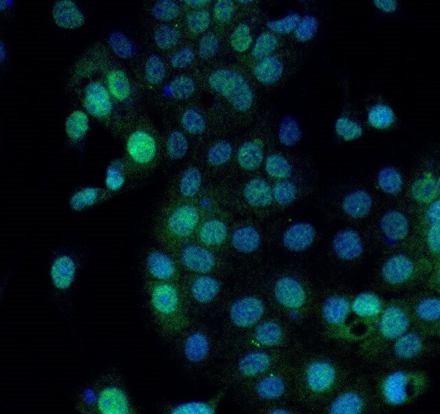
Mxi1 in LNCaP Human Cell Line.
Mxi1 was detected in immersion fixed LNCaP human prostate cancer cell line using Goat Anti-Human Mxi1 Antigen Affinity-purified Polyclonal Antibody (Catalog # AF4185) at 15 µg/mL for 3 hours at room temperature. Cells were stained using the NorthernLights™ 557-conjugated Anti-Goat IgG Secondary Antibody (red; Catalog # NL001) and counterstained with DAPI (blue). Specific staining was localized to nuclei. View our protocol for Fluorescent ICC Staining of Cells on Coverslips.Applications for Human Mxi1 Antibody
Application
Recommended Usage
Immunocytochemistry
5-15 µg/mL
Sample: Immersion fixed LNCaP human prostate cancer cell line and U937 human histiocytic lymphoma cell line treated with PMA
Sample: Immersion fixed LNCaP human prostate cancer cell line and U937 human histiocytic lymphoma cell line treated with PMA
Reviewed Applications
Read 1 review rated 5 using AF4185 in the following applications:
Formulation, Preparation, and Storage
Purification
Antigen Affinity-purified
Reconstitution
Reconstitute at 0.2 mg/mL in sterile PBS. For liquid material, refer to CoA for concentration.
Loading...
Formulation
Lyophilized from a 0.2 μm filtered solution in PBS with Trehalose. *Small pack size (SP) is supplied either lyophilized or as a 0.2 µm filtered solution in PBS.
Shipping
Lyophilized product is shipped at ambient temperature. Liquid small pack size (-SP) is shipped with polar packs. Upon receipt, store immediately at the temperature recommended below.
Stability & Storage
Use a manual defrost freezer and avoid repeated freeze-thaw cycles.
- 12 months from date of receipt, -20 to -70 °C as supplied.
- 1 month, 2 to 8 °C under sterile conditions after reconstitution.
- 6 months, -20 to -70 °C under sterile conditions after reconstitution.
Calculators
Background: Mxi1
(aa 69‑80) and a bHLH domain (aa 81‑120). Multiple splice variants of Mxi-1 appear to exist. They may incorporate an alternate start site at Met37, a 92 aa substitution for the first 26 N-terminal amino acids, or a deletion of the C-terminal 59 or 74 amino acids. Over aa 81‑228, human Mxi-1 is 95% aa identical to mouse Mxi-1.
Long Name
MAX Interacting Protein 1
Alternate Names
MAD2, MXD2, MXI, MXI-WR
Gene Symbol
MXI1
UniProt
Additional Mxi1 Products
Product Documents for Human Mxi1 Antibody
Certificate of Analysis
To download a Certificate of Analysis, please enter a lot or batch number in the search box below.
Note: Certificate of Analysis not available for kit components.
Product Specific Notices for Human Mxi1 Antibody
For research use only
Citations for Human Mxi1 Antibody
 Powered by Bioz
Customer Reviews for Human Mxi1 Antibody (1)
5 out of 5
1 Customer Rating
Have you used Human Mxi1 Antibody?
Submit a review and receive an Amazon gift card!
$25/€18/£15/$25CAN/¥2500 Yen for a review with an image
$10/€7/£6/$10CAN/¥1110 Yen for a review without an image
Submit a review
Customer Images
Showing
1
-
1 of
1 review
Showing All
Filter By:
-
Application: Immunocytochemistry/ImmunofluorescenceSample Tested: SK-Mel-28 human malignant melanoma cell lineSpecies: HumanVerified Customer | Posted 07/26/2021

There are no reviews that match your criteria.
Protocols
Find general support by application which include: protocols, troubleshooting, illustrated assays, videos and webinars.
- Appropriate Fixation of IHC/ICC Samples
- Cellular Response to Hypoxia Protocols
- ClariTSA™ Fluorophore Kits
- Detection & Visualization of Antibody Binding
- ICC Cell Smear Protocol for Suspension Cells
- ICC Immunocytochemistry Protocol Videos
- ICC for Adherent Cells
- Immunocytochemistry (ICC) Protocol
- Immunocytochemistry Troubleshooting
- Immunofluorescence of Organoids Embedded in Cultrex Basement Membrane Extract
- Immunohistochemistry (IHC) and Immunocytochemistry (ICC) Protocols
- Preparing Samples for IHC/ICC Experiments
- Preventing Non-Specific Staining (Non-Specific Binding)
- Primary Antibody Selection & Optimization
- Protocol for VisUCyte™ HRP Polymer Detection Reagent
- Protocol for the Fluorescent ICC Staining of Cell Smears - Graphic
- Protocol for the Fluorescent ICC Staining of Cultured Cells on Coverslips - Graphic
- Protocol for the Preparation and Fluorescent ICC Staining of Cells on Coverslips
- Protocol for the Preparation and Fluorescent ICC Staining of Non-adherent Cells
- Protocol for the Preparation and Fluorescent ICC Staining of Stem Cells on Coverslips
- Protocol for the Preparation of a Cell Smear for Non-adherent Cell ICC - Graphic
- TUNEL and Active Caspase-3 Detection by IHC/ICC Protocol
- The Importance of IHC/ICC Controls
- View all Protocols, Troubleshooting, Illustrated assays and Webinars
Loading...