Human MYH7 Antibody (2021A)
R&D Systems | Catalog # MAB90961

Key Product Details
Species Reactivity
Validated:
Cited:
Applications
Validated:
Cited:
Label
Antibody Source
Product Specifications
Immunogen
Accession # P12883
Specificity
Clonality
Host
Isotype
Scientific Data Images for Human MYH7 Antibody (2021A)
Detection of MYH7 by Western Blot.
Western blot shows lysates of human heart (ventricle) tissue. PVDF membrane was probed with 0.5 µg/mL of Rabbit Anti-Human MYH7 Monoclonal Antibody (Catalog # MAB90961) followed by HRP-conjugated Anti-Rabbit IgG Secondary Antibody (Catalog # HAF008). A specific band was detected for MYH7 at approximately 230 kDa (as indicated). This experiment was conducted under reducing conditions and using Immunoblot Buffer Group 1.
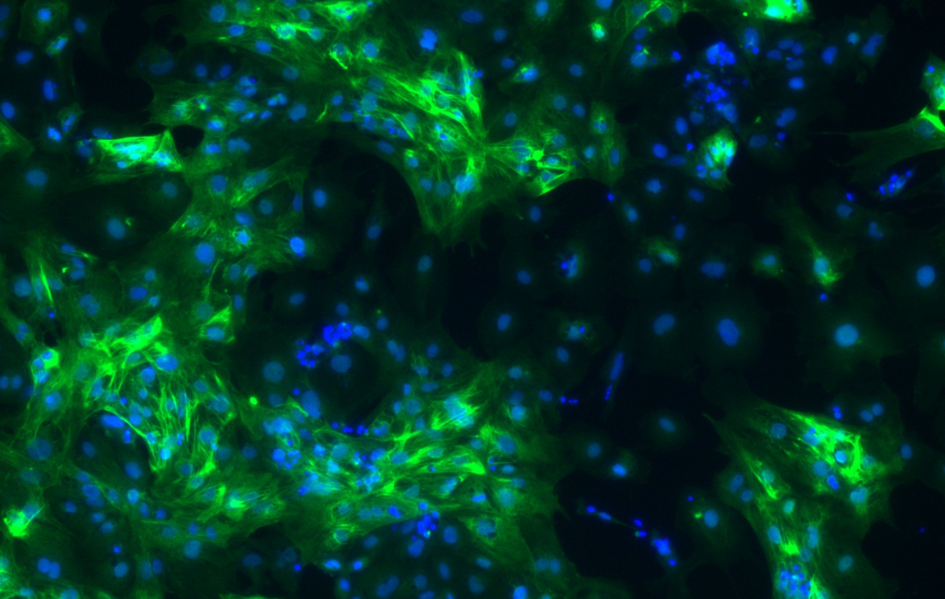
MYH7 in Human Cardiomyocytes.
MYH7 was detected in immersion fixed BG01V human embryonic stem cells differentiated into cardiomyocytes using Rabbit Anti-Human MYH7 Monoclonal Antibody (Catalog # MAB90961) at 10 µg/mL for 3 hours at room temperature. Cells were stained using the NorthernLights™ 557-conjugated Anti-Rabbit IgG Secondary Antibody (red; Catalog # NL004) and counterstained with DAPI (blue). Specific staining was localized to cytoplasm. View our protocol for Fluorescent ICC Staining of Stem Cells on Coverslips.Applications for Human MYH7 Antibody (2021A)
Immunocytochemistry
Sample: Immersion fixed BG01V human embryonic stem cells differentiated into cardiomyocytes
Western Blot
Sample: Human heart (ventricle) tissue
Reviewed Applications
Read 1 review rated 4 using MAB90961 in the following applications:
Formulation, Preparation, and Storage
Purification
Reconstitution
Reconstitute at 0.5 mg/mL in sterile PBS. For liquid material, refer to CoA for concentration.
Formulation
Shipping
Stability & Storage
- 12 months from date of receipt, -20 to -70 °C as supplied.
- 1 month, 2 to 8 °C under sterile conditions after reconstitution.
- 6 months, -20 to -70 °C under sterile conditions after reconstitution.
Calculators
Background: MYH7
Long Name
Alternate Names
Gene Symbol
UniProt
Additional MYH7 Products
Product Documents for Human MYH7 Antibody (2021A)
Certificate of Analysis
To download a Certificate of Analysis, please enter a lot or batch number in the search box below.
Note: Certificate of Analysis not available for kit components.
Product Specific Notices for Human MYH7 Antibody (2021A)
Contains <0.1% Sodium Azide, which is not hazardous at this concentration according to GHS classifications. Refer to SDS for additional information and handling instructions.
For research use only
Related Research Areas
Citations for Human MYH7 Antibody (2021A)
 Powered by Bioz
Customer Reviews for Human MYH7 Antibody (2021A) (1)
Have you used Human MYH7 Antibody (2021A)?
Submit a review and receive an Amazon gift card!
$25/€18/£15/$25CAN/¥2500 Yen for a review with an image
$10/€7/£6/$10CAN/¥1110 Yen for a review without an image
Submit a review
Customer Images
-
Application: Immunocytochemistry/ImmunofluorescenceSample Tested: human iPSC-derived cardiomyocytesSpecies: HumanVerified Customer | Posted 03/05/2019the antibody was used for immunofluorescence staining on day 15 human iPSC-derived cardiomyocytes (the day of initiation of differentiation from iPSCs is counted as day 0).

There are no reviews that match your criteria.
Protocols
Find general support by application which include: protocols, troubleshooting, illustrated assays, videos and webinars.
- Appropriate Fixation of IHC/ICC Samples
- Cellular Response to Hypoxia Protocols
- ClariTSA™ Fluorophore Kits
- Detection & Visualization of Antibody Binding
- ICC Cell Smear Protocol for Suspension Cells
- ICC Immunocytochemistry Protocol Videos
- ICC for Adherent Cells
- Immunocytochemistry (ICC) Protocol
- Immunocytochemistry Troubleshooting
- Immunofluorescence of Organoids Embedded in Cultrex Basement Membrane Extract
- Immunohistochemistry (IHC) and Immunocytochemistry (ICC) Protocols
- Preparing Samples for IHC/ICC Experiments
- Preventing Non-Specific Staining (Non-Specific Binding)
- Primary Antibody Selection & Optimization
- Protocol for VisUCyte™ HRP Polymer Detection Reagent
- Protocol for the Fluorescent ICC Staining of Cell Smears - Graphic
- Protocol for the Fluorescent ICC Staining of Cultured Cells on Coverslips - Graphic
- Protocol for the Preparation and Fluorescent ICC Staining of Cells on Coverslips
- Protocol for the Preparation and Fluorescent ICC Staining of Non-adherent Cells
- Protocol for the Preparation and Fluorescent ICC Staining of Stem Cells on Coverslips
- Protocol for the Preparation of a Cell Smear for Non-adherent Cell ICC - Graphic
- R&D Systems Quality Control Western Blot Protocol
- TUNEL and Active Caspase-3 Detection by IHC/ICC Protocol
- The Importance of IHC/ICC Controls
- Troubleshooting Guide: Western Blot Figures
- Western Blot Conditions
- Western Blot Protocol
- Western Blot Protocol for Cell Lysates
- Western Blot Troubleshooting
- Western Blot Troubleshooting Guide
- View all Protocols, Troubleshooting, Illustrated assays and Webinars