OSM R beta is a 150‑180 kDa member of the IL-6 receptor family. It associates with gp130 to form the type II OSM receptor that is responsive to OSM. The gp130 subunit is shared by other IL-6 family cytokine receptors (1, 2, 3, 4), and OSM R beta associates with gp130-like receptor (GPL) to form a receptor complex responsive to IL-31 (5, 6). The human OSM R beta cDNA encodes a 979 amino acid (aa) precursor that includes a 27 aa signal sequence, a 712 aa extracellular domain (ECD), a 22 aa transmembrane segment, and a 218 aa cytoplasmic domain. The ECD contains one partial and one complete hematopoietin domain, an Ig-like domain, and three fibronectin type-III domains. The cytoplasmic domain contains box1, 2, and 3 motifs (7). Within the ECD, human OSM R beta shares 55%, 58%, 61%, and 72% aa sequence identity with mouse, rat, bovine, and canine OSM R beta, respectively. It also shares 31% aa sequence identity with human LIF R, but less than 20% aa sequence identity with human CNTF R alpha, G-CSF R, IL-6 R, IL-11 R alpha, and TCCR. OSM R beta does not bind cytokines directly, but increases the affinity of gp130 for OSM, and GPL for IL-31 (7, 8). OSM R beta, gp130, and GPL each initiate signaling events following ligand stimulation (9, 10). Jak/STAT and MAPK pathways are activated by OSM R beta -containing receptors (9, 11, 12, 13), including STAT5b and SHC which are not activated by other IL-6 family receptors (10, 13). In mice, the loss of OSM R beta expression blocks erythroid progenitor development in bone marrow, and dramatically reduces the number of circulating platelets and erythrocytes (14). The type II OSM receptor is the only IL-6 family receptor that promotes osteoblast differentiation in calvaria cell cultures (15).

Key Product Details
Validated by
Knockout/Knockdown
Species Reactivity
Human
Applications
Knockout Validated, Western Blot, Flow Cytometry, CyTOF-ready
Label
Unconjugated
Antibody Source
Polyclonal Goat IgG
Loading...
Product Specifications
Immunogen
Mouse myeloma cell line NS0-derived recombinant human OSM R beta
Glu28-Ser739
Accession # Q99650
Glu28-Ser739
Accession # Q99650
Specificity
Detects human OSM R beta in direct ELISAs and Western blots. In Western blots, less than 5% cross-reactivity with recombinant mouse OSM R beta is observed.
Clonality
Polyclonal
Host
Goat
Isotype
IgG
Scientific Data Images for Human OSMR beta Antibody
Detection of OSM R beta in HeLa Human Cell Line by Flow Cytometry.
HeLa human cervical epithelial carcinoma cell line was stained with Goat Anti-Human OSM R beta Antigen Affinity-purified Polyclonal Antibody (Catalog # AF4389, filled histogram) or isotype control antibody (Catalog # AB-108-C, open histogram), followed by Phycoerythrin-conjugated Anti-Goat IgG Secondary Antibody (Catalog # F0107).
OSM R beta Specificity is Shown by Flow Cytometry in Knockout Cell Line.
OSM R beta knockout HeLa epithelial carcinoma cell line was stained with Mouse Anti-Human OSM R beta Affinity Purified Polyclonal Antibody (Catalog # AF4389, filled histogram) or isotype control antibody (Catalog # AB-108-C, open histogram) followed by PE-conjugated anti-Goat IgG Secondary Antibody (Catalog # F0107). No staining in the OSM R beta knockout HeLa cell line was observed. View our protocol for Staining Membrane-associated Proteins.Applications for Human OSMR beta Antibody
Application
Recommended Usage
CyTOF-ready
Ready to be labeled using established conjugation methods. No BSA or other carrier proteins that could interfere with conjugation.
Flow Cytometry
0.25 µg/106 cells
Sample: HeLa human cervical epithelial carcinoma cell line
Sample: HeLa human cervical epithelial carcinoma cell line
Knockout Validated
0.25 µg/106 cells
Sample: OSM R beta is specifically detected in HeLa human carcinoma parental cell line but is not detectable in OSM R beta knockout HeLa cell line.
Sample: OSM R beta is specifically detected in HeLa human carcinoma parental cell line but is not detectable in OSM R beta knockout HeLa cell line.
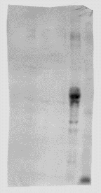
Western Blot
0.1 µg/mL
Sample: Recombinant Human OSM R beta (Catalog # 4389-OR)
Sample: Recombinant Human OSM R beta (Catalog # 4389-OR)
Reviewed Applications
Read 1 review rated 2 using AF4389 in the following applications:
Flow Cytometry Panel Builder
Bio-Techne Knows Flow Cytometry
Save time and reduce costly mistakes by quickly finding compatible reagents using the Panel Builder Tool.
Advanced Features
- Spectra Viewer - Custom analysis of spectra from multiple fluorochromes
- Spillover Popups - Visualize the spectra of individual fluorochromes
- Antigen Density Selector - Match fluorochrome brightness with antigen density

Formulation, Preparation, and Storage
Purification
Antigen Affinity-purified
Reconstitution
Reconstitute at 0.2 mg/mL in sterile PBS. For liquid material, refer to CoA for concentration.
Loading...
Formulation
Lyophilized from a 0.2 μm filtered solution in PBS with Trehalose. *Small pack size (SP) is supplied either lyophilized or as a 0.2 µm filtered solution in PBS.
Shipping
Lyophilized product is shipped at ambient temperature. Liquid small pack size (-SP) is shipped with polar packs. Upon receipt, store immediately at the temperature recommended below.
Stability & Storage
Use a manual defrost freezer and avoid repeated freeze-thaw cycles.
- 12 months from date of receipt, -20 to -70 °C as supplied.
- 1 month, 2 to 8 °C under sterile conditions after reconstitution.
- 6 months, -20 to -70 °C under sterile conditions after reconstitution.
Calculators
Background: OSMR beta
References
- Chen, S.-H. and E.N. Benveniste (2004) Cytokine Growth Factor Rev. 15:379.
- Heinrich, P.C. et al. (2003) Biochem. J. 374:1.
- Tanaka, M. and A. Miyajima (2003) Rev. Physiol. Biochem. Pharmacol. 149:39.
- Gearing, D.P. et al. (1992) Science 255:1434.
- Dillon, S.R. et al. (2004) Nat. Immunol. 5:752.
- Diveu, C. et al. (2003) J. Biol. Chem. 278:49850.
- Mosley, B. et al. (1996) J. Biol. Chem. 271:32635.
- Diveu, C. et al. (2004) Eur. Cytokine Netw. 15:291.
- Dreuw, A. et al. (2004) J. Biol. Chem. 279:36112.
- Wang, Y. et al. (2000) J. Biol. Chem. 275:25273.
- Hermanns, H.M. et al. (2000) J. Biol. Chem. 275:40742.
- Kuropatwinski, K.K. et al. (1997) J. Biol. Chem. 272:15135.
- Auguste, P. et al. (1997) J. Biol. Chem. 272:15760.
- Tanaka, M. et al. (2003) Blood 102:3154.
- Malaval, L. et al. (2005) J. Cell. Physiol. 204:585.
Long Name
Oncostatin M Receptor beta
Alternate Names
OSM R beta, OSMR, OSMRB
Gene Symbol
OSMR
UniProt
Additional OSMR beta Products
Product Documents for Human OSMR beta Antibody
Certificate of Analysis
To download a Certificate of Analysis, please enter a lot or batch number in the search box below.
Note: Certificate of Analysis not available for kit components.
Product Specific Notices for Human OSMR beta Antibody
For research use only
Related Research Areas
Customer Reviews for Human OSMR beta Antibody (1)
2 out of 5
1 Customer Rating
Have you used Human OSMR beta Antibody?
Submit a review and receive an Amazon gift card!
$25/€18/£15/$25CAN/¥2500 Yen for a review with an image
$10/€7/£6/$10CAN/¥1110 Yen for a review without an image
Submit a review
Customer Images
Showing
1
-
1 of
1 review
Showing All
Filter By:
-
Application: Western BlotSample Tested: Cell Lysates and Tissue LysatesSpecies: HumanVerified Customer | Posted 05/06/2021Tried with various conditions on cells lysates without success by Western blot. However, when I tried an OSMR antibody from a different company on the same samples alongside, that worked no problem. The antibody was only tried for Western blot.

There are no reviews that match your criteria.
Protocols
Find general support by application which include: protocols, troubleshooting, illustrated assays, videos and webinars.
- 7-Amino Actinomycin D (7-AAD) Cell Viability Flow Cytometry Protocol
- Cellular Response to Hypoxia Protocols
- Extracellular Membrane Flow Cytometry Protocol
- Flow Cytometry Protocol for Cell Surface Markers
- Flow Cytometry Protocol for Staining Membrane Associated Proteins
- Flow Cytometry Staining Protocols
- Flow Cytometry Troubleshooting Guide
- Intracellular Flow Cytometry Protocol Using Alcohol (Methanol)
- Intracellular Flow Cytometry Protocol Using Detergents
- Intracellular Nuclear Staining Flow Cytometry Protocol Using Detergents
- Intracellular Staining Flow Cytometry Protocol Using Alcohol Permeabilization
- Intracellular Staining Flow Cytometry Protocol Using Detergents to Permeabilize Cells
- Propidium Iodide Cell Viability Flow Cytometry Protocol
- Protocol for Liperfluo
- Protocol for the Characterization of Human Th22 Cells
- Protocol for the Characterization of Human Th9 Cells
- Protocol: Annexin V and PI Staining by Flow Cytometry
- Protocol: Annexin V and PI Staining for Apoptosis by Flow Cytometry
- R&D Systems Quality Control Western Blot Protocol
- Troubleshooting Guide: Fluorokine Flow Cytometry Kits
- Troubleshooting Guide: Western Blot Figures
- Western Blot Conditions
- Western Blot Protocol
- Western Blot Protocol for Cell Lysates
- Western Blot Troubleshooting
- Western Blot Troubleshooting Guide
- View all Protocols, Troubleshooting, Illustrated assays and Webinars
Loading...