Best Seller
Human PDX-1/IPF1 Antibody
R&D Systems | Catalog # AF2419

Key Product Details
Species Reactivity
Validated:
Human
Cited:
Human, Mouse, Rat, Porcine, Xenograft
Applications
Validated:
Immunohistochemistry, Western Blot, Dual RNAscope ISH-IHC Compatible, Immunocytochemistry, Simple Western
Cited:
Immunohistochemistry, Immunohistochemistry-Paraffin, Immunohistochemistry-Frozen, Western Blot, Flow Cytometry, Immunocytochemistry, Immunoprecipitation, Mass Cytometry
Label
Unconjugated
Antibody Source
Polyclonal Goat IgG
Loading...
Product Specifications
Immunogen
E. coli-derived recombinant human PDX‑1/IPF1
Ala91-Arg283
Accession # P52945
Ala91-Arg283
Accession # P52945
Specificity
Detects human PDX‑1/IPF1 in direct ELISAs and Western blots. In direct ELISAs, approximately 45% cross-reactivity with recombinant mouse PDX-1 is observed.
Clonality
Polyclonal
Host
Goat
Isotype
IgG
Scientific Data Images for Human PDX-1/IPF1 Antibody
Detection of Human PDX‑1/IPF1 by Western Blot.
Western blot shows lysates of beta TC-6 mouse beta cell insulinoma cell line. PVDF membrane was probed with 1 µg/mL of Goat Anti-Human PDX-1/IPF1 Antigen Affinity-purified Polyclonal Antibody (Catalog # AF2419) followed by HRP-conjugated Anti-Goat IgG Secondary Antibody (Catalog # HAF019). A specific band was detected for PDX-1/IPF1 at approximately 45 kDa (as indicated). This experiment was conducted under reducing conditions and using Immunoblot Buffer Group 8.
Detection of Mouse PDX‑1/IPF1 by Simple WesternTM.
Simple Western lane view shows lysates of beta TC-6 mouse beta cell insulinoma cell line, loaded at 0.2 mg/mL. A specific band was detected for PDX-1/IPF1 at approximately 48 kDa (as indicated) using 10 µg/mL of Goat Anti-Human PDX-1/IPF1 Antigen Affinity-purified Polyclonal Antibody (Catalog # AF2419) followed by 1:50 dilution of HRP-conjugated Anti-Goat IgG Secondary Antibody (Catalog # HAF109). This experiment was conducted under reducing conditions and using the 12-230 kDa separation system.
PDX‑1/IPF1 in BG01V Human Embryonic Stem Cells.
PDX-1/IPF1 was detected in immersion fixed BG01V human embryonic stem cells differentiated into pancreatic progenitor cells using Goat Anti-Human PDX-1/IPF1 Antigen Affinity-purified Polyclonal Antibody (Catalog # AF2419) at 10 µg/mL for 3 hours at room temperature. Cells were stained using the NorthernLights™ 493-conjugated Anti-Goat IgG Secondary Antibody (green; Catalog # NL003) and counterstained with DAPI (blue). Specific staining was localized to nuclei. View our protocol for Fluorescent ICC Staining of Stem Cells on Coverslips.
PDX‑1/IPF1 in Human Pancreatic Cancer Tissue.
PDX-1/IPF1 was detected in immersion fixed paraffin-embedded sections of human pancreatic cancer tissue using Goat Anti-Human PDX-1/IPF1 Antigen Affinity-purified Polyclonal Antibody (Catalog # AF2419) at 15 µg/mL overnight at 4 °C. Tissue was stained using the Anti-Goat HRP-DAB Cell & Tissue Staining Kit (brown; Catalog # CTS008) and counterstained with hematoxylin (blue). Specific staining was localized to nuclei in cancer cells. View our protocol for Chromogenic IHC Staining of Paraffin-embedded Tissue Sections.
Detection of Human PDX-1/IPF1 by Immunocytochemistry/Immunofluorescence
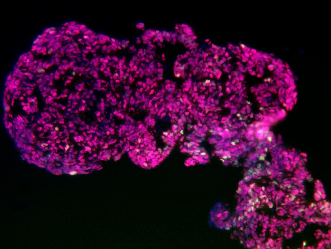
Differentiation of MODY3-iPSCs into pancreatic beta cells.(A) scheme of 6 step differentiation protocol (B), expression pattern of HNF1A mRNA during differentiation process, (C) Phase-contrast image of spheroids of differentiated MODY3-iPS-beta cells, (D, E) Immunocytochemistry of differentiated MODY3-iPS-beta cells for pancreatic beta cell markers (D) C-peptide, PDX1, NKX6.1 and DAPI staining, (E) HNF1A and DAPI staining. Image collected and cropped by CiteAb from the following publication (https://pubmed.ncbi.nlm.nih.gov/31145732), licensed under a CC-BY license. Not internally tested by R&D Systems.
Detection of PDX‑1/IPF1 in Human Pancreas.
Formalin-fixed paraffin-embedded tissue sections of human pancreas were probed for PDX-1 mRNA (ACD RNAScope Probe, catalog #437088; Fast Red chromogen, ACD catalog # 322750). Adjacent tissue section was processed for immunohistochemistry using goat anti-human PDX-1 polyclonal antibody (R&D Systems catalog # AF2419) at 1ug/mL with overnight incubation at 4 degrees Celsius followed by incubation with anti-goat IgG VisUCyte HRP Polymer Antibody (Catalog # VC004) and DAB chromogen (yellow-brown). Tissue was counterstained with hematoxylin (blue). Specific staining was localized to islet cells and exocrine glands.
Detection of Human Human PDX-1/IPF1 Antibody by Flow Cytometry
GP2 and CD142 comparative profiles. a–j Flow cytometry analyses of H1-derived day 13 PP cultures obtained using 10 mM nicotinamide (a–e, optimal differentiation) and 3.3 mM nicotinamide (f–j, suboptimal differentiation) during stage 4 of differentiation. Cells were stained with anti-GP2 and anti-CD142 and then fixed and stained for the intracellular markers PDX1 and NKX6-1. IgG controls are shown on the left of each panel. Green boxes highlight the low percentage of GP2+/PDX− cells (c–h), red box highlights the presence of CD142+PDX1− cells (j). Abbreviations: GP2, pancreatic secretory granule membrane major glycoprotein 2; CD142, tissue factor; NKX6-1, NK6 homeobox 1; PDX1, pancreatic, and duodenal homeobox 1 Image collected and cropped by CiteAb from the following publication (https://pubmed.ncbi.nlm.nih.gov/28835709), licensed under a CC-BY license. Not internally tested by R&D Systems.
Detection of Human Human PDX-1/IPF1 Antibody by Flow Cytometry
MAC-sorted GP2+ cells give rise to ‘ beta -like’ cells in vitro. a Flow plots showing the GP2 profile at day 13 of differentiation of H9 cells. Cells were analyzed either before MACS sorting (presort), after GP2 enrichment from a positive selection column (GP2+) or in the flow through from a depletion column. Top right plot shows NKX6-1 and PDX1 expression by flow cytometry in day 13 unsorted (presort) H9 cells. b Following MACS sorting for GP2 at day 13, cells were cultured generate beta -like cells up to day 23. Representative flow cytometry plots of NKX6-1 and C-PEPTIDE (CPEP) expression at day 23 of differentiation from either unsorted (PRESORT), enriched for GP2 using a MACS positive selection column (GP2+) or in the flow through cell population (Flow-). The bar graph shows the average percentage of NKX6-1+/C-PEPTIDE+ cells at Day 23. N = 4, error bars indicate s.e.m. **p < 0.01, One-way ANOVA. c Model depicting the in vivo and in vitro equivalent of the human multipotent pancreatic progenitor (MPC) expressing PTF1A/GP2/NKX6-1. The MPC residing at the tip of the developing human pancreas has the potential to develop into acinar (PTF1A+/GP2+) and ductal/endocrine (NKX6-1+/GP2−) progenitors. Abbreviations: GP2, pancreatic secretory granule membrane major glycoprotein 2; D13, day 13; NKX6-1, NK6 homeobox 1; PDX1, pancreatic and duodenal homeobox 1; PTF1A, pancreas specific transcription factor 1a Image collected and cropped by CiteAb from the following publication (https://pubmed.ncbi.nlm.nih.gov/28835709), licensed under a CC-BY license. Not internally tested by R&D Systems.
Detection of Human Human PDX-1/IPF1 Antibody by Flow Cytometry
GP2 and CD142 comparative profiles. a–j Flow cytometry analyses of H1-derived day 13 PP cultures obtained using 10 mM nicotinamide (a–e, optimal differentiation) and 3.3 mM nicotinamide (f–j, suboptimal differentiation) during stage 4 of differentiation. Cells were stained with anti-GP2 and anti-CD142 and then fixed and stained for the intracellular markers PDX1 and NKX6-1. IgG controls are shown on the left of each panel. Green boxes highlight the low percentage of GP2+/PDX− cells (c–h), red box highlights the presence of CD142+PDX1− cells (j). Abbreviations: GP2, pancreatic secretory granule membrane major glycoprotein 2; CD142, tissue factor; NKX6-1, NK6 homeobox 1; PDX1, pancreatic, and duodenal homeobox 1 Image collected and cropped by CiteAb from the following publication (https://pubmed.ncbi.nlm.nih.gov/28835709), licensed under a CC-BY license. Not internally tested by R&D Systems.
Detection of Human Human PDX-1/IPF1 Antibody by Flow Cytometry
GP2 and CD142 comparative profiles. a–j Flow cytometry analyses of H1-derived day 13 PP cultures obtained using 10 mM nicotinamide (a–e, optimal differentiation) and 3.3 mM nicotinamide (f–j, suboptimal differentiation) during stage 4 of differentiation. Cells were stained with anti-GP2 and anti-CD142 and then fixed and stained for the intracellular markers PDX1 and NKX6-1. IgG controls are shown on the left of each panel. Green boxes highlight the low percentage of GP2+/PDX− cells (c–h), red box highlights the presence of CD142+PDX1− cells (j). Abbreviations: GP2, pancreatic secretory granule membrane major glycoprotein 2; CD142, tissue factor; NKX6-1, NK6 homeobox 1; PDX1, pancreatic, and duodenal homeobox 1 Image collected and cropped by CiteAb from the following publication (https://pubmed.ncbi.nlm.nih.gov/28835709), licensed under a CC-BY license. Not internally tested by R&D Systems.
Detection of Human PDX-1/IPF1 by Immunocytochemistry/ Immunofluorescence
(A) Differentiation markers for the endoderm (FOXA2, PDX1), mesoderm ( alpha -actin, brachyury), and ectoderm ( beta 3 Tubulin, Otx2) were detected for the MUCG01, MUCG02, and MUCG03 clinical-grade hESC lines by immunocytochemistry. (B) Standard karyotypes without chromosomal aberration were detected for all hESC lines MUCG01: 46,XX; MUCG02: 46,XX; and MUCG03: 46,XY. (C) Pluripotency markers Oct3/4, Sox2, and Nanog were detected for the MUCG01, MUCG02, and MUCG03 clinical-grade hESC lines by immunocytochemistry. Image collected and cropped by CiteAb from the following open publication (https://pubmed.ncbi.nlm.nih.gov/36293356), licensed under a CC-BY license. Not internally tested by R&D Systems.
Detection of PDX-1/IPF1 by Flow Cytometry
PP formation is affected by the loss of DSC2 and SLC22A1. KO cell lines were differentiated to (A) pancreatic endoderm stage followed by (B–D) pancreatic progenitor stage. Differentiation efficiency was analysed by flow cytometry staining of PDX1 (PE) or PDX1 and NKX6-1 (PP) (n = 3 independent experiments, 2 different clones per genotype and 3 different WT clones, each clone in technical duplicates, dots represent means of these duplicates). (B,D) To better unveil differences in differentiation efficiencies, cells were differentiated without indolactam V (ILV) (n = 1 experiment, at least 2 clones per genotype each in technical duplicates). (B) Representative FACS plot. Mann–Whitney test was used for statistical analysis; error bars represent mean ± SD, ** p < 0.01, * p < 0.05. Image collected and cropped by CiteAb from the following open publication (https://pubmed.ncbi.nlm.nih.gov/35159392), licensed under a CC-BY license. Not internally tested by R&D Systems.Applications for Human PDX-1/IPF1 Antibody
Application
Recommended Usage
Dual RNAscope ISH-IHC Compatible
5-15 µg/mL
Sample: Immersion fixed paraffin-embedded sections of human pancreas
Sample: Immersion fixed paraffin-embedded sections of human pancreas
Immunocytochemistry
5-15 µg/mL
Sample: Immersion fixed BG01V human embryonic stem cells differentiated into pancreatic progenitor cells
Sample: Immersion fixed BG01V human embryonic stem cells differentiated into pancreatic progenitor cells
Immunohistochemistry
5-15 µg/mL
Sample: Immersion fixed paraffin-embedded sections of human pancreatic cancer tissue
Sample: Immersion fixed paraffin-embedded sections of human pancreatic cancer tissue
Simple Western
10 µg/mL
Sample: beta TC‑6 mouse beta cell insulinoma cell line
Sample: beta TC‑6 mouse beta cell insulinoma cell line
Western Blot
1 µg/mL
Sample: beta TC‑6 mouse beta cell insulinoma cell line
Sample: beta TC‑6 mouse beta cell insulinoma cell line
Reviewed Applications
Read 3 reviews rated 5 using AF2419 in the following applications:
Formulation, Preparation, and Storage
Purification
Antigen Affinity-purified
Reconstitution
Reconstitute at 0.2 mg/mL in sterile PBS. For liquid material, refer to CoA for concentration.
Loading...
Formulation
Lyophilized from a 0.2 μm filtered solution in PBS with Trehalose. See Certificate of Analysis for details.
*Small pack size (-SP) is supplied either lyophilized or as a 0.2 µm filtered solution in PBS.
*Small pack size (-SP) is supplied either lyophilized or as a 0.2 µm filtered solution in PBS.
Shipping
Lyophilized product is shipped at ambient temperature. Liquid small pack size (-SP) is shipped with polar packs. Upon receipt, store immediately at the temperature recommended below.
Stability & Storage
Use a manual defrost freezer and avoid repeated freeze-thaw cycles.
- 12 months from date of receipt, -20 to -70 °C as supplied.
- 1 month, 2 to 8 °C under sterile conditions after reconstitution.
- 6 months, -20 to -70 °C under sterile conditions after reconstitution.
Calculators
Background: PDX-1/IPF1
Long Name
Pancreas/Duodenum Homeobox-1
Alternate Names
IDX1, IPF1
Gene Symbol
PDX1
UniProt
Additional PDX-1/IPF1 Products
Product Documents for Human PDX-1/IPF1 Antibody
Certificate of Analysis
To download a Certificate of Analysis, please enter a lot or batch number in the search box below.
Note: Certificate of Analysis not available for kit components.
Product Specific Notices for Human PDX-1/IPF1 Antibody
For research use only
Citations for Human PDX-1/IPF1 Antibody
 Powered by Bioz
Customer Reviews for Human PDX-1/IPF1 Antibody (3)
5 out of 5
3 Customer Ratings
Have you used Human PDX-1/IPF1 Antibody?
Submit a review and receive an Amazon gift card!
$25/€18/£15/$25CAN/¥2500 Yen for a review with an image
$10/€7/£6/$10CAN/¥1110 Yen for a review without an image
Submit a review
Customer Images
Showing
1
-
3 of
3 reviews
Showing All
Filter By:
-
Application: Immunocytochemistry/ImmunofluorescenceSample Tested: iPS2 human induced pluripotent stem cellsSpecies: HumanVerified Customer | Posted 04/17/2018

-
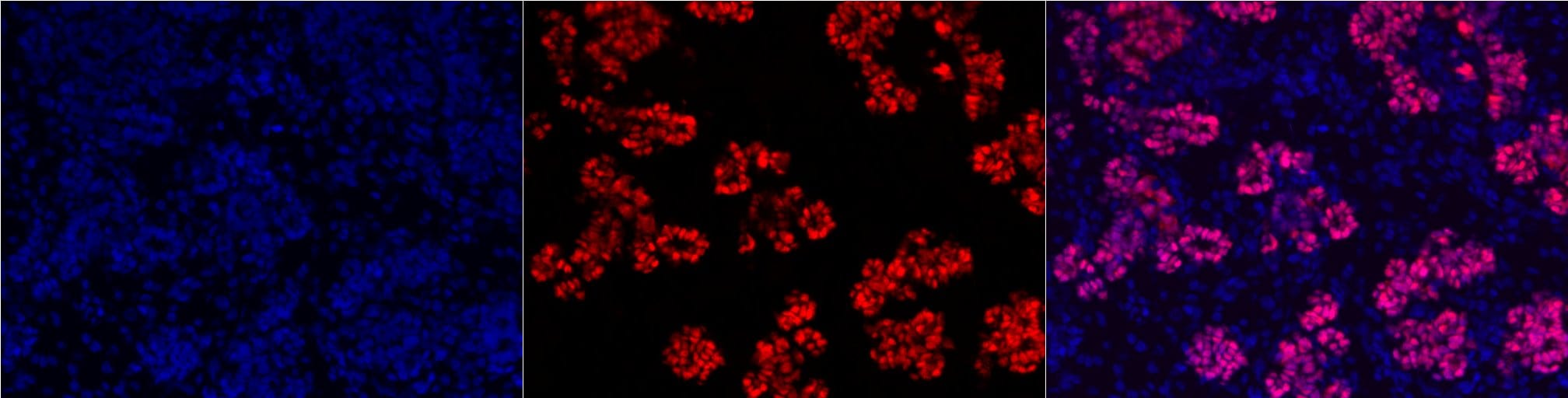
Application: ImmunohistochemistrySample Tested: Fetal pancreasSpecies: HumanVerified Customer | Posted 07/08/20161:50 on paraffin sections PDX1 (Red) and DAPI nuclear stain
-
Application: ImmunohistochemistrySample Tested: Pancreas tissueSpecies: MouseVerified Customer | Posted 06/29/2016
There are no reviews that match your criteria.
Protocols
Find general support by application which include: protocols, troubleshooting, illustrated assays, videos and webinars.
- Antigen Retrieval Protocol (PIER)
- Antigen Retrieval for Frozen Sections Protocol
- Appropriate Fixation of IHC/ICC Samples
- Cellular Response to Hypoxia Protocols
- Chromogenic IHC Staining of Formalin-Fixed Paraffin-Embedded (FFPE) Tissue Protocol
- Chromogenic Immunohistochemistry Staining of Frozen Tissue
- ClariTSA™ Fluorophore Kits
- Detection & Visualization of Antibody Binding
- Fluorescent IHC Staining of Frozen Tissue Protocol
- Graphic Protocol for Heat-induced Epitope Retrieval
- Graphic Protocol for the Preparation and Fluorescent IHC Staining of Frozen Tissue Sections
- Graphic Protocol for the Preparation and Fluorescent IHC Staining of Paraffin-embedded Tissue Sections
- Graphic Protocol for the Preparation of Gelatin-coated Slides for Histological Tissue Sections
- ICC Cell Smear Protocol for Suspension Cells
- ICC Immunocytochemistry Protocol Videos
- ICC for Adherent Cells
- IHC Sample Preparation (Frozen sections vs Paraffin)
- ISH-IHC Protocol for Chromogenic Detection on Formalin Fixed Paraffin Embedded (FFPE) Tissue
- Immunocytochemistry (ICC) Protocol
- Immunocytochemistry Troubleshooting
- Immunofluorescence of Organoids Embedded in Cultrex Basement Membrane Extract
- Immunofluorescent IHC Staining of Formalin-Fixed Paraffin-Embedded (FFPE) Tissue Protocol
- Immunohistochemistry (IHC) and Immunocytochemistry (ICC) Protocols
- Immunohistochemistry Frozen Troubleshooting
- Immunohistochemistry Paraffin Troubleshooting
- Preparing Samples for IHC/ICC Experiments
- Preventing Non-Specific Staining (Non-Specific Binding)
- Primary Antibody Selection & Optimization
- Protocol for Heat-Induced Epitope Retrieval (HIER)
- Protocol for Making a 4% Formaldehyde Solution in PBS
- Protocol for VisUCyte™ HRP Polymer Detection Reagent
- Protocol for the Fluorescent ICC Staining of Cell Smears - Graphic
- Protocol for the Fluorescent ICC Staining of Cultured Cells on Coverslips - Graphic
- Protocol for the Preparation & Fixation of Cells on Coverslips
- Protocol for the Preparation and Chromogenic IHC Staining of Frozen Tissue Sections
- Protocol for the Preparation and Chromogenic IHC Staining of Frozen Tissue Sections - Graphic
- Protocol for the Preparation and Chromogenic IHC Staining of Paraffin-embedded Tissue Sections
- Protocol for the Preparation and Chromogenic IHC Staining of Paraffin-embedded Tissue Sections - Graphic
- Protocol for the Preparation and Fluorescent ICC Staining of Cells on Coverslips
- Protocol for the Preparation and Fluorescent ICC Staining of Non-adherent Cells
- Protocol for the Preparation and Fluorescent ICC Staining of Stem Cells on Coverslips
- Protocol for the Preparation and Fluorescent IHC Staining of Frozen Tissue Sections
- Protocol for the Preparation and Fluorescent IHC Staining of Paraffin-embedded Tissue Sections
- Protocol for the Preparation of Gelatin-coated Slides for Histological Tissue Sections
- Protocol for the Preparation of a Cell Smear for Non-adherent Cell ICC - Graphic
- R&D Systems Quality Control Western Blot Protocol
- TUNEL and Active Caspase-3 Detection by IHC/ICC Protocol
- The Importance of IHC/ICC Controls
- Troubleshooting Guide: Immunohistochemistry
- Troubleshooting Guide: Western Blot Figures
- Western Blot Conditions
- Western Blot Protocol
- Western Blot Protocol for Cell Lysates
- Western Blot Troubleshooting
- Western Blot Troubleshooting Guide
- View all Protocols, Troubleshooting, Illustrated assays and Webinars
Loading...
Associated Pathways
