Mitogen- and stress-activated protein kinase 1 (MSK1), also known as ribosomal protein S6 kinase 5 (RPS6KA5), belongs to the AGC family of kinases. Both MSK1 and the related MSK2, also known as RSKB and RPS6KA4, have two kinase domains connected by a regulatory linker region, and are activated by the mitogen-activated protein kinases ERK1, ERK2, and p38. Nuclear MSK phosphorylates and activates a number of transcription factors, including ATF1 and CREB.
Human phospho-MSK1 (S212) Antibody
R&D Systems | Catalog # AF1036
 by Western Blot.")
Key Product Details
Validated by
Biological Validation
Species Reactivity
Validated:
Human
Cited:
Mouse
Applications
Validated:
Immunohistochemistry, Western Blot
Cited:
Western Blot
Label
Unconjugated
Antibody Source
Polyclonal Rabbit IgG
Loading...
Product Specifications
Immunogen
Phosphopeptide containing human MSK1 S212 site
Specificity
Detects human MSK1 when phosphorylated at S212.
Clonality
Polyclonal
Host
Rabbit
Isotype
IgG
Scientific Data Images for Human phospho-MSK1 (S212) Antibody
Detection of Human Phospho-MSK1 (S212) by Western Blot.
Western blot shows lysates of HeLa human cervical epithelial carcinoma cell line untreated (-) or treated (+) with 1 mM H2O2for 1 hour or 300 mM sorbitol for 30 minutes. PVDF membrane was probed with 0.2 µg/mL of Rabbit Anti-Human Phospho-MSK1 (S212) Antigen Affinity-purified Polyclonal Antibody (Catalog # AF1036), followed by HRP-conjugated Anti-Rabbit IgG Secondary Antibody (Catalog # HAF008). A specific band was detected for Phospho-MSK1 (S212) at approximately 90 kDa (as indicated). This experiment was conducted under reducing conditions and using Immunoblot Buffer Group 1.
MSK1 in Human Kidney Cancer Tissue.
MSK1 phosphorylated at S212 was detected in immersion fixed paraffin-embedded sections of human kidney cancer tissue using Rabbit Anti-Human Phospho-MSK1 (S212) Antigen Affinity-purified Polyclonal Antibody (Catalog # AF1036) at 15 µg/mL overnight at 4 °C. Tissue was stained using the Anti-Rabbit HRP-DAB Cell & Tissue Staining Kit (brown; Catalog # CTS005) and counterstained with hematoxylin (blue). View our protocol for Chromogenic IHC Staining of Paraffin-embedded Tissue Sections.Applications for Human phospho-MSK1 (S212) Antibody
Application
Recommended Usage
Immunohistochemistry
5-15 µg/mL
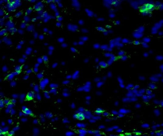
Sample: Perfusion fixed frozen sections of rat dorsal root ganglia and immersion fixed paraffin-embedded sections of human kidney cancer tissue
Sample: Perfusion fixed frozen sections of rat dorsal root ganglia and immersion fixed paraffin-embedded sections of human kidney cancer tissue
Western Blot
0.2 µg/mL
Sample: H2O2 or sorbitol-treated HeLa human cervical epithelial carcinoma cell line
Sample: H2O2 or sorbitol-treated HeLa human cervical epithelial carcinoma cell line
Reviewed Applications
Read 3 reviews rated 4.3 using AF1036 in the following applications:
Formulation, Preparation, and Storage
Purification
Antigen Affinity-purified
Reconstitution
Reconstitute at 0.2 mg/mL in sterile PBS. For liquid material, refer to CoA for concentration.
Loading...
Formulation
Lyophilized from a 0.2 μm filtered solution in PBS with Trehalose. *Small pack size (SP) is supplied either lyophilized or as a 0.2 µm filtered solution in PBS.
Shipping
Lyophilized product is shipped at ambient temperature. Liquid small pack size (-SP) is shipped with polar packs. Upon receipt, store immediately at the temperature recommended below.
Stability & Storage
Use a manual defrost freezer and avoid repeated freeze-thaw cycles.
- 12 months from date of receipt, -20 to -70 °C as supplied.
- 1 month, 2 to 8 °C under sterile conditions after reconstitution.
- 6 months, -20 to -70 °C under sterile conditions after reconstitution.
Calculators
Background: MSK1
Long Name
Mitogen- and Stress-activated Protein Kinase 1
Alternate Names
RPS6KA5
Gene Symbol
RPS6KA5
Additional MSK1 Products
Product Documents for Human phospho-MSK1 (S212) Antibody
Certificate of Analysis
To download a Certificate of Analysis, please enter a lot or batch number in the search box below.
Note: Certificate of Analysis not available for kit components.
Product Specific Notices for Human phospho-MSK1 (S212) Antibody
For research use only
Related Research Areas
Citations for Human phospho-MSK1 (S212) Antibody
 Powered by Bioz
Customer Reviews for Human phospho-MSK1 (S212) Antibody (3)
4.3 out of 5
3 Customer Ratings
Have you used Human phospho-MSK1 (S212) Antibody?
Submit a review and receive an Amazon gift card!
$25/€18/£15/$25CAN/¥2500 Yen for a review with an image
$10/€7/£6/$10CAN/¥1110 Yen for a review without an image
Submit a review
Customer Images
Showing
1
-
3 of
3 reviews
Showing All
Filter By:
-
Application: ImmunohistochemistrySample Tested: Melanoma tissueSpecies: HumanVerified Customer | Posted 04/06/2022Tissue was cryopreserved

-
Application: MicroarraysSample Tested: EDTA PlasmaSpecies: HumanVerified Customer | Posted 03/11/2019
-
Application: MicroarraySample Tested: EDTA PlasmaSpecies: HumanVerified Customer | Posted 10/09/2018
There are no reviews that match your criteria.
Protocols
Find general support by application which include: protocols, troubleshooting, illustrated assays, videos and webinars.
- Antigen Retrieval Protocol (PIER)
- Antigen Retrieval for Frozen Sections Protocol
- Appropriate Fixation of IHC/ICC Samples
- Cellular Response to Hypoxia Protocols
- Chromogenic IHC Staining of Formalin-Fixed Paraffin-Embedded (FFPE) Tissue Protocol
- Chromogenic Immunohistochemistry Staining of Frozen Tissue
- ClariTSA™ Fluorophore Kits
- Detection & Visualization of Antibody Binding
- Fluorescent IHC Staining of Frozen Tissue Protocol
- Graphic Protocol for Heat-induced Epitope Retrieval
- Graphic Protocol for the Preparation and Fluorescent IHC Staining of Frozen Tissue Sections
- Graphic Protocol for the Preparation and Fluorescent IHC Staining of Paraffin-embedded Tissue Sections
- Graphic Protocol for the Preparation of Gelatin-coated Slides for Histological Tissue Sections
- IHC Sample Preparation (Frozen sections vs Paraffin)
- Immunofluorescent IHC Staining of Formalin-Fixed Paraffin-Embedded (FFPE) Tissue Protocol
- Immunohistochemistry (IHC) and Immunocytochemistry (ICC) Protocols
- Immunohistochemistry Frozen Troubleshooting
- Immunohistochemistry Paraffin Troubleshooting
- Preparing Samples for IHC/ICC Experiments
- Preventing Non-Specific Staining (Non-Specific Binding)
- Primary Antibody Selection & Optimization
- Protocol for Heat-Induced Epitope Retrieval (HIER)
- Protocol for Making a 4% Formaldehyde Solution in PBS
- Protocol for VisUCyte™ HRP Polymer Detection Reagent
- Protocol for the Preparation & Fixation of Cells on Coverslips
- Protocol for the Preparation and Chromogenic IHC Staining of Frozen Tissue Sections
- Protocol for the Preparation and Chromogenic IHC Staining of Frozen Tissue Sections - Graphic
- Protocol for the Preparation and Chromogenic IHC Staining of Paraffin-embedded Tissue Sections
- Protocol for the Preparation and Chromogenic IHC Staining of Paraffin-embedded Tissue Sections - Graphic
- Protocol for the Preparation and Fluorescent IHC Staining of Frozen Tissue Sections
- Protocol for the Preparation and Fluorescent IHC Staining of Paraffin-embedded Tissue Sections
- Protocol for the Preparation of Gelatin-coated Slides for Histological Tissue Sections
- R&D Systems Quality Control Western Blot Protocol
- TUNEL and Active Caspase-3 Detection by IHC/ICC Protocol
- The Importance of IHC/ICC Controls
- Troubleshooting Guide: Immunohistochemistry
- Troubleshooting Guide: Western Blot Figures
- Western Blot Conditions
- Western Blot Protocol
- Western Blot Protocol for Cell Lysates
- Western Blot Troubleshooting
- Western Blot Troubleshooting Guide
- View all Protocols, Troubleshooting, Illustrated assays and Webinars
Loading...
Associated Pathways
