
Key Product Details
Species Reactivity
Validated:
Human
Cited:
Human
Applications
Validated:
Western Blot, Immunocytochemistry, Simple Western, Immunoprecipitation
Cited:
Western Blot, Immunoprecipitation
Label
Unconjugated
Antibody Source
Monoclonal Mouse IgG1 Clone # HL71/10
Loading...
Product Specifications
Immunogen
Protein kinase fraction from interferon-treated Daudi human Burkitt's lymphoma cell line. Laurent, A.G. et al. (1985) Proc. Natl. Acad. Sci. 82:4341.
Specificity
Detects human PKR in Western blots.
Clonality
Monoclonal
Host
Mouse
Isotype
IgG1
Scientific Data Images for Human PKR Antibody (HL71/10)
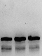
Detection of Human PKR by Western Blot.
Western blot shows lysates of HeLa human cervical epithelial carcinoma cell line, K562 human chronic myelogenous leukemia cell line, and HEK293 human embryonic kidney cell line. PVDF membrane was probed with 1 µg/mL of Mouse Anti-Human PKR Monoclonal Antibody (Catalog # MAB1980) followed by HRP-conjugated Anti-Mouse IgG Secondary Antibody (Catalog # HAF018). A specific band was detected for PKR at approximately 74 kDa (as indicated). This experiment was conducted under reducing conditions and using Immunoblot Buffer Group 1.
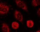
PKR in HeLa Human Cell Line.
PKR was detected in immersion fixed HeLa human cervical epithelial carcinoma cell line stimulated with rhIFN-alpha (Catalog # 11110-1) using Mouse Anti-Human PKR Monoclonal Antibody (Catalog # MAB1980) at 10 µg/mL for 3 hours at room temperature. Cells were stained using the NorthernLights™ 557-conjugated Anti-Mouse IgG Secondary Antibody (yellow; Catalog # NL007) and counterstained with DAPI (blue). View our protocol for Fluorescent ICC Staining of Cells on Coverslips.
Detection of Human PKR by Simple WesternTM.
Simple Western lane view shows lysates of K562 human chronic myelogenous leukemia cell line, loaded at 0.2 mg/mL. A specific band was detected for PKR at approximately 73 kDa (as indicated) using 10 µg/mL of Mouse Anti-Human PKR Monoclonal Antibody (Catalog # MAB1980). This experiment was conducted under reducing conditions and using the 12-230 kDa separation system. Non-specific interaction with the 230 kDa Simple Western standard may be seen with this antibody.Applications for Human PKR Antibody (HL71/10)
Application
Recommended Usage
Immunocytochemistry
8-25 µg/mL
Sample: Immersion fixed HeLa human cervical epithelial carcinoma cell line treated with universal IFN-alpha (Catalog # 11200-1) or recombinant human IFN-alpha (Catalog # 11100-1). Besse, S. et al. (1998) Exp. Cell Res. 239:379.
Sample: Immersion fixed HeLa human cervical epithelial carcinoma cell line treated with universal IFN-alpha (Catalog # 11200-1) or recombinant human IFN-alpha (Catalog # 11100-1). Besse, S. et al. (1998) Exp. Cell Res. 239:379.
Immunoprecipitation
Laurent, A.G. et al. (1985) Proc. Natl. Acad. Sci. 82:4341.
Simple Western
10 µg/mL
Sample: K562 human chronic myelogenous leukemia cell line
Sample: K562 human chronic myelogenous leukemia cell line
Western Blot
1 µg/mL
Sample: HeLa human cervical epithelial carcinoma cell line, K562 human chronic myelogenous leukemia cell line, and HEK293 human embryonic kidney cell line
Sample: HeLa human cervical epithelial carcinoma cell line, K562 human chronic myelogenous leukemia cell line, and HEK293 human embryonic kidney cell line
Reviewed Applications
Read 3 reviews rated 5 using MAB1980 in the following applications:
Formulation, Preparation, and Storage
Purification
Protein A or G purified from hybridoma culture supernatant
Reconstitution
Reconstitute at 0.5 mg/mL in sterile PBS. For liquid material, refer to CoA for concentration.
Loading...
Formulation
Lyophilized from a 0.2 μm filtered solution in PBS with Trehalose. *Small pack size (SP) is supplied either lyophilized or as a 0.2 µm filtered solution in PBS.
Shipping
Lyophilized product is shipped at ambient temperature. Liquid small pack size (-SP) is shipped with polar packs. Upon receipt, store immediately at the temperature recommended below.
Stability & Storage
Use a manual defrost freezer and avoid repeated freeze-thaw cycles.
- 12 months from date of receipt, -20 to -70 °C as supplied.
- 1 month, 2 to 8 °C under sterile conditions after reconstitution.
- 6 months, -20 to -70 °C under sterile conditions after reconstitution.
Calculators
Background: PKR
References
- Bonnet, M.C. et al. (2000) Mol. Cell Biol. 20:4532.
Long Name
Protein Kinase R
Alternate Names
EIF2AK2, P68 Kinase, PPP1R83, PRKR, Protein Kinase R
Gene Symbol
EIF2AK2
Additional PKR Products
Product Documents for Human PKR Antibody (HL71/10)
Certificate of Analysis
To download a Certificate of Analysis, please enter a lot or batch number in the search box below.
Note: Certificate of Analysis not available for kit components.
Product Specific Notices for Human PKR Antibody (HL71/10)
For research use only
Related Research Areas
Citations for Human PKR Antibody (HL71/10)
 Powered by Bioz
Customer Reviews for Human PKR Antibody (HL71/10) (3)
5 out of 5
3 Customer Ratings
Have you used Human PKR Antibody (HL71/10)?
Submit a review and receive an Amazon gift card!
$25/€18/£15/$25CAN/¥2500 Yen for a review with an image
$10/€7/£6/$10CAN/¥1110 Yen for a review without an image
Submit a review
Customer Images
Showing
1
-
3 of
3 reviews
Showing All
Filter By:
-
Application: Immunocytochemistry/ImmunofluorescenceSample Tested: Mitotic cellsSpecies: HumanVerified Customer | Posted 10/20/2021

-
Application: Western BlotSample Tested: HeLa cellsSpecies: HumanVerified Customer | Posted 07/25/2021
-
Application: Western BlotSample Tested: HeLa cellsSpecies: HumanVerified Customer | Posted 11/18/2019
There are no reviews that match your criteria.
Protocols
Find general support by application which include: protocols, troubleshooting, illustrated assays, videos and webinars.
- Appropriate Fixation of IHC/ICC Samples
- Cellular Response to Hypoxia Protocols
- ClariTSA™ Fluorophore Kits
- Detection & Visualization of Antibody Binding
- ICC Cell Smear Protocol for Suspension Cells
- ICC Immunocytochemistry Protocol Videos
- ICC for Adherent Cells
- Immunocytochemistry (ICC) Protocol
- Immunocytochemistry Troubleshooting
- Immunofluorescence of Organoids Embedded in Cultrex Basement Membrane Extract
- Immunohistochemistry (IHC) and Immunocytochemistry (ICC) Protocols
- Immunoprecipitation Protocol
- Preparing Samples for IHC/ICC Experiments
- Preventing Non-Specific Staining (Non-Specific Binding)
- Primary Antibody Selection & Optimization
- Protocol for VisUCyte™ HRP Polymer Detection Reagent
- Protocol for the Fluorescent ICC Staining of Cell Smears - Graphic
- Protocol for the Fluorescent ICC Staining of Cultured Cells on Coverslips - Graphic
- Protocol for the Preparation and Fluorescent ICC Staining of Cells on Coverslips
- Protocol for the Preparation and Fluorescent ICC Staining of Non-adherent Cells
- Protocol for the Preparation and Fluorescent ICC Staining of Stem Cells on Coverslips
- Protocol for the Preparation of a Cell Smear for Non-adherent Cell ICC - Graphic
- R&D Systems Quality Control Western Blot Protocol
- TUNEL and Active Caspase-3 Detection by IHC/ICC Protocol
- The Importance of IHC/ICC Controls
- Troubleshooting Guide: Western Blot Figures
- Western Blot Conditions
- Western Blot Protocol
- Western Blot Protocol for Cell Lysates
- Western Blot Troubleshooting
- Western Blot Troubleshooting Guide
- View all Protocols, Troubleshooting, Illustrated assays and Webinars
Loading...