Key Product Details

Validated by

Knockout/Knockdown, Biological Validation

Species Reactivity

Validated:

Human, Mouse, Rat

Cited:

Human, Mouse, Rat, Hamster, Transgenic Mouse

Applications

Validated:

Immunohistochemistry, Western Blot, Blockade of Receptor-ligand Interaction

Cited:

Immunohistochemistry, Immunohistochemistry-Paraffin, Immunohistochemistry-Frozen, Western Blot, Neutralization, Flow Cytometry, Immunocytochemistry, Blocking, ELISA Development

Label

Unconjugated

Antibody Source

Polyclonal Goat IgG
Loading...

Product Specifications

Immunogen

Mouse myeloma cell line NS0-derived recombinant human RAGE
Gln24-Ala344
Accession # Q15109

Specificity

Detects human RAGE in direct ELISAs and Western blots.

Clonality

Polyclonal

Host

Goat

Isotype

IgG

Endotoxin Level

<0.10 EU per 1 μg of the antibody by the LAL method.

Scientific Data Images for Human/Mouse/Rat RAGE/AGER Antibody

Detection of Human, Mouse, and Rat RAGE antibody by Western Blot.

Detection of Human, Mouse, and Rat RAGE by Western Blot.

Western blot shows lysates of human, mouse, and rat lung tissue. PVDF membrane was probed with 1 µg/mL of Goat Anti-Human/Mouse/Rat RAGE Antigen Affinity-purified Polyclonal Antibody (Catalog # AF1145) followed by HRP-conjugated Anti-Goat IgG Secondary Antibody (Catalog # HAF017). Specific bands were detected for RAGE at approximately 45-55 kDa (as indicated). This experiment was conducted under reducing conditions and using Immunoblot Buffer Group 1.

Detection of Human RAGE antibody by Western Blot.

Detection of Human RAGE by Western Blot.

Western blot shows lysates of human lung tissue and HEK001 human epidermal keratinocyte cell line. PVDF Membrane was probed with 1 µg/mL of Goat Anti-Human/Mouse/Rat RAGE Antigen Affinity-purified Polyclonal Antibody (Catalog # AF1145) followed by HRP-conjugated Anti-Goat IgG Secondary Antibody (Catalog # HAF017). Specific bands were detected for RAGE at approximately 43 kDa under non-reducing (NR) conditions and 50 kDa under reducing (R) conditions (as indicated). This experiment was conducted using Immunoblot Buffer Group 5.

RAGE antibody in Human Alzheimer's Disease Brain by Immunohistochemistry (IHC-P).

RAGE in Human Alzheimer's Disease Brain.

RAGE was detected in immersion fixed paraffin-embedded sections of human Alzheimer's disease brain (cerebellum) using 15 µg/mL Goat Anti-Human/Mouse/Rat RAGE Antigen Affinity-purified Polyclonal Antibody (Catalog # AF1145) overnight at 4 °C. Tissue was stained with the Anti-Goat HRP-DAB Cell & Tissue Staining Kit (brown; Catalog # CTS008) and counterstained with hematoxylin (blue). View our protocol for Chromogenic IHC Staining of Paraffin-embedded Tissue Sections.

RAGE antibody in Human Lung by Immunohistochemistry (IHC-P).

RAGE in Human Lung.

RAGE was detected in immersion fixed paraffin-embedded sections of human lung using Goat Anti-Human/Mouse/Rat RAGE Antigen Affinity-purified Polyclonal Antibody (Catalog # AF1145) at 0.5 µg/mL for 1 hour at room temperature followed by incubation with the Anti-Goat IgG VisUCyte™ HRP Polymer Antibody (Catalog # VC004). Tissue was stained using DAB (brown) and counterstained with hematoxylin (blue). Specific staining was localized to cell membranes. View our protocol for IHC Staining with VisUCyte HRP Polymer Detection Reagents.

Detection of Mouse AGER by Western Blot

Detection of Mouse AGER by Western Blot

RAGE expression mediates cell aggregation through homophilic interactions.(A) Western blot analysis on control preB 300.19 cells (preB/pCAGS) or expressing FL-RAGE (preB/FL-RAGE) using alpha RAGE N-term1 antibody. Forty µg of cell lysate were loaded and detection of GAPDH was used as loading control. (B) Cell aggregation assay. preB/pCAGS or preB/FL-RAGE cells were cultured for 2, 6 or 24 hours before being photographed at 20× magnification in phase contrast (left panel). Bar corresponds to 50 µm. Quantification of cell aggregates area (right panel). Results are displayed as means±SEM (*, P<0.05; **, P<0.01; ***, P<0.0001). (C) Mixed cell aggregation assay. Red preB (Red-preB) or preB/FL-RAGE (expressing GFP; preB/FL-RAGE-GFP) cells were grown as single cell line suspension or mixed in equal number for 24 hours. Red-preB/FL-RAGE-GFP were mixed with preB/FL-RAGE-GFP for the same time. Bar corresponds to 50 µm. Image collected and cropped by CiteAb from the following publication (https://pubmed.ncbi.nlm.nih.gov/24475194), licensed under a CC-BY license. Not internally tested by R&D Systems.
Detection of Rat AGER by Western Blot

Detection of Rat AGER by Western Blot

RAGE expression enhances cell-matrix adhesion.(A) Western blot analysis on rat lung lysate and R3/1 cells using three different antibodies recognizing RAGE extracellular (anti-RAGE N-term1 and anti-RAGE N-term2) or intracellular (anti-RAGE C-term) domains. Asterisk (*) indicates nonspecific bands. Forty µg of cell lysate were loaded and detection of GAPDH was used as loading control. (B) Western blot analysis on R3/1-pLXSN and R3/1-FL-RAGE cells using anti-RAGE N-term1 antibody. Forty µg of cell lysate were loaded and detection of GAPDH was used as loading control. (C, D) Cell-matrix adhesion assay. Adhesion of R3/1-pLXSN or R3/1-FL-RAGE cells onto culture dishes coated with 10 µg/ml ECM proteins. Adhesion to PBS, collagen I (Coll I), Fibronectin (FN), or Laminin (Lam) was assayed for 15 minutes (C) or 45 minutes (D). One representative experiment out of three is shown. Results from triplicate wells are displayed as means±SEM (ns, not significant; *, P<0.05; **, P<0.01). Image collected and cropped by CiteAb from the following publication (https://pubmed.ncbi.nlm.nih.gov/24475194), licensed under a CC-BY license. Not internally tested by R&D Systems.
Detection of Human AGER by Immunohistochemistry

Detection of Human AGER by Immunohistochemistry

Differential expression of RAGE and S100A8/A9 on protein level in whole skin.The expression level was analysed by immunochistochemistry using specific anti human S100A8/A9 and anti human RAGE antibodies. The expression was tested in 5 patient samples per group (IC and OTR with in situ or invasive SCC). a: Expression of RAGE and S100A8/A9 in IC and OTR patients with in situ SCC in comparison to normal skin. b: Expression of RAGE and S100A8/A9 in IC and OTR patient with invasive SCC in comparison to normal skin. Image collected and cropped by CiteAb from the following publication (https://dx.plos.org/10.1371/journal.pone.0120971), licensed under a CC-BY license. Not internally tested by R&D Systems.
Detection of Rat AGER by Western Blot

Detection of Rat AGER by Western Blot

RAGE expression enhances cell-matrix adhesion.(A) Western blot analysis on rat lung lysate and R3/1 cells using three different antibodies recognizing RAGE extracellular (anti-RAGE N-term1 and anti-RAGE N-term2) or intracellular (anti-RAGE C-term) domains. Asterisk (*) indicates nonspecific bands. Forty µg of cell lysate were loaded and detection of GAPDH was used as loading control. (B) Western blot analysis on R3/1-pLXSN and R3/1-FL-RAGE cells using anti-RAGE N-term1 antibody. Forty µg of cell lysate were loaded and detection of GAPDH was used as loading control. (C, D) Cell-matrix adhesion assay. Adhesion of R3/1-pLXSN or R3/1-FL-RAGE cells onto culture dishes coated with 10 µg/ml ECM proteins. Adhesion to PBS, collagen I (Coll I), Fibronectin (FN), or Laminin (Lam) was assayed for 15 minutes (C) or 45 minutes (D). One representative experiment out of three is shown. Results from triplicate wells are displayed as means±SEM (ns, not significant; *, P<0.05; **, P<0.01). Image collected and cropped by CiteAb from the following publication (https://pubmed.ncbi.nlm.nih.gov/24475194), licensed under a CC-BY license. Not internally tested by R&D Systems.
Detection of Human AGER by Immunohistochemistry

Detection of Human AGER by Immunohistochemistry

Differential expression of RAGE and S100A8/A9 on protein level in whole skin.The expression level was analysed by immunochistochemistry using specific anti human S100A8/A9 and anti human RAGE antibodies. The expression was tested in 5 patient samples per group (IC and OTR with in situ or invasive SCC). a: Expression of RAGE and S100A8/A9 in IC and OTR patients with in situ SCC in comparison to normal skin. b: Expression of RAGE and S100A8/A9 in IC and OTR patient with invasive SCC in comparison to normal skin. Image collected and cropped by CiteAb from the following publication (https://dx.plos.org/10.1371/journal.pone.0120971), licensed under a CC-BY license. Not internally tested by R&D Systems.
Detection of Human AGER by Flow Cytometry

Detection of Human AGER by Flow Cytometry

Exogenous S100A8/A9 induces keratinocyte proliferation.a: Assessment of the proliferation by BrdU assay. To assess the role of S100A8/A9 in normal primary, AK and SCC cultures, they were treated with purified S100A8/A9 for 24 hours and proliferation was assessed by BrdU incorporation. The induction of cellular proliferation was between 30–70% (2 way Anova, p<0.0001). b: Assessment of the proliferation by the incorporation of BrdU analysed by flow cytrometry. Normal keratinocytes were treated with S100A8/A9 (10ng/ml) and BrdU for 24 hours. Afterwards cells were fixed in 4% PFA, permeabilized with 0.5% Triton and stained for RAGE and BrdU using the antibodies mentioned above. The differential BrdU incorporation was compared to untreated cell. Image collected and cropped by CiteAb from the following publication (https://dx.plos.org/10.1371/journal.pone.0120971), licensed under a CC-BY license. Not internally tested by R&D Systems.
Detection of Human AGER by Western Blot

Detection of Human AGER by Western Blot

RAGE expression contributes to cell-cell adhesion.(A) Western blot analysis on control HEK cells (HEK/pcDNA) or expressing FL-RAGE (HEK/FL-RAGE) using anti-RAGE N-term1 antibody. Forty µg of cell lysate were loaded and detection of GAPDH was used as loading control. (B) A dissociation assay was performed by applying mechanical force on monolayers of HEK/pcDNA or HEK/FL-RAGE cells. Photographs were taken at 40× magnification in phase contrast. Bar corresponds to 50 µm. The dissociation index is the means±SEM of three independent experiments (**, P<0.01). (C) Localization of FL-RAGE on HEK/pcDNA, HEK/FL-RAGE or a mix of both cell lines (orange). Nuclei are stained in blue. Red arrows indicate HEK/pcDNA cells in contact with HEK/FL-RAGE. Green arrows indicate HEK/FL-RAGE cells in contact with each other. Bar corresponds to 10 µm. (D) Localization of FL-RAGE on R3/1-pLXSN, R3/1-FL-RAGE or a mix of both cell lines (orange). Nuclei are stained in blue. Red arrows indicate R3/1-pLXSN cells in contact with R3/1-FL-RAGE. Green arrows indicates R3/1-FL-RAGE cells in contact with each other. Red arrows indicate cells not expressing RAGE in contact with cells expressing RAGE. Bar corresponds to 10 µm. Image collected and cropped by CiteAb from the following publication (https://pubmed.ncbi.nlm.nih.gov/24475194), licensed under a CC-BY license. Not internally tested by R&D Systems.
Detection of Human AGER by Western Blot

Detection of Human AGER by Western Blot

The receptor RAGE critically mediates the cellular response to S100A8/A9.a: Direct blockade by a specific RAGE blocking antibody reduces cellular proliferation. RAGE dependent proliferation was analyzed in normal primary, AK and SCC cells. Cells were incubated for 1 hour with a blocking anti-RAGE antibody (80μg/ml, as recommended by manufacturer) followed by S100A8/A9 stimulation for additional 24 hours. The differences in the proliferation after the blockade were assessed by BrdU incorporation (1 way Anova, Bonferroni`s Multiple test, **p<0.05, ***p<0.05). b: Knockdown of RAGE using shRNA reduces cellular proliferation. The knockdown studies were performed using specific lentiviral shRNA against RAGE. Primary keratinocytes were infected by shRAGE and sh control viral particles. Selection of positive clones was performed by puromycine selection. Cells were grown to optimal confluence and were stimulated with 10ng/ml S100A8/A9.Cells with RAGE knockdown showed a delay in proliferation in comparison to control as assessed by BrdU (70%) (1 way Anova, Bonferroni`s Multiple test p****<0.0001) and microscope images. Exogenous S100A8/A9 did not induce proliferation of shRAGE keratinocytes, but only in the control. c: Blocking TLR4 using specific blocking antibody (HTA125) does not impair cellular proliferation. AK cells were treated with a specific blocking TLR4 antibody (HTA125). AK cells were grown for 24 hours in the presence of HTA125 antibody (1μg/ml) and S100A8/A9 (10ng/ml). For detection of the cellular proliferation rate BrdU proliferation assay was performed. Image collected and cropped by CiteAb from the following publication (https://dx.plos.org/10.1371/journal.pone.0120971), licensed under a CC-BY license. Not internally tested by R&D Systems.
Detection of Mouse AGER by Immunohistochemistry

Detection of Mouse AGER by Immunohistochemistry

Immunohistochemical analysis of murine pancreatic tumors generated from KPC #5508 cells. (A) Staining for HMGB1. (B) Staining for RAGE. (C) Control staining where the primary antibody was omitted. In (A–C), the regions defined in the yellow rectangles are shown at higher magnification. Arrows indicate (A) staining for HMGB1 in the luminal part of the cells and (B) cell surface staining for RAGE. All slides come from the same tumor block. Scale bar: 50 µm. Image collected and cropped by CiteAb from the following open publication (https://pubmed.ncbi.nlm.nih.gov/33915939), licensed under a CC-BY license. Not internally tested by R&D Systems.
RAGE in Rat Lung.

RAGE in Rat Lung.

Native rat lung tissue stained for RAGE/AGER (red) and Acta2 (green). Image from a verified customer review.

Applications for Human/Mouse/Rat RAGE/AGER Antibody

Application
Recommended Usage

Blockade of Receptor-ligand Interaction

In a functional ELISA, 8-40 µg/mL of this antibody will block 50% of the binding of 500 ng/mL of biotinylated AGE‑BSA to immobilized Recombinant Human RAGE Fc Chimera (Catalog # 1145-RG) coated at 5 µg/mL (100 µL/well). At 100 μg/mL, this antibody will block >90% of the binding.

Immunohistochemistry

0.5-15 µg/mL
Sample: Immersion fixed paraffin-embedded sections of human Alzheimer's disease brain (cerebellum) and immersion fixed paraffin-embedded sections of human lung

Western Blot

1 µg/mL
Sample: Human, mouse, and rat lung tissue and HEK001 human epidermal keratinocyte cell line

Reviewed Applications

Read 5 reviews rated 4.6 using AF1145 in the following applications:

Formulation, Preparation, and Storage

Purification

Antigen Affinity-purified

Reconstitution

Reconstitute at 0.2 mg/mL in sterile PBS. For liquid material, refer to CoA for concentration.


Loading...

Formulation

Lyophilized from a 0.2 μm filtered solution in PBS with Trehalose. *Small pack size (SP) is supplied either lyophilized or as a 0.2 µm filtered solution in PBS.

Shipping

Lyophilized product is shipped at ambient temperature. Liquid small pack size (-SP) is shipped with polar packs. Upon receipt, store immediately at the temperature recommended below.

Stability & Storage

Use a manual defrost freezer and avoid repeated freeze-thaw cycles.
  • 12 months from date of receipt, -20 to -70 °C as supplied.
  • 1 month, 2 to 8 °C under sterile conditions after reconstitution.
  • 6 months, -20 to -70 °C under sterile conditions after reconstitution.

Calculators

The reconstitution calculator allows you to quickly calculate the volume of a reagent to reconstitute your vial. Simply enter the mass of reagent and the target concentration and the calculator will determine the rest.

=
÷

Background: RAGE/AGER

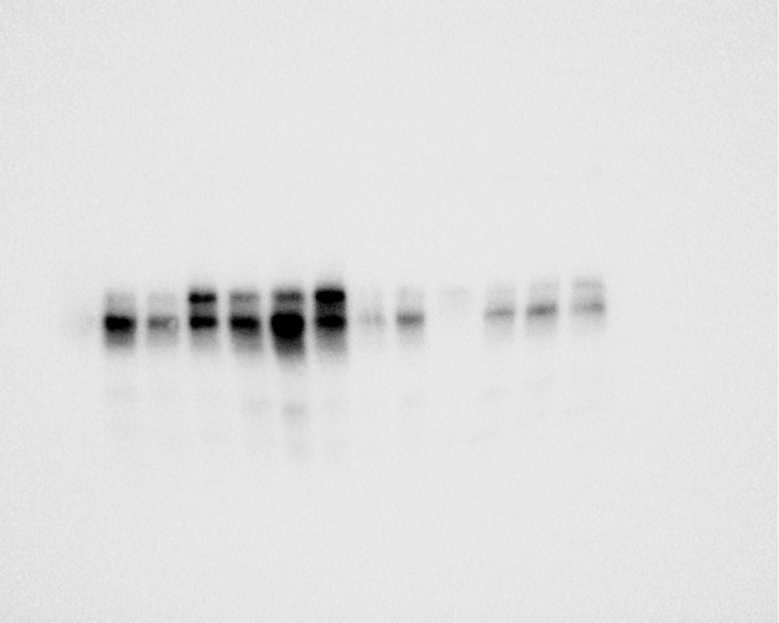
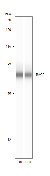
Advanced glycation endproducts (AGE) are adducts formed by the non-enzymatic glycation or oxidation of macromolecules. AGE forms during aging and its formation is accelerated under pathophysiologic states such as diabetes, Alzheimer’s disease, renal failure and immune/inflammatory disorders. Receptor for Advanced Glycation Endoproducts (RAGE), named for its ability to bind AGE, is a multi-ligand receptor belonging the immunoglobulin (Ig) superfamily. Besides AGE, RAGE binds amyloid beta -peptide, S100/calgranulin family proteins, high mobility group B1 (HMGB1, also know as amphoterin) and leukocyte integrins.

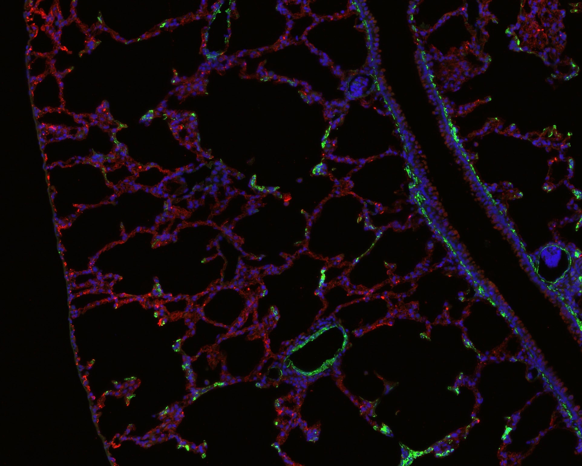
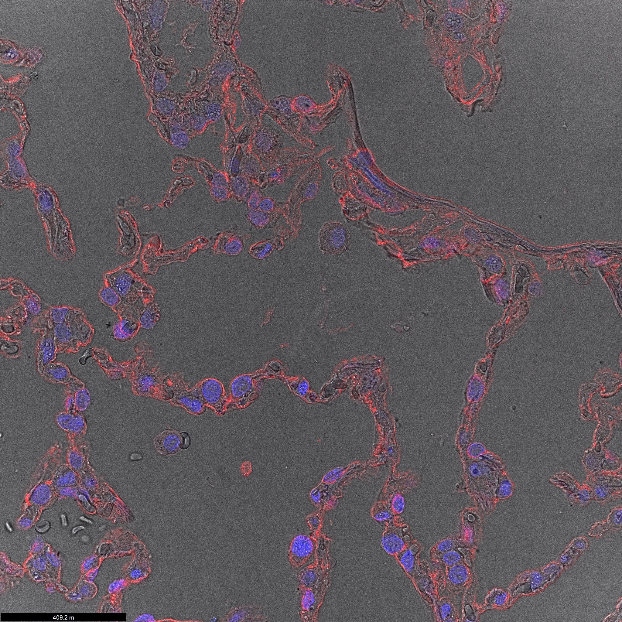
The human RAGE gene encodes a 404 amino acid residues (aa) type I transmembrane glycoprotein with a 22 aa signal peptide, a 320 aa extracellular domain containing an Ig-like V-type domain and two Ig-like Ce-type domains, a 21 aa transmembrane domain and a 41 aa cytoplasmic domain. The V-type domain and the cytoplasmic domain are important for ligand binding and for intracellular signaling, respectively. Two alternative splice variants, lacking the V-type domain or the cytoplasmic tail, are known. RAGE is highly expressed in the embryonic central nervous system. In adult tissues, RAGE is expressed at low levels in multiple tissues including endothelial and smooth muscle cells, mononuclear phagocytes, pericytes, microglia, neurons, cardiac myocytes, and hepatocytes. The expression of RAGE is upregulated upon ligand interaction. Depending on the cellular context and interacting ligand, RAGE activation can trigger differential signaling pathways that affect divergent pathways of gene expression. RAGE activation modulates varied essential cellular responses (including inflammation, immunity, proliferation, cellular adhesion, and migration) that contribute to cellular dysfunction associated with chronic diseases such as diabetes, cancer, amyloidoses, and immune or inflammatory disorders.

Long Name

Receptor for Advanced Glycation End Products

Alternate Names

AGER, SCARJ1

Entrez Gene IDs

177 (Human); 11596 (Mouse); 81722 (Rat); 403168 (Canine)

Gene Symbol

AGER

UniProt

Additional RAGE/AGER Products

Product Documents for Human/Mouse/Rat RAGE/AGER Antibody

Certificate of Analysis

To download a Certificate of Analysis, please enter a lot or batch number in the search box below.

Note: Certificate of Analysis not available for kit components.

Product Specific Notices for Human/Mouse/Rat RAGE/AGER Antibody

For research use only

Citations for Human/Mouse/Rat RAGE/AGER Antibody

Customer Reviews for Human/Mouse/Rat RAGE/AGER Antibody (5)

4.6 out of 5
5 Customer Ratings
5 Stars
60%
4 Stars
40%
3 Stars
0%
2 Stars
0%
1 Stars
0%

Have you used Human/Mouse/Rat RAGE/AGER Antibody?

Submit a review and receive an Amazon gift card!

$25/€18/£15/$25CAN/¥2500 Yen for a review with an image

$10/€7/£6/$10CAN/¥1110 Yen for a review without an image

Submit a review
Amazon Gift Card

Customer Images


Showing  1 - 5 of 5 reviews Showing All
Filter By:
  • RAGE protein expression in human PCLS
    Name: Anonymous
    Application: Western Blot
    Sample Tested: PCLS
    Species: Human
    Verified Customer | Posted 09/08/2025
    RAGE protein expression in human PCLS samples using western blot
    I treated the human PCLS with elastase and checked the RAGE expression in ctrl and treated
    Human/Mouse/Rat RAGE/AGER Antibody AF1145
  • Human/Mouse/Rat RAGE/AGER Antibody
    Name: Anonymous
    Application: Native rat lung tissue stained for RAGE/AGER (red) and Acta2 (green)
    Sample Tested: Lung tissue
    Species: Rat
    Verified Customer | Posted 07/19/2025
    Human/Mouse/Rat RAGE/AGER Antibody AF1145
  • RAGE in human lungs
    Name: Anonymous
    Application: Simple Western
    Sample Tested: Lung tissue
    Species: Human
    Verified Customer | Posted 11/19/2024
    RAGE expression in human lungs
    Dilutions 1:10 and 1:20 in milk-free antibody diluent. 1h incubation in primary antibodies.
    Human/Mouse/Rat RAGE/AGER Antibody AF1145
    Bio-Techne Response
    This review reflects a new species or application tested on a primary antibody.
  • RAGE in human lungs
    Name: Anonymous
    Application: Immunofluorescence
    Sample Tested: Lung tissue
    Species: Human
    Verified Customer | Posted 08/28/2024
    Nucleus stained in blue RAGE in red
    primary antibody - goat anti-RAGE (1:200, overnight incubation at +4°C) secondary antibody - donkey anti-goat 647 (1:400, 3h at RT)
    Human/Mouse/Rat RAGE/AGER Antibody AF1145
  • Human/Mouse/Rat RAGE Antibody
    Name: Balaji Mahender
    Application: ELISA
    Sample Tested: EDTA Plasma, Platelet-poor EDTA Plasma and Heparin Plasma
    Species: Human
    Verified Customer | Posted 12/06/2017

There are no reviews that match your criteria.

Showing  1 - 5 of 5 reviews Showing All

Protocols

Find general support by application which include: protocols, troubleshooting, illustrated assays, videos and webinars.

FAQs

No product specific FAQs exist for this product.

View all FAQs for Antibodies