
Key Product Details
Species Reactivity
Validated:
Human
Cited:
Human, Mouse, Primate - Callithrix jacchus (Common Marmoset)
Applications
Validated:
Immunocytochemistry
Cited:
Immunohistochemistry, Immunohistochemistry-Frozen, Western Blot, Flow Cytometry, Immunocytochemistry, Immunocytochemistry/ Immunofluorescence
Label
Unconjugated
Antibody Source
Monoclonal Mouse IgG3 Clone # 245013
Loading...
Product Specifications
Immunogen
E. coli-derived recombinant human SOX17
Asp177-Val414
Accession # Q9H6I2
Asp177-Val414
Accession # Q9H6I2
Specificity
Detects human SOX17 in direct ELISAs. In direct ELISAs, this antibody does not cross-react with recombinant human SOX2, 3, 7, or 10.
Clonality
Monoclonal
Host
Mouse
Isotype
IgG3
Scientific Data Images for Human SOX17 Antibody (245013)
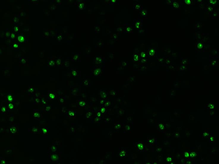
SOX17 in B16 Mouse Cell Line.
SOX17 was detected in immersion fixed B16 mouse melanoma cell line using Mouse Anti-Human SOX17 Monoclonal Antibody (Catalog # MAB1924) at 10 µg/mL for 3 hours at room temperature. Cells were stained using the NorthernLights™ 557-conjugated Anti-Mouse IgG Secondary Antibody (yellow, upper panel; Catalog # NL007) and counterstained with DAPI (blue, lower panel). View our protocol for Fluorescent ICC Staining of Cells on Coverslips.
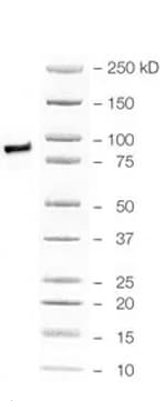
Detection of SOX17 by Western Blot
VAMP5 KO doesn’t impair the stemness of ESCs.a–d Representative western blot images of VAMP5 KO potentially impair the related markers expression in hESCs (a) and also after differentiated into mesoderm (MES) (b), endoderm (END), (c) and ectoderm (ECT) (d). Data are represented as mean ± SEM, n = 3 biological replicates in each group. Student’s two tailed t test was used. No adjustments for multiple comparisons were made, as only two groups were compared. *p < 0.05, **p < 0.01, ***p < 0.001, ****p < 0.0001. e PCA of the transcriptomic comparison of VAMP5 WT and KO in hESCs and followed differentiated three germ layers, n = 3 in each cell type. f Representative heatmap of hESCs and three germ layers related markers comparison of VAMP5 WT and KO in hESCs and followed differentiated three germ layers. g Immunofluorescence staining for lineage differentiation from VAMP5 WT and KO hESCs to mesoderm derived smooth muscles cells (markers of alpha -SMA and Transgelin), endoderm derived alveolar type II cells (markers of EpCAM and SP-B), and ectoderm derived neuron cells ( beta 3 tubulin and MAP2). The results are representative of three independent experiments. Scale bars, 50 μm. KO, knock-out; WT, wild-type. Image collected and cropped by CiteAb from the following open publication (https://pubmed.ncbi.nlm.nih.gov/40624080), licensed under a CC-BY license. Not internally tested by R&D Systems.Applications for Human SOX17 Antibody (245013)
Application
Recommended Usage
Immunocytochemistry
8-25 µg/mL
Sample: Immersion fixed human embryonic stem cell derived endodermal progenitors and B16 mouse melanoma cell line
Sample: Immersion fixed human embryonic stem cell derived endodermal progenitors and B16 mouse melanoma cell line
Reviewed Applications
Read 4 reviews rated 5 using MAB1924 in the following applications:
Formulation, Preparation, and Storage
Purification
Protein A or G purified from hybridoma culture supernatant
Reconstitution
Reconstitute at 0.5 mg/mL in sterile PBS. For liquid material, refer to CoA for concentration.
Loading...
Formulation
Lyophilized from a 0.2 μm filtered solution in PBS with Trehalose. *Small pack size (SP) is supplied either lyophilized or as a 0.2 µm filtered solution in PBS.
Shipping
Lyophilized product is shipped at ambient temperature. Liquid small pack size (-SP) is shipped with polar packs. Upon receipt, store immediately at the temperature recommended below.
Stability & Storage
Use a manual defrost freezer and avoid repeated freeze-thaw cycles.
- 12 months from date of receipt, -20 to -70 °C as supplied.
- 1 month, 2 to 8 °C under sterile conditions after reconstitution.
- 6 months, -20 to -70 °C under sterile conditions after reconstitution.
Calculators
Background: SOX17
References
- Kanai-Azuma, M. et al. (2002) Development 129:2367.
- Katoh, M. et al. (2002) Int. J. Mol. Med. 9:153.
Long Name
Transcription Factor SOX17
Alternate Names
FLJ22252, SRY (sex determining region Y)-box 17, SRY-related HMG-box transcription factor SOX17, transcription factor SOX-17, VUR3
Entrez Gene IDs
64321 (Human)
Gene Symbol
SOX17
UniProt
Additional SOX17 Products
Product Documents for Human SOX17 Antibody (245013)
Certificate of Analysis
To download a Certificate of Analysis, please enter a lot or batch number in the search box below.
Note: Certificate of Analysis not available for kit components.
Product Specific Notices for Human SOX17 Antibody (245013)
For research use only
Related Research Areas
Citations for Human SOX17 Antibody (245013)
 Powered by Bioz
Customer Reviews for Human SOX17 Antibody (245013) (4)
5 out of 5
4 Customer Ratings
Have you used Human SOX17 Antibody (245013)?
Submit a review and receive an Amazon gift card!
$25/€18/£15/$25CAN/¥2500 Yen for a review with an image
$10/€7/£6/$10CAN/¥1110 Yen for a review without an image
Submit a review
Customer Images
Showing
1
-
4 of
4 reviews
Showing All
Filter By:
-
Application: Western BlotSample Tested: MCF-7 human breast cancer cell lineSpecies: HumanVerified Customer | Posted 01/01/2026Whole cell lysate of human adipocytes and MCF7 coculture

-
Application: Western BlotSample Tested: HCT-116 human colorectal carcinoma cell lineSpecies: HumanVerified Customer | Posted 07/10/2025
Bio-Techne ResponseThis review reflects a new species or application tested on a primary antibody.
-
Application: Immunocytochemistry/ImmunofluorescenceSample Tested: iPS2 human induced pluripotent stem cellsSpecies: HumanVerified Customer | Posted 08/23/2021It worked well in 1:200 dilution
-
Application: ImmunofluorescenceSample Tested: See PMID 23041313Species: MouseVerified Customer | Posted 02/12/2015
There are no reviews that match your criteria.
Protocols
Find general support by application which include: protocols, troubleshooting, illustrated assays, videos and webinars.
- Appropriate Fixation of IHC/ICC Samples
- Cellular Response to Hypoxia Protocols
- ClariTSA™ Fluorophore Kits
- Detection & Visualization of Antibody Binding
- ICC Cell Smear Protocol for Suspension Cells
- ICC Immunocytochemistry Protocol Videos
- ICC for Adherent Cells
- Immunocytochemistry (ICC) Protocol
- Immunocytochemistry Troubleshooting
- Immunofluorescence of Organoids Embedded in Cultrex Basement Membrane Extract
- Immunohistochemistry (IHC) and Immunocytochemistry (ICC) Protocols
- Preparing Samples for IHC/ICC Experiments
- Preventing Non-Specific Staining (Non-Specific Binding)
- Primary Antibody Selection & Optimization
- Protocol for VisUCyte™ HRP Polymer Detection Reagent
- Protocol for the Fluorescent ICC Staining of Cell Smears - Graphic
- Protocol for the Fluorescent ICC Staining of Cultured Cells on Coverslips - Graphic
- Protocol for the Preparation and Fluorescent ICC Staining of Cells on Coverslips
- Protocol for the Preparation and Fluorescent ICC Staining of Non-adherent Cells
- Protocol for the Preparation and Fluorescent ICC Staining of Stem Cells on Coverslips
- Protocol for the Preparation of a Cell Smear for Non-adherent Cell ICC - Graphic
- TUNEL and Active Caspase-3 Detection by IHC/ICC Protocol
- The Importance of IHC/ICC Controls
- View all Protocols, Troubleshooting, Illustrated assays and Webinars
Loading...
Associated Pathways
