Human TRAIL R1, also called DR4, is a type 1, TNF R family, membrane protein which is a receptor for TRAIL (APO2 ligand). In the TNF superfamily nomenclature, TRAIL RI is referred to as TNFRSF10A. TRAIL R1 cDNA encodes a 468 amino acid precursor protein containing extracellular cysteine-rich domains, a transmembrane domain and a cytoplasmic death domain. Among the TNF receptor family proteins, TRAIL R1 is most closely related to TRAIL R2/DR5, sharing 55% amino acid sequence identity. Binding of trimeric TRAIL to TRAIL R1 induces apoptosis. The induction of apoptosis likely requires oligomerization of the receptor. The human TRAIL R1/Fc chimera neutralizes the ability of TRAIL to induce apoptosis. Besides TRAIL R1, an additional TRAIL R2/DR5, which tranduces apoptosis signal, and two TRAIL decoy receptors, which antagonize TRAIL-induced apoptosis, have been reported.
Human TRAILR1/TNFRSF10A Antibody (69036)
R&D Systems | Catalog # MAB347

Key Product Details
Species Reactivity
Validated:
Human
Cited:
Human, Mouse, Rat
Applications
Validated:
Immunohistochemistry, Flow Cytometry, CyTOF-ready
Cited:
Immunohistochemistry, Immunohistochemistry-Paraffin, Flow Cytometry, Immunocytochemistry, Functional Assay
Label
Unconjugated
Antibody Source
Monoclonal Mouse IgG1 Clone # 69036
Loading...
Product Specifications
Immunogen
S. frugiperda insect ovarian cell line Sf 21-derived recombinant human TRAIL R1/TNFRSF10A
Ala24-Asn239
Accession # AAC51226
Ala24-Asn239
Accession # AAC51226
Specificity
Detects recombinant human TRAIL R1/TNFRSF10A in direct ELISAs. In direct ELISAs, no cross-reactivity with recombinant human (rh) TRAIL R2, rhTRAIL R3, rhTRAIL R4, or rhDcR3 is observed.
Clonality
Monoclonal
Host
Mouse
Isotype
IgG1
Scientific Data Images for Human TRAILR1/TNFRSF10A Antibody (69036)
Detection of Human TRAILR1/TNFRSF10A by Flow Cytometry
Flow cytometry studies of scFv-Fc-scTRAIL molecules and Fc-scTRAIL on Colo205 and HCT116 cells(A) Expression levels of EGFR, HER2, HER3, EpCAM, and TRAIL-receptors were investigated after treatment of the cells with medium or 250 ng/ml BZB for 16 h. (B) Serial dilutions of the molecules were analyzed. (C) 20 nM scTRAIL units were analyzed after preincubation of the cells with PBA or a 200-fold molar excess of the respective blocking antibody. Binding of Fc-scTRAIL was measured in the presence of all blocking antibodies separately and is here represented as the mean of blocking with all different antibodies. Bound molecules were detected via anti-FLAG-PE. relative MFI, relative median fluorescence intensity. Pairwise comparisons were performed by unpaired t test (two-tailed; *P < 0.05; **P < 0.01; ***P < 0.001; ns, P > 0.05). Image collected and cropped by CiteAb from the following publication (https://www.oncotarget.com/lookup/doi/10.18632/oncotarget.24379), licensed under a CC-BY license. Not internally tested by R&D Systems.
Detection of TRAILR1/TNFRSF10A in Human Breast.
TRAILR1/TNFRSF10A was detected in immersion fixed paraffin-embedded sections of human breast using Mouse Anti-Human TRAILR1/TNFRSF10A Monoclonal Antibody (Catalog # MAB347) at 15 µg/ml for 1 hour at room temperature followed by incubation with the Anti-Mouse IgG VisUCyte™ HRP Polymer Antibody (Catalog # VC001) or the HRP-conjugated Anti-Mouse IgG Secondary Antibody (Catalog # HAF007). Before incubation with the primary antibody, tissue was subjected to heat-induced epitope retrieval using VisUCyte Antigen Retrieval Reagent-Basic (Catalog # VCTS021). Tissue was stained using DAB (brown) and counterstained with hematoxylin (blue). Specific staining was localized to the membrane. View our protocol for Chromogenic IHC Staining of Paraffin-embedded Tissue Sections.Applications for Human TRAILR1/TNFRSF10A Antibody (69036)
Application
Recommended Usage
CyTOF-ready
Ready to be labeled using established conjugation methods. No BSA or other carrier proteins that could interfere with conjugation.
Flow Cytometry
2.5 µg/106 cells
Sample: HeLa human cell line
Sample: HeLa human cell line
Immunohistochemistry
1-25 µg/mL
Sample: Immersion fixed paraffin-embedded sections of human breast tissue
Sample: Immersion fixed paraffin-embedded sections of human breast tissue
Reviewed Applications
Read 1 review rated 4 using MAB347 in the following applications:
Flow Cytometry Panel Builder
Bio-Techne Knows Flow Cytometry
Save time and reduce costly mistakes by quickly finding compatible reagents using the Panel Builder Tool.
Advanced Features
- Spectra Viewer - Custom analysis of spectra from multiple fluorochromes
- Spillover Popups - Visualize the spectra of individual fluorochromes
- Antigen Density Selector - Match fluorochrome brightness with antigen density

Formulation, Preparation, and Storage
Purification
Protein A or G purified from hybridoma culture supernatant
Reconstitution
Reconstitute at 0.5 mg/mL in sterile PBS. For liquid material, refer to CoA for concentration.
Loading...
Formulation
Lyophilized from a 0.2 μm filtered solution in PBS with Trehalose. See Certificate of Analysis for details.
*Small pack size (-SP) is supplied either lyophilized or as a 0.2 µm filtered solution in PBS.
*Small pack size (-SP) is supplied either lyophilized or as a 0.2 µm filtered solution in PBS.
Shipping
Lyophilized product is shipped at ambient temperature. Liquid small pack size (-SP) is shipped with polar packs. Upon receipt, store immediately at the temperature recommended below.
Stability & Storage
Use a manual defrost freezer and avoid repeated freeze-thaw cycles.
- 12 months from date of receipt, -20 to -70 °C as supplied.
- 1 month, 2 to 8 °C under sterile conditions after reconstitution.
- 6 months, -20 to -70 °C under sterile conditions after reconstitution.
Calculators
Background: TRAILR1/TNFRSF10A
References
- Pan, G. et al. (1997) Science 276:111.
- Golstein, P. (1997) Curr. Biol. 7:R750.
Long Name
TRAIL Receptor 1
Alternate Names
CD261, DR4, TNFRSF10A, TRAIL R1
Entrez Gene IDs
8797 (Human)
Gene Symbol
TNFRSF10A
UniProt
Additional TRAILR1/TNFRSF10A Products
Product Documents for Human TRAILR1/TNFRSF10A Antibody (69036)
Certificate of Analysis
To download a Certificate of Analysis, please enter a lot or batch number in the search box below.
Note: Certificate of Analysis not available for kit components.
Product Specific Notices for Human TRAILR1/TNFRSF10A Antibody (69036)
For research use only
Citations for Human TRAILR1/TNFRSF10A Antibody (69036)
 Powered by Bioz
Customer Reviews for Human TRAILR1/TNFRSF10A Antibody (69036) (1)
4 out of 5
1 Customer Rating
Have you used Human TRAILR1/TNFRSF10A Antibody (69036)?
Submit a review and receive an Amazon gift card!
$25/€18/£15/$25CAN/¥2500 Yen for a review with an image
$10/€7/£6/$10CAN/¥1110 Yen for a review without an image
Submit a review
Customer Images
Showing
1
-
1 of
1 review
Showing All
Filter By:
-
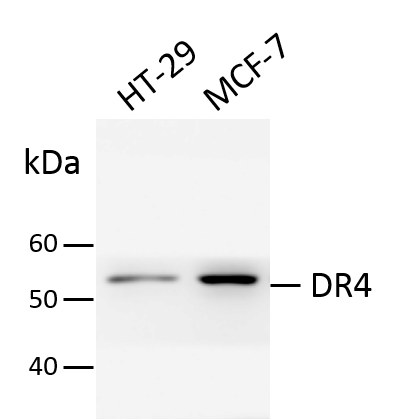
Application: Western BlotSample Tested: MCF-7 human breast cancer cell line and HT-29 human colon adenocarcinoma cell lineSpecies: HumanVerified Customer | Posted 07/23/2018Total cell lysates from HT29 and MCF-7 were subjected to western blot. PVDF membrane were probed with 1 um/ml Human DR4 Antibody (MAB347). A specific band was detected for DR4 at approximately 55 kDa. This experiment was conducted under reducing conditions.

There are no reviews that match your criteria.
Protocols
Find general support by application which include: protocols, troubleshooting, illustrated assays, videos and webinars.
- 7-Amino Actinomycin D (7-AAD) Cell Viability Flow Cytometry Protocol
- Antigen Retrieval Protocol (PIER)
- Antigen Retrieval for Frozen Sections Protocol
- Appropriate Fixation of IHC/ICC Samples
- Cellular Response to Hypoxia Protocols
- Chromogenic IHC Staining of Formalin-Fixed Paraffin-Embedded (FFPE) Tissue Protocol
- Chromogenic Immunohistochemistry Staining of Frozen Tissue
- ClariTSA™ Fluorophore Kits
- Detection & Visualization of Antibody Binding
- Extracellular Membrane Flow Cytometry Protocol
- Flow Cytometry Protocol for Cell Surface Markers
- Flow Cytometry Protocol for Staining Membrane Associated Proteins
- Flow Cytometry Staining Protocols
- Flow Cytometry Troubleshooting Guide
- Fluorescent IHC Staining of Frozen Tissue Protocol
- Graphic Protocol for Heat-induced Epitope Retrieval
- Graphic Protocol for the Preparation and Fluorescent IHC Staining of Frozen Tissue Sections
- Graphic Protocol for the Preparation and Fluorescent IHC Staining of Paraffin-embedded Tissue Sections
- Graphic Protocol for the Preparation of Gelatin-coated Slides for Histological Tissue Sections
- IHC Sample Preparation (Frozen sections vs Paraffin)
- Immunofluorescent IHC Staining of Formalin-Fixed Paraffin-Embedded (FFPE) Tissue Protocol
- Immunohistochemistry (IHC) and Immunocytochemistry (ICC) Protocols
- Immunohistochemistry Frozen Troubleshooting
- Immunohistochemistry Paraffin Troubleshooting
- Intracellular Flow Cytometry Protocol Using Alcohol (Methanol)
- Intracellular Flow Cytometry Protocol Using Detergents
- Intracellular Nuclear Staining Flow Cytometry Protocol Using Detergents
- Intracellular Staining Flow Cytometry Protocol Using Alcohol Permeabilization
- Intracellular Staining Flow Cytometry Protocol Using Detergents to Permeabilize Cells
- Preparing Samples for IHC/ICC Experiments
- Preventing Non-Specific Staining (Non-Specific Binding)
- Primary Antibody Selection & Optimization
- Propidium Iodide Cell Viability Flow Cytometry Protocol
- Protocol for Heat-Induced Epitope Retrieval (HIER)
- Protocol for Liperfluo
- Protocol for Making a 4% Formaldehyde Solution in PBS
- Protocol for VisUCyte™ HRP Polymer Detection Reagent
- Protocol for the Characterization of Human Th22 Cells
- Protocol for the Characterization of Human Th9 Cells
- Protocol for the Preparation & Fixation of Cells on Coverslips
- Protocol for the Preparation and Chromogenic IHC Staining of Frozen Tissue Sections
- Protocol for the Preparation and Chromogenic IHC Staining of Frozen Tissue Sections - Graphic
- Protocol for the Preparation and Chromogenic IHC Staining of Paraffin-embedded Tissue Sections
- Protocol for the Preparation and Chromogenic IHC Staining of Paraffin-embedded Tissue Sections - Graphic
- Protocol for the Preparation and Fluorescent IHC Staining of Frozen Tissue Sections
- Protocol for the Preparation and Fluorescent IHC Staining of Paraffin-embedded Tissue Sections
- Protocol for the Preparation of Gelatin-coated Slides for Histological Tissue Sections
- Protocol: Annexin V and PI Staining by Flow Cytometry
- Protocol: Annexin V and PI Staining for Apoptosis by Flow Cytometry
- TUNEL and Active Caspase-3 Detection by IHC/ICC Protocol
- The Importance of IHC/ICC Controls
- Troubleshooting Guide: Fluorokine Flow Cytometry Kits
- Troubleshooting Guide: Immunohistochemistry
- View all Protocols, Troubleshooting, Illustrated assays and Webinars
Loading...
Associated Pathways

