

Scientific images may be resized, cropped, or adjusted for brightness or contrast for presentation purposes. These updates are limited to presentation and do not affect the underlying scientific interpretation of the data. Where available, additional source data may be provided upon request.
Key Product Details
Species Reactivity
Validated:
Cited:
Applications
Validated:
Cited:
Label
Antibody Source
Product Specifications
Immunogen
Accession # Q9BZM4
Specificity
Clonality
Host
Isotype
Scientific Data Images for Human ULBP-3 Antibody (166514)
Detection of ULBP-3 by Flow Cytometry
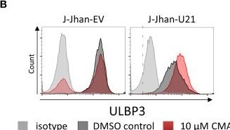
Viral U20 and U21 proteins localize to the secretory pathway. (A) Localization of U20, U21 and U23 in 293T cells determined by FLAG tag staining (yellow). PDI was co-stained to assess the localization to the endoplasmic reticulum (red, left); CD107a served as a marker for late endosomes (red, right). Nuclei were counterstained with DAPI (blue). Pearson’s correlation coefficients describe overlap of FLAG staining and organelle marker staining. (B) Flow cytometric analysis of ULBP3 on J-Jhan and 293T transfected with U21 or controls treated for 6 hours with Concanamycin A that inhibits lysosomal degradation of proteins. A representative plot is presented for ULBP3 expression on J-Jhan transfectants. (C) Bar histograms show quantification of one of two independent experiments, *p < 0.05 in student’s t test for U21-transfectants in J-Jhan and 293T cells. Differences in all other transfectants are non-significant. Image collected and cropped by CiteAb from the following open publication (https://pubmed.ncbi.nlm.nih.gov/34721381), licensed under a CC-BY license. Not internally tested by R&D Systems.Applications for Human ULBP-3 Antibody (166514)
Western Blot
Sample: Recombinant Human ULBP‑3 Fc Chimera (Catalog # 1517-UL)
Reviewed Applications
Read 1 review rated 5 using MAB15171 in the following applications:
Formulation, Preparation, and Storage
Purification
Reconstitution
Reconstitute at 0.5 mg/mL in sterile PBS. For liquid material, refer to CoA for concentration.
Formulation
Shipping
Stability & Storage
- 12 months from date of receipt, -20 to -70 °C as supplied.
- 1 month, 2 to 8 °C under sterile conditions after reconstitution.
- 6 months, -20 to -70 °C under sterile conditions after reconstitution.
Calculators
Background: ULBP-3
Long Name
Alternate Names
Entrez Gene IDs
Gene Symbol
UniProt
Additional ULBP-3 Products
Product Documents for Human ULBP-3 Antibody (166514)
Certificate of Analysis
To download a Certificate of Analysis, please enter a lot or batch number in the search box below.
Note: Certificate of Analysis not available for kit components.
Product Specific Notices for Human ULBP-3 Antibody (166514)
For research use only
Related Research Areas
Citations for Human ULBP-3 Antibody (166514)
 Powered by Bioz
Customer Reviews for Human ULBP-3 Antibody (166514) (1)
Have you used Human ULBP-3 Antibody (166514)?
Submit a review and receive an Amazon gift card!
$25/€18/£15/$25CAN/¥2500 for a review with an image
$10/€7/£6/$10CAN/¥1110 for a review without an image
Submit a review
-
Application: Block/NeutralizeSample Tested: Pancreatic cancer cellsSpecies: MouseVerified Customer | Posted 11/16/2018
There are no reviews that match your criteria.
Protocols
Find general support by application which include: protocols, troubleshooting, illustrated assays, videos and webinars.
- Cellular Response to Hypoxia Protocols
- R&D Systems Quality Control Western Blot Protocol
- Troubleshooting Guide: Western Blot Figures
- Western Blot Conditions
- Western Blot Protocol
- Western Blot Protocol for Cell Lysates
- Western Blot Troubleshooting
- Western Blot Troubleshooting Guide
- View all Protocols, Troubleshooting, Illustrated assays and Webinars