
Key Product Details
Species Reactivity
Validated:
Human
Cited:
Mouse
Applications
Validated:
Immunohistochemistry, Neutralization
Cited:
Immunocytochemistry
Label
Unconjugated
Antibody Source
Monoclonal Mouse IgG1 Clone # 117318
Loading...
Product Specifications
Immunogen
E. coli-derived recombinant human XIAP
Met1-Ser497
Accession # P98170
Met1-Ser497
Accession # P98170
Specificity
Detects human XIAP in direct ELISAs. In direct ELISAs, no cross-reactivity with recombinant human (rh) cIAP-1 or rh cIAP-2 is observed. Neutralizes human XIAP and human XIAP-BIR2 bioactivity. Does not neutralize XIAP-BIR3 or Livin bioactivity.
Clonality
Monoclonal
Host
Mouse
Isotype
IgG1
Scientific Data Images for Human XIAP Antibody (117318)
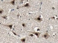
XIAP in Human Brain.
XIAP was detected in immersion fixed paraffin-embedded sections of human brain (cortex) using 25 µg/mL Mouse Anti-Human XIAP Monoclonal Antibody (Catalog # MAB8221) overnight at 4 °C. Tissue was stained with the Anti-Mouse HRP-DAB Cell & Tissue Staining Kit (brown; Catalog # CTS002) and counter-stained with hematoxylin (blue). View our protocol for Chromogenic IHC Staining of Paraffin-embedded Tissue Sections.
Neutralization of XIAP Activity by Human XIAP Antibody.
Recombinant Human Caspase-7 (0.012 µg/mL, Catalog # 823-C7) activity is measured in the presence of Recombinant Human XIAP (2.75 µg/mL, Catalog # 822-XF) that has been preincubated with increasing concentrations of Mouse Anti-Human XIAP Antibody (Catalog # MAB8221) The ND50 is typically 10 µg/mL.Applications for Human XIAP Antibody (117318)
Application
Recommended Usage
Immunohistochemistry
8-25 µg/mL
Sample: Immersion fixed paraffin-embedded sections of human brain (cortex)
Sample: Immersion fixed paraffin-embedded sections of human brain (cortex)
Reviewed Applications
Read 2 reviews rated 3 using MAB8221 in the following applications:
Formulation, Preparation, and Storage
Purification
Protein A or G purified from hybridoma culture supernatant
Reconstitution
Reconstitute at 0.5 mg/mL in sterile PBS. For liquid material, refer to CoA for concentration.
Loading...
Formulation
Lyophilized from a 0.2 μm filtered solution in PBS with Trehalose. *Small pack size (SP) is supplied either lyophilized or as a 0.2 µm filtered solution in PBS.
Shipping
Lyophilized product is shipped at ambient temperature. Liquid small pack size (-SP) is shipped with polar packs. Upon receipt, store immediately at the temperature recommended below.
Stability & Storage
Use a manual defrost freezer and avoid repeated freeze-thaw cycles.
- 6 months from date of receipt, -20 to -70 °C as supplied.
- 3 months, -20 to -70 °C under sterile conditions after reconstitution.
Calculators
Background: XIAP
Long Name
X-linked Inhibitor of Apoptosis
Alternate Names
BIRC4
Gene Symbol
XIAP
UniProt
Additional XIAP Products
Product Documents for Human XIAP Antibody (117318)
Certificate of Analysis
To download a Certificate of Analysis, please enter a lot or batch number in the search box below.
Note: Certificate of Analysis not available for kit components.
Product Specific Notices for Human XIAP Antibody (117318)
For research use only
Related Research Areas
Citations for Human XIAP Antibody (117318)
 Powered by Bioz
Customer Reviews for Human XIAP Antibody (117318) (2)
3 out of 5
2 Customer Ratings
Have you used Human XIAP Antibody (117318)?
Submit a review and receive an Amazon gift card!
$25/€18/£15/$25CAN/¥2500 Yen for a review with an image
$10/€7/£6/$10CAN/¥1110 Yen for a review without an image
Submit a review
Customer Images
Showing
1
-
2 of
2 reviews
Showing All
Filter By:
-
Application: Immunohistochemistry-ParaffinSample Tested: Brain tissueSpecies: HumanVerified Customer | Posted 04/03/2022Brain tissue25 ug/mL dilution for ihc

-
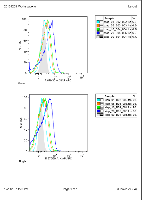
Application: Flow CytometrySample Tested: Blood mononuclear cells (PBMCs)Species: HumanVerified Customer | Posted 12/11/2016This antibody DOES NOT work with flow cytometry. Intracellularly stained at dilutions of 0, 1:300, 1:60, 1:30, and 1:15. Shifts seen are similar to those expected from background staining.Fixed with BD Fix/perm Intracellular stained with BD perm/wash Top graph shows stainings with various dilutions in gated monocytes Bottom graph shows bulk lymphocytes. Grey filled histogram represents no antibody staining.
There are no reviews that match your criteria.
Protocols
Find general support by application which include: protocols, troubleshooting, illustrated assays, videos and webinars.
- Antigen Retrieval Protocol (PIER)
- Antigen Retrieval for Frozen Sections Protocol
- Appropriate Fixation of IHC/ICC Samples
- Cellular Response to Hypoxia Protocols
- Chromogenic IHC Staining of Formalin-Fixed Paraffin-Embedded (FFPE) Tissue Protocol
- Chromogenic Immunohistochemistry Staining of Frozen Tissue
- ClariTSA™ Fluorophore Kits
- Detection & Visualization of Antibody Binding
- Fluorescent IHC Staining of Frozen Tissue Protocol
- Graphic Protocol for Heat-induced Epitope Retrieval
- Graphic Protocol for the Preparation and Fluorescent IHC Staining of Frozen Tissue Sections
- Graphic Protocol for the Preparation and Fluorescent IHC Staining of Paraffin-embedded Tissue Sections
- Graphic Protocol for the Preparation of Gelatin-coated Slides for Histological Tissue Sections
- IHC Sample Preparation (Frozen sections vs Paraffin)
- Immunofluorescent IHC Staining of Formalin-Fixed Paraffin-Embedded (FFPE) Tissue Protocol
- Immunohistochemistry (IHC) and Immunocytochemistry (ICC) Protocols
- Immunohistochemistry Frozen Troubleshooting
- Immunohistochemistry Paraffin Troubleshooting
- Preparing Samples for IHC/ICC Experiments
- Preventing Non-Specific Staining (Non-Specific Binding)
- Primary Antibody Selection & Optimization
- Protocol for Heat-Induced Epitope Retrieval (HIER)
- Protocol for Making a 4% Formaldehyde Solution in PBS
- Protocol for VisUCyte™ HRP Polymer Detection Reagent
- Protocol for the Preparation & Fixation of Cells on Coverslips
- Protocol for the Preparation and Chromogenic IHC Staining of Frozen Tissue Sections
- Protocol for the Preparation and Chromogenic IHC Staining of Frozen Tissue Sections - Graphic
- Protocol for the Preparation and Chromogenic IHC Staining of Paraffin-embedded Tissue Sections
- Protocol for the Preparation and Chromogenic IHC Staining of Paraffin-embedded Tissue Sections - Graphic
- Protocol for the Preparation and Fluorescent IHC Staining of Frozen Tissue Sections
- Protocol for the Preparation and Fluorescent IHC Staining of Paraffin-embedded Tissue Sections
- Protocol for the Preparation of Gelatin-coated Slides for Histological Tissue Sections
- TUNEL and Active Caspase-3 Detection by IHC/ICC Protocol
- The Importance of IHC/ICC Controls
- Troubleshooting Guide: Immunohistochemistry
- View all Protocols, Troubleshooting, Illustrated assays and Webinars
Loading...
Associated Pathways

