IL-1 beta/IL-1F2 Antibody - Azide and BSA Free
Novus Biologicals | Catalog # NB600-633
![Western Blot: IL-1 beta/IL-1F2 Antibody [NB600-633]](https://resources.rndsystems.com/images/products/IL-1-beta-IL-1F2-Antibody-Western-Blot-NB600-633-img0012.jpg "Western Blot: IL-1 beta/IL-1F2 Antibody [NB600-633]")
Loading...
Key Product Details
Validated by
Biological Validation
Species Reactivity
Validated:
Human, Mouse, Rat, Porcine, Canine, Golden Syrian Hamster, Monkey
Cited:
Human, Mouse, Rat, Porcine, Bacteria, Canine, Golden Syrian Hamster, Ovine, Primate - Macaca mulatta (Rhesus Macaque)
Applications
Validated:
Immunohistochemistry, Immunohistochemistry-Paraffin, Immunohistochemistry-Frozen, Immunohistochemistry Whole-Mount, Western Blot, ELISA, Flow Cytometry, Immunocytochemistry/ Immunofluorescence, Immunoprecipitation, Dot Blot, Electron Microscopy
Cited:
Immunohistochemistry, Immunohistochemistry-Paraffin, Immunohistochemistry-Frozen, Immunohistochemistry Whole-Mount, Western Blot, ELISA, Immunocytochemistry/ Immunofluorescence, Immunoprecipitation, IF/IHC, Electron Microscopy
Label
Unconjugated
Antibody Source
Polyclonal Rabbit IgG
Format
Azide and BSA Free
Loading...
Product Specifications
Immunogen
This IL-1 beta/IL-1F2 Antibody was prepared by repeated immunizations with recombinant human IL-1 beta/IL-1F2 produced in E.coli. The MW of the recombinant 153 aa IL-1 beta/IL-1F2 was 17 kDa with the N-terminal amino acid at position alanine 117. This cleavage site is generated by the IL-1 beta/IL-1F2 converting enzyme (ICE, capase-1). (Uniprot: P01584)
Reactivity Notes
In general, this antibody also detects primate IL-1 beta/IL-1F2 in the same formats using similar dilutions. Use in Mouse reported in scientific literature (PMID: 33731931).
Localization
Secreted
Specificity
This antibody is primarily directed against mature, 17,000 MW human IL-1 beta/IL-1F2and is useful in determining its presence in various assays. In general, this antibody also detects primate IL-1 beta/IL-1F2in the same formats using similar dilutions. The antiserum does not recognize human IL-1alpha. In ELISA formats and other immunoreactive assays, this antibody will recognize 10% of the non-denatured (native) precursor 31,000 MW IL-1 beta/IL-1F2containing samples but will primarily detect all of the 17,000 MW mature molecule. However, in immunoblot analysis of natural cell products or human body fluids, the usual procedure of hearing the sample in SDS with or without reducing agents will facilitate denaturing of the 31,000 MW IL- 1beta precursor molecule. Denatured 31,000 precursor IL-1 beta/IL-1F2will be recognized by this antibody but often migrates as a 35,000 MW band. This is due to the unfolding of the denatured precursor IL-1 beta/IL-1F2exposing epitopes not exposed in the natural state. In immunoblots, depending on the number of cells, the antibody detects the 17,000 MW band in supernatants as well as a 35,000 MW band representing the 31,000 MW IL-1 beta/IL-1F2precursor in lysates.
Clonality
Polyclonal
Host
Rabbit
Isotype
IgG
Description
This is an IgG preparation of whole rabbit serum purified by DEAE fractionation. Store vial at -20C prior to opening. Aliquot contents and freeze at -20C or below for extended storage. Avoid cycles of freezing and thawing. Centrifuge product if not completely clear after standing at room temperature. This product is stable for several weeks at 4C as an undiluted liquid. Dilute only prior to immediate use.
Scientific Data Images for IL-1 beta/IL-1F2 Antibody - Azide and BSA Free
![Immunocytochemistry/ Immunofluorescence: IL-1 beta/IL-1F2 Antibody [NB600-633]](https://resources.rndsystems.com/images/products/IL-1-beta-IL-1F2-Antibody-Immunocytochemistry-Immunofluorescence-NB600-633-img0009.jpg "Immunocytochemistry/ Immunofluorescence: IL-1 beta/IL-1F2 Antibody [NB600-633]")
Immunocytochemistry/ Immunofluorescence: IL-1 beta/IL-1F2 Antibody [NB600-633]
Immunocytochemistry/Immunofluorescence: IL-1 beta/IL-1F2 Antibody [NB600-633] - Trabecular Meshwork (TM) region of pig eyes were stained with IL-1 beta antibody (red). Image from verified customer review.![Immunohistochemistry: IL-1 beta/IL-1F2 Antibody [NB600-633]](https://resources.rndsystems.com/images/products/IL-1-beta-IL-1F2-Antibody-Immunohistochemistry-NB600-633-img0010.jpg "Immunohistochemistry: IL-1 beta/IL-1F2 Antibody [NB600-633]")
Immunohistochemistry: IL-1 beta/IL-1F2 Antibody [NB600-633]
Immunohistochemistry: IL-1 beta/IL-1F2 Antibody [NB600-633] - Analysis of: Human IL1beta antibody Secondary antibody: Peroxidase goat anti-rabbit at 1:10,000 for 45 min at RT Localization: cytoplasm Staining: Close up of medullary lymph node: positive staining in the cytoplasm of circulating macrophages. Neg Ctr (far right) normal rabbit IgG with pH 6.2 40X![Western Blot: IL-1 beta/IL-1F2 Antibody [NB600-633]](https://resources.rndsystems.com/images/products/IL-1-beta-IL-1F2-Antibody-Western-Blot-NB600-633-img0005.jpg "Western Blot: IL-1 beta/IL-1F2 Antibody [NB600-633]")
Western Blot: IL-1 beta/IL-1F2 Antibody [NB600-633]
Western Blot: IL-1 beta/IL-1F2 Antibody [NB600-633] - Analysis of IL-1 beta in Rhesus fetal membrane using anti-IL-1 beta antibody. Image from verified customer review.![Western Blot: IL-1 beta/IL-1F2 Antibody [NB600-633]](https://resources.rndsystems.com/images/products/IL-1-beta-IL-1F2-Antibody-Western-Blot-NB600-633-img0006.jpg "Western Blot: IL-1 beta/IL-1F2 Antibody [NB600-633]")
Western Blot: IL-1 beta/IL-1F2 Antibody [NB600-633]
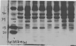
Western Blot: IL-1 beta/IL-1F2 Antibody [NB600-633] - Antibody was used at a 1:200 dilution incubated 1h at room temperature to detect dog IL-1b by Western blot. Lane 1 and 2 were loaded with 2.5 ug and 500 ng of dog IL-1b respectively. The molecular weight of the detected band is estimated by comparison to molecular weight markers (not shown). Detection occurred using a 1:3,000 dilution of IRDye (TM) 800 conjugated Donkey anti-Rabbit IgG for 1h at room temperature.![Western Blot: IL-1 beta/IL-1F2 Antibody [NB600-633]](https://resources.rndsystems.com/images/products/IL-1-beta-IL-1F2-Antibody-Western-Blot-NB600-633-img0008.jpg "Western Blot: IL-1 beta/IL-1F2 Antibody [NB600-633]")
Western Blot: IL-1 beta/IL-1F2 Antibody [NB600-633]
Western Blot: IL-1 beta/IL-1F2 Antibody [NB600-633] - pro-IL-1beta in glioblastoma cells. Image from verified customer review.![Western Blot: IL-1 beta/IL-1F2 Antibody [NB600-633]](https://resources.rndsystems.com/images/products/IL-1-beta-IL-1F2-Antibody-Western-Blot-NB600-633-img0011.jpg "Western Blot: IL-1 beta/IL-1F2 Antibody [NB600-633]")
Western Blot: IL-1 beta/IL-1F2 Antibody [NB600-633]
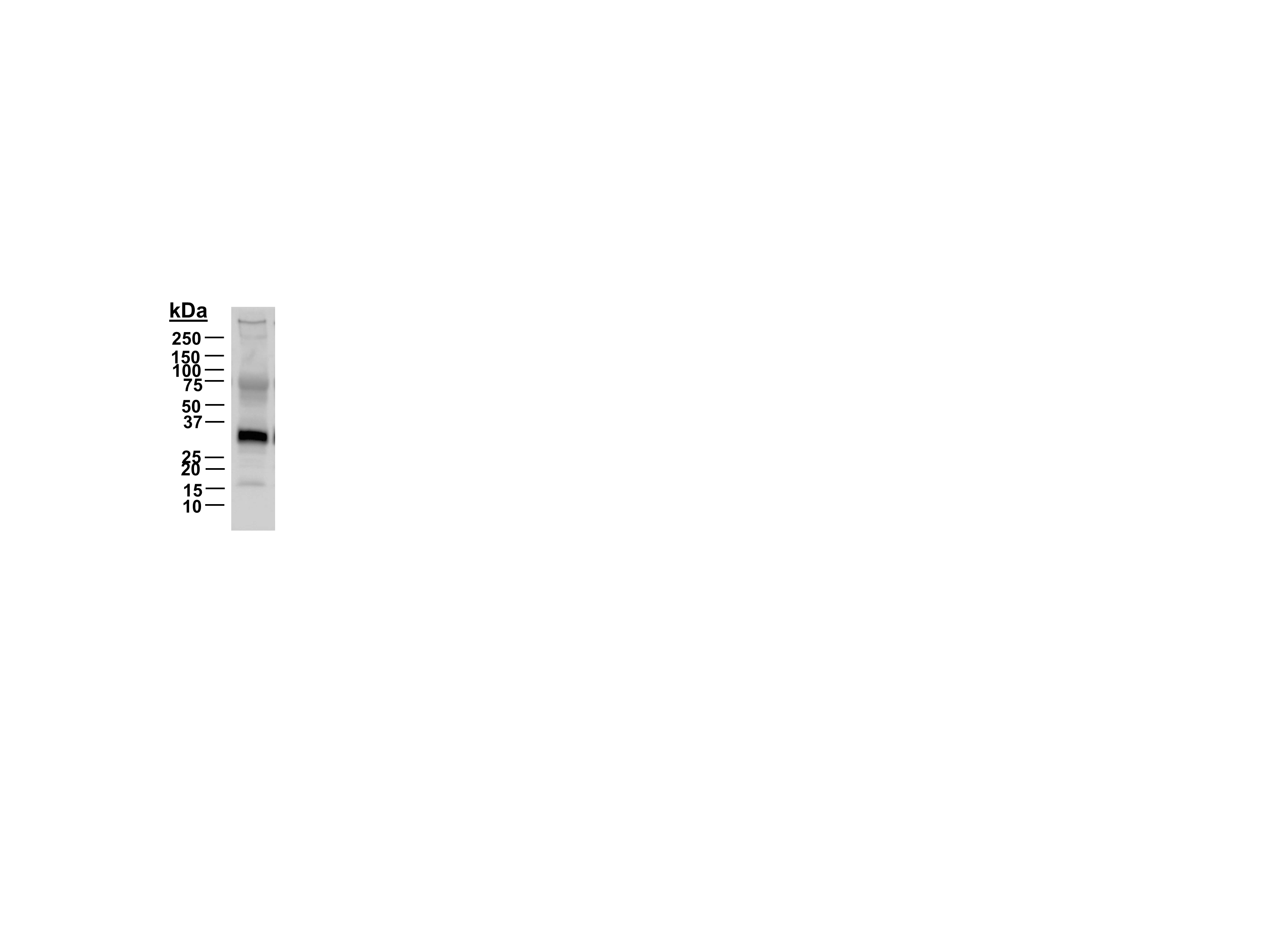
Western Blot: IL-1 beta/IL-1F2 Antibody [NB600-633] - Lane 1: Human IL-1Beta. Load: 5 ng per lane. Primary antibody: Human IL-1Beta antibody at 1:2,000 for overnight at 4C. Secondary antibody: Peroxidase rabbit secondary antibody at 1:40,000 for 30 min at RT. Block: Blocking Buffer for Fluorescent Western Blotting for 30 min at RT. Predicted/Observed size: 17 kDa, 17 kDa for Human IL-1Beta. Other band(s): Unspecific band at 35 kDa.
IL-1 beta/IL-1F2 Antibody
Western Blot of Rabbit anti-Human IL-1beta antibody. Lane 1: Human IL-1beta. Load: 5 ng per lane. Primary antibody: Human IL-1beta antibody at 1:2,000 for overnight at 4C. Secondary antibody: Peroxidase rabbit secondary antibody at 1:40,000 for 30 min at RT. Block: Blocking Buffer for Fluorescent Western Blotting () for 30 min at RT. Predicted/Observed size: 17 kDa, 17 kDa for Human IL-1beta. Other band(s): Unspecific band at ~35 kDa.
Western Blot: IL-1 beta/IL-1F2 Antibody [NB600-633] -
Western Blot: IL-1 beta/IL-1F2 Antibody [NB600-633] - Methylene blue inhibits inflammasome formation in cultured microglia. (A) Expression of Pro-Caspase-1, Caspase-1 p20, pro-IL-1 beta & mature IL-1 beta in microglia with or without stimulation in the presence or absence of methylene blue. Left: representative Western blot images. Right: statistics. N = 5 per group. The (B) expression of NLRP3, NLRC4 & Aim2 in microglia. Left: representative Western blot images. Right: statistics. N = 4 per group. (C) Co-immunoprecipitation assay showing the binding of ASC to NLRP3 or NLRC4. Left: representative Western blot images. Right: statistics. Un: no stimulation. LPS+ATP: stimulation with LPS followed by ATP treatment. V: vehicle; M: 500 nM methylene blue. IP: immunoprecipitation with the antibody against indicated protein. IB: detection of indicated protein. To analyze the relative expression of target proteins, the band intensities of target proteins were normalized to corresponding band intensities of alpha -Tubulin, followed by calculating the expression in other groups relative to “Un (V)” group. N = 4 per group. *p < 0.05; **p < 0.01; ***p < 0.001 in comparison with unstimulated cells of the vehicle group. #p < 0.05; ##p < 0.01 in comparison with stimulated cells of the vehicle group. Image collected & cropped by CiteAb from the following publication (http://journal.frontiersin.org/article/10.3389/fncel.2017.00391/full), licensed under a CC-BY license. Not internally tested by Novus Biologicals.
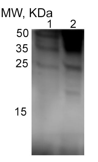
Western Blot: Rabbit Polyclonal IL-1 beta/IL-1F2 Antibody [NB600-633]
Western Blot: Rabbit Polyclonal IL-1 beta/IL-1F2 Antibody [NB600-633] - IL-1 beta/IL-1F2 Western blot of liver homogenate from control (1) and alcohol-fed mice (2). Image from a verified customer review.
Immunohistochemistry: IL-1 beta/IL-1F2 Antibody - Azide and BSA Free [NB600-633] -
Representative photomicrographs showing immunohistochemical staining of IL-1 beta (A), IBA-1 (B), GFAP (C), NeuN (D), caspase3 (E) and ApopTag (TUNEL, F) in the lateral gyrus. Arrowheads point to Caspase 3 + cells and arrows point to ApopTag (TUNEL +) cells. Scale bar = 100 μm Image collected and cropped by CiteAb from the following open publication (https://pubmed.ncbi.nlm.nih.gov/37226206), licensed under a CC-BY license. Not internally tested by Novus Biologicals.
Immunohistochemistry: IL-1 beta/IL-1F2 Antibody - Azide and BSA Free [NB600-633] -
Effects of butyrate treatment on curdlan-induced SpA mice. SKG mice were injected with 3 mg of curdlan through intraperitoneal and oral administration. One week later, sodium butyrate (100 mg/kg) was administered orally once a day for 14 weeks. (A) Arthritis score and incidence were monitored by observation twice a week. (B, C) At 15 weeks post curdlan injection, joint and spine tissue samples were collected from all groups. Histopathology was evaluated for H&E and Safranin O staining. Inflammation, bone erosion, cartilage destruction, and nucleus pulposus (NP) and annulus fibrosus (AF) scores were assessed in both groups. (D) Synovial tissues in joints were immunohistochemically stained for IL-1 beta, IL-17, and TNF-alpha. Positive area (%) was analyzed with the color deconvolution tool in ImageJ. *P < 0.05, **P < 0.01, ***P < 0.001 and ****P < 0.0001. Image collected and cropped by CiteAb from the following open publication (https://www.frontiersin.org/articles/10.3389/fimmu.2023.1096565/full), licensed under a CC-BY license. Not internally tested by Novus Biologicals.
Immunohistochemistry: IL-1 beta/IL-1F2 Antibody - Azide and BSA Free [NB600-633] -
The anti-inflammatory effects of CoQ10-micelles during OA progression.(A) Representative images of immunohistochemical staining of IL-1 beta, IL-6, and MMP13 in the joint synovia of a WT group, a vehicle group, a CoQ10-micelle group, and a celecoxib group. (B) The bar graphs show the average positive areal percentages for IL-1 beta, IL-6, and MMP13. Data are shown as means +/- S.E.Ms. Statistical significance was assessed using the Bonferroni test. *p < 0.05. Image collected and cropped by CiteAb from the following open publication (https://pubmed.ncbi.nlm.nih.gov/35749420), licensed under a CC-BY license. Not internally tested by Novus Biologicals.
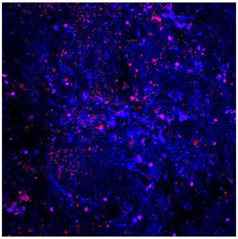
Immunocytochemistry/ Immunofluorescence: IL-1 beta/IL-1F2 Antibody - Azide and BSA Free [NB600-633] -
Expression of pyroptosis-related genes is increased by M1 macrophage conditioned media in ASCs. hASCs were incubated with M1-like macrophage-CM (MØCM) for 24 h. (A) mRNA and (B,C) protein expression levels were measured by qRT-PCR and western blotting, respectively. #p < 0.05 vs. Control, ** p < 0.01 vs. Control, * p < 0.001 vs. Control. (D) Caspase-1 and GSDMD and (E) Caspase-4 and IL-1 beta expression levels were detected by immunofluorescence. Blue indicates the nuclei. Scale bar = 50 um. Image collected and cropped by CiteAb from the following open publication (https://pubmed.ncbi.nlm.nih.gov/34360711), licensed under a CC-BY license. Not internally tested by Novus Biologicals.Applications for IL-1 beta/IL-1F2 Antibody - Azide and BSA Free
Application
Recommended Usage
ELISA
1:500-1:2000
Electron Microscopy
1:10-1:500
Immunocytochemistry/ Immunofluorescence
1:10-1:500
Immunohistochemistry
1:100-1:200
Immunohistochemistry-Paraffin
1:10-1:500
Immunoprecipitation
1:400-1:800
Western Blot
1:1000
Application Notes
This product has been tested for use in ELISA, immunohistochemistry, immunoblotting. This antibody is suitable for neutralizations, radioimmunoassays, flow cytometry, and immunoprecipitation. It recognizes the 17,000 MW mature IL-1beta. For immunoblots, typically, IL-1beta is detected from supernatants or lysates of 2 x 10E6 endotoxin-stimulated peripheral blood mononuclear cells (PBMC). PBMC are stimulated for 24 hours with 1% (v/v) serum plus 10 ng/mL E.coli LPS. For immunoprecipitation pre-clearing the preparation with a non-specific Rabbit IgG to reduce background is suggested. For immunohistochemistry either paraffin fixation or cryofixation can be used for sample preparation to stain intracellular IL-1beta. For ELISA use HRP Conjugated Anti-Rabbit IgG [H&L] (Goat) (611-1302) for detection. In ELISA formats this antibody is best used as the second antibody in combination with a monoclonal antibody as a capture antibody. This antibody is also useful for neutralization of human and primate IL-1beta activity in bioassays. It does not neutralize the biological activity IL-1alpha. It does not neutralize the biological activity of murine, rat or rabbit IL-1beta. For neutralization, it is recommended to incubate the sample with a dilution of the antibody for at least 4 hours before being tested. A control of similarly diluted normal rabbit IgG is recommended. This antibody can be used for FACS analysis. Caution should be exhibited as the F(c) domain of the rabbit IgG molecule may interact with cells non-specifically.
Use in Immunohistochemistry-Frozen reported in scientific literature (PMID: 22898394).
Use in Immunohistochemistry Whole-Mount reported in scientific literature (PMID:31399621).
Use in Immunohistochemistry-Frozen reported in scientific literature (PMID: 22898394).
Use in Immunohistochemistry Whole-Mount reported in scientific literature (PMID:31399621).
Reviewed Applications
Read 5 reviews rated 4 using NB600-633 in the following applications:
Flow Cytometry Panel Builder
Bio-Techne Knows Flow Cytometry
Save time and reduce costly mistakes by quickly finding compatible reagents using the Panel Builder Tool.
Advanced Features
- Spectra Viewer - Custom analysis of spectra from multiple fluorochromes
- Spillover Popups - Visualize the spectra of individual fluorochromes
- Antigen Density Selector - Match fluorochrome brightness with antigen density

Formulation, Preparation, and Storage
Purification
Ion exchange chromatography
Formulation
0.02 M Potassium Phosphate, 0.15 M Sodium Chloride, pH 7.2
Format
Azide and BSA Free
Preservative
No Preservative
Concentration
Please see the vial label for concentration. If unlisted please contact technical services.
Shipping
The product is shipped with polar packs. Upon receipt, store it immediately at the temperature recommended below.
Stability & Storage
Store at -20C short term. Aliquot and store at -80C long term. Avoid freeze-thaw cycles.
Background: IL-1 beta/IL-1F2
IL-1 beta binding to its receptor IL-1RI and the downstream signaling contributes to a dual pathophysiological role (3). On one hand, IL-1 beta signaling activates immune cells and drives CD4+ T cell polarization to T helper type 1 (Th1) and Th17 cells, resulting in anti-tumor responses and mediation of acute inflammation (2,3). However, IL-1 beta also supports tumor growth and metastasis driven by multiple mechanisms including chronic inflammation, an immunosuppressive tumor microenvironment (TME), and angiogenesis (3). Additionally, IL-1 beta signaling been implicated in the pathogenesis of neuroinflammatory diseases of the central nervous system (CNS) such as multiple sclerosis (MS), Alzheimer's disease, and diabetic retinopathy (DR) (2). Mouse studies have shown regression of tumors treated with IL-1 as well as protective effects of IL-1 beta in instances of induced colitis and colon carcinoma (3). Conversely, blocking IL-1 beta has also shown promising effect in cancer treatment, especially when combined with chemotherapeutics (2,3). Approved IL-1 beta monoclonal antibody canakinumab has shown significant therapeutic promise in the treatment of DR (2). Given its multifaceted role in disease, IL-1 beta is a promising therapeutic target.
References
1. Lopez-Castejon G, Brough D. Understanding the mechanism of IL-1beta secretion. Cytokine Growth Factor Rev. 2011;22(4):189-195. https://doi.org/10.1016/j.cytogfr.2011.10.001
2. Mendiola AS, Cardona AE. The IL-1beta phenomena in neuroinflammatory diseases. J Neural Transm (Vienna). 2018;125(5):781-795. https://doi.org/10.1007/s00702-017-1732-9
3. Bent R, Moll L, Grabbe S, Bros M. Interleukin-1 Beta-A Friend or Foe in Malignancies?. Int J Mol Sci. 2018;19(8):2155. https://doi.org/doi:10.3390/ijms19082155
4. Krumm B, Xiang Y, Deng J. Structural biology of the IL-1 superfamily: key cytokines in the regulation of immune and inflammatory responses. Protein Sci. 2014;23(5):526-538. https://doi.org/10.1002/pro.2441
5. He Y, Hara H, Nunez G. Mechanism and Regulation of NLRP3 Inflammasome Activation. Trends Biochem Sci. 2016;41(12):1012-1021. https://doi.org/10.1016/j.tibs.2016.09.002
6. Uniprot (P01584)
Long Name
Interleukin 1 beta
Alternate Names
IL-1b, IL-1F2, IL1 beta, IL1B
Gene Symbol
IL1B
UniProt
Additional IL-1 beta/IL-1F2 Products
Product Documents for IL-1 beta/IL-1F2 Antibody - Azide and BSA Free
Certificate of Analysis
To download a Certificate of Analysis, please enter a lot or batch number in the search box below.
Product Specific Notices for IL-1 beta/IL-1F2 Antibody - Azide and BSA Free
This product is for research use only and is not approved for use in humans or in clinical diagnosis. Primary Antibodies are guaranteed for 1 year from date of receipt.
Related Research Areas
Citations for IL-1 beta/IL-1F2 Antibody - Azide and BSA Free
 Powered by Bioz
Customer Reviews for IL-1 beta/IL-1F2 Antibody - Azide and BSA Free (5)
4 out of 5
5 Customer Ratings
Have you used IL-1 beta/IL-1F2 Antibody - Azide and BSA Free?
Submit a review and receive an Amazon gift card!
$25/€18/£15/$25CAN/¥2500 Yen for a review with an image
$10/€7/£6/$10CAN/¥1110 Yen for a review without an image
Submit a review
Customer Images
-(01-mg)_NB600-633_7081.jpg)
Showing
1
-
5 of
5 reviews
Showing All
Filter By:
-
Application: Western BlotSample Tested: Liver homogenates sampleSpecies: MouseVerified Customer | Posted 11/20/2024IL-1b W-B of liver homogenate from control (1) and alcohol-fed mice (2)

-
Application: Western BlotSample Tested: cellSpecies: raw264.7Verified Customer | Posted 04/24/2018
-
Application: ImmunofluorescenceSample Tested: trabecular meshwork (TM) region of pig eyesSpecies: OtherVerified Customer | Posted 01/07/2016Pig TM tissue stained with IL-1 beta antibody
-
Application: Western BlotSample Tested: Fetal membranesSpecies: OtherVerified Customer | Posted 11/25/2014IL-1b antibody
-
Application: Western BlotSample Tested: glioblastoma cell lysatesSpecies: HumanVerified Customer | Posted 04/28/2014pro-IL-1beta in glioblastoma cells
There are no reviews that match your criteria.
Protocols
Find general support by application which include: protocols, troubleshooting, illustrated assays, videos and webinars.
- 7-Amino Actinomycin D (7-AAD) Cell Viability Flow Cytometry Protocol
- Antigen Retrieval Protocol (PIER)
- Antigen Retrieval for Frozen Sections Protocol
- Appropriate Fixation of IHC/ICC Samples
- Cellular Response to Hypoxia Protocols
- Chromogenic IHC Staining of Formalin-Fixed Paraffin-Embedded (FFPE) Tissue Protocol
- Chromogenic Immunohistochemistry Staining of Frozen Tissue
- ClariTSA™ Fluorophore Kits
- Detection & Visualization of Antibody Binding
- ELISA Sample Preparation & Collection Guide
- ELISA Troubleshooting Guide
- Extracellular Membrane Flow Cytometry Protocol
- Flow Cytometry Protocol for Cell Surface Markers
- Flow Cytometry Protocol for Staining Membrane Associated Proteins
- Flow Cytometry Staining Protocols
- Flow Cytometry Troubleshooting Guide
- Fluorescent IHC Staining of Frozen Tissue Protocol
- Graphic Protocol for Heat-induced Epitope Retrieval
- Graphic Protocol for the Preparation and Fluorescent IHC Staining of Frozen Tissue Sections
- Graphic Protocol for the Preparation and Fluorescent IHC Staining of Paraffin-embedded Tissue Sections
- Graphic Protocol for the Preparation of Gelatin-coated Slides for Histological Tissue Sections
- How to Run an R&D Systems DuoSet ELISA
- How to Run an R&D Systems Quantikine ELISA
- How to Run an R&D Systems Quantikine™ QuicKit™ ELISA
- ICC Cell Smear Protocol for Suspension Cells
- ICC Immunocytochemistry Protocol Videos
- ICC for Adherent Cells
- IHC Sample Preparation (Frozen sections vs Paraffin)
- Immunocytochemistry (ICC) Protocol
- Immunocytochemistry Troubleshooting
- Immunofluorescence of Organoids Embedded in Cultrex Basement Membrane Extract
- Immunofluorescent IHC Staining of Formalin-Fixed Paraffin-Embedded (FFPE) Tissue Protocol
- Immunohistochemistry (IHC) and Immunocytochemistry (ICC) Protocols
- Immunohistochemistry Frozen Troubleshooting
- Immunohistochemistry Paraffin Troubleshooting
- Immunoprecipitation Protocol
- Intracellular Flow Cytometry Protocol Using Alcohol (Methanol)
- Intracellular Flow Cytometry Protocol Using Detergents
- Intracellular Nuclear Staining Flow Cytometry Protocol Using Detergents
- Intracellular Staining Flow Cytometry Protocol Using Alcohol Permeabilization
- Intracellular Staining Flow Cytometry Protocol Using Detergents to Permeabilize Cells
- Preparing Samples for IHC/ICC Experiments
- Preventing Non-Specific Staining (Non-Specific Binding)
- Primary Antibody Selection & Optimization
- Propidium Iodide Cell Viability Flow Cytometry Protocol
- Protocol for Heat-Induced Epitope Retrieval (HIER)
- Protocol for Liperfluo
- Protocol for Making a 4% Formaldehyde Solution in PBS
- Protocol for VisUCyte™ HRP Polymer Detection Reagent
- Protocol for the Characterization of Human Th22 Cells
- Protocol for the Characterization of Human Th9 Cells
- Protocol for the Fluorescent ICC Staining of Cell Smears - Graphic
- Protocol for the Fluorescent ICC Staining of Cultured Cells on Coverslips - Graphic
- Protocol for the Preparation & Fixation of Cells on Coverslips
- Protocol for the Preparation and Chromogenic IHC Staining of Frozen Tissue Sections
- Protocol for the Preparation and Chromogenic IHC Staining of Frozen Tissue Sections - Graphic
- Protocol for the Preparation and Chromogenic IHC Staining of Paraffin-embedded Tissue Sections
- Protocol for the Preparation and Chromogenic IHC Staining of Paraffin-embedded Tissue Sections - Graphic
- Protocol for the Preparation and Fluorescent ICC Staining of Cells on Coverslips
- Protocol for the Preparation and Fluorescent ICC Staining of Non-adherent Cells
- Protocol for the Preparation and Fluorescent ICC Staining of Stem Cells on Coverslips
- Protocol for the Preparation and Fluorescent IHC Staining of Frozen Tissue Sections
- Protocol for the Preparation and Fluorescent IHC Staining of Paraffin-embedded Tissue Sections
- Protocol for the Preparation of Gelatin-coated Slides for Histological Tissue Sections
- Protocol for the Preparation of a Cell Smear for Non-adherent Cell ICC - Graphic
- Protocol: Annexin V and PI Staining by Flow Cytometry
- Protocol: Annexin V and PI Staining for Apoptosis by Flow Cytometry
- Quantikine HS ELISA Kit Assay Principle, Alkaline Phosphatase
- Quantikine HS ELISA Kit Principle, Streptavidin-HRP Polymer
- R&D Systems Quality Control Western Blot Protocol
- Sandwich ELISA (Colorimetric) – Biotin/Streptavidin Detection Protocol
- Sandwich ELISA (Colorimetric) – Direct Detection Protocol
- TUNEL and Active Caspase-3 Detection by IHC/ICC Protocol
- The Importance of IHC/ICC Controls
- Troubleshooting Guide: ELISA
- Troubleshooting Guide: Fluorokine Flow Cytometry Kits
- Troubleshooting Guide: Immunohistochemistry
- Troubleshooting Guide: Western Blot Figures
- Western Blot Conditions
- Western Blot Protocol
- Western Blot Protocol for Cell Lysates
- Western Blot Troubleshooting
- Western Blot Troubleshooting Guide
- View all Protocols, Troubleshooting, Illustrated assays and Webinars
Loading...
Associated Pathways


Innate Lymphoid Cell Differentiation Pathways
 NOD-like Receptor Signaling Pathways
 Th17 Differentiation Pathway
 Toll-Like Receptor Signaling Pathways
