
Mouse Angiopoietin-like Protein 2/ANGPTL2 Antibody
R&D Systems | Catalog # AF1444


Scientific images may be resized, cropped, or adjusted for brightness or contrast for presentation purposes. These updates are limited to presentation and do not affect the underlying scientific interpretation of the data. Where available, additional source data may be provided upon request.
Key Product Details
Species Reactivity
Validated:
Cited:
Applications
Validated:
Cited:
Label
Antibody Source
Product Specifications
Immunogen
Pro23-His493
Accession # Q9R045
Specificity
Clonality
Host
Isotype
Scientific Data Images for Mouse Angiopoietin-like Protein 2/ANGPTL2 Antibody
Detection of Mouse Angiopoietin‑like Protein 2/ANGPTL2 by Western Blot.
Western blot shows lysates of mouse uterus tissue and MEF mouse embryonic feeder cells. PVDF membrane was probed with 2 µg/mL of Goat Anti-Mouse Angiopoietin-like Protein 2/ANGPTL2 Antigen Affinity-purified Polyclonal Antibody (Catalog # AF1444) followed by HRP-conjugated Anti-Goat IgG Secondary Antibody (Catalog # HAF017). A specific band was detected for Angiopoietin-like Protein 2/ANGPTL2 at approximately 60 kDa (as indicated). This experiment was conducted under reducing conditions and using Immunoblot Buffer Group 1.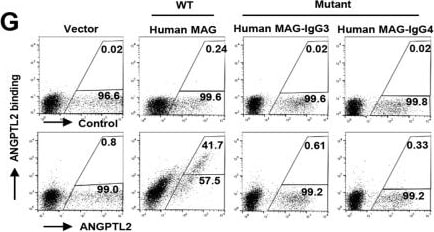
Detection of Angiopoietin-like 2 by Flow Cytometry
Human MAG is a new receptor for ANGPTL2. A Flow cytometric analysis of conditioned medium with/without ANGPTL2-Flag binding to 293T cells with transient transfection of plasmids of N1-hMAG-EGFP (human MAG, full-length) or N1-EGFP (vector). Percentage of binding/unbinding cells to ANGPTL2 in EGFP+ cells are shown. B Quantitative data of percentage of binding cells in Panel A (n = 3). C ANGPLT2 bound to the ECD of human MAG (human MAG-ECD-FC) in the conditioned medium (CM) of cotransfected 293T cells using co-immunoprecipitation. Tie-2-ECD-FC served as a negative control. D Representative flow cytometric analysis showing that purified ANGPTL2-Flag protein enhanced GFP expression in human MAG chimeric reporter cells (Human MAG). Control reporter cells (control) without human MAG-ECD were examined. E Quantitative data in Panel D (n = 3). F Binding kinetics of ANGPTL2-Flag to human MAG-ECD were measured using a biolayer interferometry instrument (Octed RED 96/96S). G–H Flow cytometric plots for the binding of ANGPTL2-Flag to full-length, mutant IgG 3 domain or IgG 4 domain of human MAG expressed 293T cells. Percentage of binding/unbinding cells to ANGPTL2 in EGFP+ cells (G) and quantitative data are shown (H) (n = 3). (***p < 0.001) Image collected and cropped by CiteAb from the following open publication (https://pubmed.ncbi.nlm.nih.gov/36855057), licensed under a CC-BY license. Not internally tested by R&D Systems.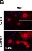
Detection of Angiopoietin-like 2 by Immunocytochemistry/ Immunofluorescence
ANGPTL2 promotes oligodendrocyte differentiation. A Cell morphology of HCN cells after treatment with IGF1 for the differentiation to oligodendrocytes in vitro with/without ANGPTL2 at the indicated time points. B Immunofluorescence staining for MBP in HCN cells 72 h after induction in Panel A. C Quantitative data in Panel B are shown; A total of 22 or 23 sections were counted (n = 4). D Representative electron microscopy images in the optic nerve fiber and corpus callosum of the brains from Angptl2+/+ and Angptl2−/− mice on day 15; red arrow indicates the normal myelin, green arrow indicates uncompacted myelin lamella; yellow arrow indicates redundant compact myelin; purple indicates excess of cytoplasm in the periaxonal space. E Shown are the quantification data of abnormally myelinated axons including uncompacted myelin lamella, redundant compact myelin and excess of cytoplasm in the periaxonal space, in optic nerve fibers and corpus callosum. 100–410 axons in each mouse were counted (n = 3). F–I Quantification of the G ratios of the myelinated axons of the optic nerve fiber (F) or corpus callosum (H) in D. A total of 120 ~ 200 axons in each mouse were counted (n = 3). The scatter plots for the individual G-ratio values and axonal size distribution in the optic nerve fibers (G) or corpus callosum (I) in Panel D are shown. (***p < 0.001) Image collected and cropped by CiteAb from the following open publication (https://pubmed.ncbi.nlm.nih.gov/36855057), licensed under a CC-BY license. Not internally tested by R&D Systems.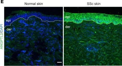
Detection of Angiopoietin-like 2 by Immunohistochemistry
ANGPTL2 is necessary for fibrotic vasculopathy.(A) qPCR of Angptl2 mRNA in WT and Snail-tg mouse keratinocytes (mKT). (B) Secreted ANGPTL2 (in green) in WT and Snail-tg skin at P3. The dotted line denotes the basement membrane separating the epidermis (epi) from the underlying dermis (der). (C) ELISA for secreted ANGPTL2 in WT and Snail-tg mKT conditioned media (CM). (D) Expression of Angptl2 in keloid lesion skin (n = 3) compared with nonlesion skin (n = 3) (from GSE92566) and in healthy (n = 44) compared with affected forearm skin from diffuse (n = 180) and localized SSc (n = 115) patients (from GSE181549). The expression values are from the GEO2R algorithm’s output. (E) Secreted ANGPTL2 (in green) in normal and SSc skin. Representative of n = 5 SSc samples and normal skin controls. (F) qPCR of vasculopathy and endoMT-related genes in SVEC4-10 endothelial cells treated with WT, Snail-tg, and Snail-tg/Angptl2-KO mKT conditioned media. (G) Quantification of collagen contraction assay in SVEC4-10 endothelial cells treated with WT, Snail-tg, and Snail-tg/Angptl2-KO mKT conditioned media. Nuclei are marked in blue. Scale bars: 50 μm. Data are shown as mean ± SEM; P values were calculated using paired Student’s t test (A), unpaired Welch’s t test (D), and 1-way ANOVA followed by Tukey’s post hoc analysis (F and G). *P < 0.05, **P < 0.01, ****P < 0.0001, NS > 0.05. All experiments except (D and E) are n = 3 biological replicates. Image collected and cropped by CiteAb from the following open publication (https://www.jci.org/articles/view/177123), licensed under a CC-BY license. Not internally tested by R&D Systems.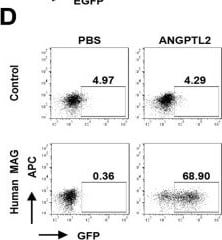
Detection of Angiopoietin-like 2 by Flow Cytometry
Human MAG is a new receptor for ANGPTL2. A Flow cytometric analysis of conditioned medium with/without ANGPTL2-Flag binding to 293T cells with transient transfection of plasmids of N1-hMAG-EGFP (human MAG, full-length) or N1-EGFP (vector). Percentage of binding/unbinding cells to ANGPTL2 in EGFP+ cells are shown. B Quantitative data of percentage of binding cells in Panel A (n = 3). C ANGPLT2 bound to the ECD of human MAG (human MAG-ECD-FC) in the conditioned medium (CM) of cotransfected 293T cells using co-immunoprecipitation. Tie-2-ECD-FC served as a negative control. D Representative flow cytometric analysis showing that purified ANGPTL2-Flag protein enhanced GFP expression in human MAG chimeric reporter cells (Human MAG). Control reporter cells (control) without human MAG-ECD were examined. E Quantitative data in Panel D (n = 3). F Binding kinetics of ANGPTL2-Flag to human MAG-ECD were measured using a biolayer interferometry instrument (Octed RED 96/96S). G–H Flow cytometric plots for the binding of ANGPTL2-Flag to full-length, mutant IgG 3 domain or IgG 4 domain of human MAG expressed 293T cells. Percentage of binding/unbinding cells to ANGPTL2 in EGFP+ cells (G) and quantitative data are shown (H) (n = 3). (***p < 0.001) Image collected and cropped by CiteAb from the following open publication (https://pubmed.ncbi.nlm.nih.gov/36855057), licensed under a CC-BY license. Not internally tested by R&D Systems.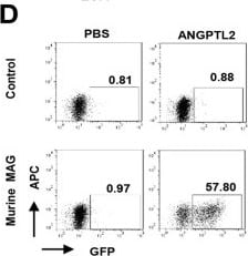
Detection of Angiopoietin-like 2 by Flow Cytometry
Murine homologue of human MAG binds to ANGPTL2. A Flow cytometric analysis of conditioned medium with/without ANGPTL2-Flag binding to 293 T cells with transient transfection of plasmids of N1-mMAG-EGFP (murine MAG, full-length) or N1-EGFP (vector). Percentage of binding/unbinding cells to ANGPTL2 in EGFP+ cells are shown. B Quantitative data of percentage of binding cells in Panel A (n = 3). C ANGPLT2 bound to the ECD of murine MAG (murine MAG-ECD) in the conditioned medium (CM) of 293 T cotransfected cells using co-immunoprecipitation. Tie-2-ECD-FC served as a negative control. D Representative flow cytometric analysis showing that purified ANGPTL2-Flag protein enhanced GFP expression in murine MAG chimeric reporter cells (Murine MAG). Control reporter cells (control) without murine MAG-ECD were examined. E Quantitative data in Panel D (n = 3). F Binding kinetics of ANGPTL2-Flag to murine MAG-ECD were measured using a biolayer interferometry instrument (Octed RED 96/96S). (***p < 0.001) Image collected and cropped by CiteAb from the following open publication (https://pubmed.ncbi.nlm.nih.gov/36855057), licensed under a CC-BY license. Not internally tested by R&D Systems.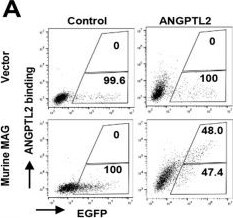
Detection of Angiopoietin-like 2 by Flow Cytometry
Murine homologue of human MAG binds to ANGPTL2. A Flow cytometric analysis of conditioned medium with/without ANGPTL2-Flag binding to 293 T cells with transient transfection of plasmids of N1-mMAG-EGFP (murine MAG, full-length) or N1-EGFP (vector). Percentage of binding/unbinding cells to ANGPTL2 in EGFP+ cells are shown. B Quantitative data of percentage of binding cells in Panel A (n = 3). C ANGPLT2 bound to the ECD of murine MAG (murine MAG-ECD) in the conditioned medium (CM) of 293 T cotransfected cells using co-immunoprecipitation. Tie-2-ECD-FC served as a negative control. D Representative flow cytometric analysis showing that purified ANGPTL2-Flag protein enhanced GFP expression in murine MAG chimeric reporter cells (Murine MAG). Control reporter cells (control) without murine MAG-ECD were examined. E Quantitative data in Panel D (n = 3). F Binding kinetics of ANGPTL2-Flag to murine MAG-ECD were measured using a biolayer interferometry instrument (Octed RED 96/96S). (***p < 0.001) Image collected and cropped by CiteAb from the following open publication (https://pubmed.ncbi.nlm.nih.gov/36855057), licensed under a CC-BY license. Not internally tested by R&D Systems.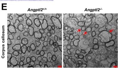
Detection of Angiopoietin-like 2 by Immunohistochemistry
ANGPTL2 promotes oligodendrocyte differentiation and remyelination under pathological conditions. A Representative images of the corpus callosum region stained with oil red O in Angptl2+/+ and Angptl2−/− mice after the withdrawal of cuprizone for two additional weeks. Myelin was stained in red color. Meanwhile, more nonspecific oil red O positive deposits (a phenomenon that oil red O deposits in damaged myelin region) were observed in Angptl2−/− mice (arrows). B Quantitative data of the demyelinating areas in Panel A; six sections from each mouse were analyzed (n = 7). C Immunofluorescence staining for MBP, MOG, MAG and GFAP in the corpus callosum region from Angptl2+/+ and Angptl2−/− mice after the withdrawal of cuprizone for two additional weeks. D Quantification of the fluorescence intensity of MBP, MOG, MAG and GFAP in the corpus callosum region in Panel C. Four sections from each mouse were analyzed (n = 6–7). E Representative electron microscopy images of the corpus callosum from Angptl2+/+ and Angptl2−/− mice after the withdrawal of cuprizone for two additional weeks; red arrow indicates uncompacted myelin lamella; F Quantification of the G ratios of the remyelinated axons of the corpus callosum in Panel E. Approximately 70 axons were counted per mouse (n = 3). G The scatter plot for the individual G-ratio values and axonal size distribution of the corpus callosum in Panel E. H Motor coordination of Angptl2+/+ and Angptl2−/− mice after cuprizone (Cupz) treatment for five weeks and cuprizone withdrawal for two additional weeks in the rotarod test; 7–8 mice were used for each group. (*p < 0.05, **p < 0. 01, ***p < 0.001) Image collected and cropped by CiteAb from the following open publication (https://pubmed.ncbi.nlm.nih.gov/36855057), licensed under a CC-BY license. Not internally tested by R&D Systems.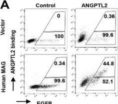
Detection of Angiopoietin-like 2 by Flow Cytometry
Human MAG is a new receptor for ANGPTL2. A Flow cytometric analysis of conditioned medium with/without ANGPTL2-Flag binding to 293T cells with transient transfection of plasmids of N1-hMAG-EGFP (human MAG, full-length) or N1-EGFP (vector). Percentage of binding/unbinding cells to ANGPTL2 in EGFP+ cells are shown. B Quantitative data of percentage of binding cells in Panel A (n = 3). C ANGPLT2 bound to the ECD of human MAG (human MAG-ECD-FC) in the conditioned medium (CM) of cotransfected 293T cells using co-immunoprecipitation. Tie-2-ECD-FC served as a negative control. D Representative flow cytometric analysis showing that purified ANGPTL2-Flag protein enhanced GFP expression in human MAG chimeric reporter cells (Human MAG). Control reporter cells (control) without human MAG-ECD were examined. E Quantitative data in Panel D (n = 3). F Binding kinetics of ANGPTL2-Flag to human MAG-ECD were measured using a biolayer interferometry instrument (Octed RED 96/96S). G–H Flow cytometric plots for the binding of ANGPTL2-Flag to full-length, mutant IgG 3 domain or IgG 4 domain of human MAG expressed 293T cells. Percentage of binding/unbinding cells to ANGPTL2 in EGFP+ cells (G) and quantitative data are shown (H) (n = 3). (***p < 0.001) Image collected and cropped by CiteAb from the following open publication (https://pubmed.ncbi.nlm.nih.gov/36855057), licensed under a CC-BY license. Not internally tested by R&D Systems.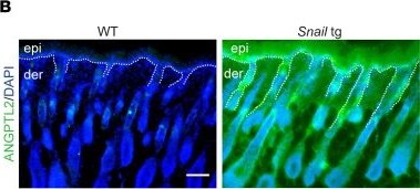
Detection of Angiopoietin-like 2 by Immunohistochemistry
ANGPTL2 is necessary for fibrotic vasculopathy.(A) qPCR of Angptl2 mRNA in WT and Snail-tg mouse keratinocytes (mKT). (B) Secreted ANGPTL2 (in green) in WT and Snail-tg skin at P3. The dotted line denotes the basement membrane separating the epidermis (epi) from the underlying dermis (der). (C) ELISA for secreted ANGPTL2 in WT and Snail-tg mKT conditioned media (CM). (D) Expression of Angptl2 in keloid lesion skin (n = 3) compared with nonlesion skin (n = 3) (from GSE92566) and in healthy (n = 44) compared with affected forearm skin from diffuse (n = 180) and localized SSc (n = 115) patients (from GSE181549). The expression values are from the GEO2R algorithm’s output. (E) Secreted ANGPTL2 (in green) in normal and SSc skin. Representative of n = 5 SSc samples and normal skin controls. (F) qPCR of vasculopathy and endoMT-related genes in SVEC4-10 endothelial cells treated with WT, Snail-tg, and Snail-tg/Angptl2-KO mKT conditioned media. (G) Quantification of collagen contraction assay in SVEC4-10 endothelial cells treated with WT, Snail-tg, and Snail-tg/Angptl2-KO mKT conditioned media. Nuclei are marked in blue. Scale bars: 50 μm. Data are shown as mean ± SEM; P values were calculated using paired Student’s t test (A), unpaired Welch’s t test (D), and 1-way ANOVA followed by Tukey’s post hoc analysis (F and G). *P < 0.05, **P < 0.01, ****P < 0.0001, NS > 0.05. All experiments except (D and E) are n = 3 biological replicates. Image collected and cropped by CiteAb from the following open publication (https://www.jci.org/articles/view/177123), licensed under a CC-BY license. Not internally tested by R&D Systems.Applications for Mouse Angiopoietin-like Protein 2/ANGPTL2 Antibody
Immunohistochemistry
Sample: Immersion fixed frozen sections of mouse embryo (E15), skeletal muscle, and thymus
Western Blot
Sample: Mouse uterus tissue and MEF mouse embryonic feeder cells
Formulation, Preparation, and Storage
Purification
Reconstitution
Reconstitute at 0.2 mg/mL in sterile PBS. For liquid material, refer to CoA for concentration.
Formulation
Shipping
Stability & Storage
- 12 months from date of receipt, -20 to -70 °C as supplied.
- 1 month, 2 to 8 °C under sterile conditions after reconstitution.
- 6 months, -20 to -70 °C under sterile conditions after reconstitution.
Calculators
Background: Angiopoietin-like Protein 2/ANGPTL2
Alternate Names
Gene Symbol
UniProt
Additional Angiopoietin-like Protein 2/ANGPTL2 Products
- All Products for Angiopoietin-like Protein 2/ANGPTL2
- Angiopoietin-like Protein 2/ANGPTL2 cDNA Clones
- Angiopoietin-like Protein 2/ANGPTL2 ELISA Kits
- Angiopoietin-like Protein 2/ANGPTL2 Lysates
- Angiopoietin-like Protein 2/ANGPTL2 Primary Antibodies
- Angiopoietin-like Protein 2/ANGPTL2 Proteins and Enzymes
Product Documents for Mouse Angiopoietin-like Protein 2/ANGPTL2 Antibody
Certificate of Analysis
To download a Certificate of Analysis, please enter a lot or batch number in the search box below.
Note: Certificate of Analysis not available for kit components.
Product Specific Notices for Mouse Angiopoietin-like Protein 2/ANGPTL2 Antibody
For research use only
Related Research Areas
Citations for Mouse Angiopoietin-like Protein 2/ANGPTL2 Antibody
 Powered by Bioz
Customer Reviews for Mouse Angiopoietin-like Protein 2/ANGPTL2 Antibody
There are currently no reviews for this product. Be the first to review Mouse Angiopoietin-like Protein 2/ANGPTL2 Antibody and earn rewards!
Have you used Mouse Angiopoietin-like Protein 2/ANGPTL2 Antibody?
Submit a review and receive an Amazon gift card!
$25/€18/£15/$25CAN/¥2500 for a review with an image
$10/€7/£6/$10CAN/¥1110 for a review without an image
Submit a review
Protocols
Find general support by application which include: protocols, troubleshooting, illustrated assays, videos and webinars.
- Antigen Retrieval Protocol (PIER)
- Antigen Retrieval for Frozen Sections Protocol
- Appropriate Fixation of IHC/ICC Samples
- Cellular Response to Hypoxia Protocols
- Chromogenic IHC Staining of Formalin-Fixed Paraffin-Embedded (FFPE) Tissue Protocol
- Chromogenic Immunohistochemistry Staining of Frozen Tissue
- ClariTSA™ Fluorophore Kits
- Detection & Visualization of Antibody Binding
- Fluorescent IHC Staining of Frozen Tissue Protocol
- Graphic Protocol for Heat-induced Epitope Retrieval
- Graphic Protocol for the Preparation and Fluorescent IHC Staining of Frozen Tissue Sections
- Graphic Protocol for the Preparation and Fluorescent IHC Staining of Paraffin-embedded Tissue Sections
- Graphic Protocol for the Preparation of Gelatin-coated Slides for Histological Tissue Sections
- IHC Sample Preparation (Frozen sections vs Paraffin)
- Immunofluorescent IHC Staining of Formalin-Fixed Paraffin-Embedded (FFPE) Tissue Protocol
- Immunohistochemistry (IHC) and Immunocytochemistry (ICC) Protocols
- Immunohistochemistry Frozen Troubleshooting
- Immunohistochemistry Paraffin Troubleshooting
- Preparing Samples for IHC/ICC Experiments
- Preventing Non-Specific Staining (Non-Specific Binding)
- Primary Antibody Selection & Optimization
- Protocol for Heat-Induced Epitope Retrieval (HIER)
- Protocol for Making a 4% Formaldehyde Solution in PBS
- Protocol for VisUCyte™ HRP Polymer Detection Reagent
- Protocol for the Preparation & Fixation of Cells on Coverslips
- Protocol for the Preparation and Chromogenic IHC Staining of Frozen Tissue Sections
- Protocol for the Preparation and Chromogenic IHC Staining of Frozen Tissue Sections - Graphic
- Protocol for the Preparation and Chromogenic IHC Staining of Paraffin-embedded Tissue Sections
- Protocol for the Preparation and Chromogenic IHC Staining of Paraffin-embedded Tissue Sections - Graphic
- Protocol for the Preparation and Fluorescent IHC Staining of Frozen Tissue Sections
- Protocol for the Preparation and Fluorescent IHC Staining of Paraffin-embedded Tissue Sections
- Protocol for the Preparation of Gelatin-coated Slides for Histological Tissue Sections
- R&D Systems Quality Control Western Blot Protocol
- TUNEL and Active Caspase-3 Detection by IHC/ICC Protocol
- The Importance of IHC/ICC Controls
- Troubleshooting Guide: Immunohistochemistry
- Troubleshooting Guide: Western Blot Figures
- Western Blot Conditions
- Western Blot Protocol
- Western Blot Protocol for Cell Lysates
- Western Blot Troubleshooting
- Western Blot Troubleshooting Guide
- View all Protocols, Troubleshooting, Illustrated assays and Webinars
Associated Pathways
