Mouse CD83 is a 30‑35 kDa member of the Siglec (or sialic-acid-binding immunoglobulin-like lectin) family of transmembrane proteins (1, 2, 3). CD83 is synthesized as a type I transmembrane glycoprotein that contains a 114 amino acid (aa) extracellular region, a 22 aa transmembrane segment, and a 39 aa cytoplasmic domain. It contains one V type Ig-like domain in the extracellular region with no inhibitory cytoplasmic motif(s). In the extracellular region, mouse and human CD83 are 66% aa identical (1, 2, 4). Relative to mouse, human CD83 has an 11 aa insertion in its extracellular domain and is expressed as a 45‑55 kDa protein (1, 4, 5, 6). No alternate splice variants have been reported for mouse. In human, however, one soluble splice form has been reported and proteolytic processing is suggested to generate a second circulating isoform (6, 7). Notably, although soluble CD83 has the potential to exist as either a monomer or disulfide-linked dimer, both show immunosuppressive activity (4, 8, 9). Membrane CD83, by contrast, is immunostimulatory (10). CD83 is a primary marker for dendritic cells (3, 5, 6). It is also found on B cells (6, 11), neutrophils (12), monocytes and macrophages (13). Except for dendritic cells, CD83 expression is often transient. CD83 binds to sialic acids on monocytes (3). The function of CD83 is only now becoming clear. As noted, membrane-immobilized CD83 appears to promote T cell proliferation, particularly of CD8+ cytotoxic T cells (14). On monocytes, CD83 may also drive monocytes into a fibrocyte phenotype (14). And a lack of membrane-expressed CD83 leads to an unusual IL-4/IL-10 producing CD4+ T cell phenotype (15).

Key Product Details
Species Reactivity
Validated:
Mouse
Cited:
Mouse
Applications
Validated:
Western Blot, Adhesion Blockade, Flow Cytometry, Immunocytochemistry, CyTOF-ready
Cited:
Western Blot, Flow Cytometry, ELISA Development (Capture)
Label
Unconjugated
Antibody Source
Polyclonal Goat IgG
Loading...
Product Specifications
Immunogen
Mouse myeloma cell line NS0-derived recombinant mouse CD83
Met22-Ala134
Accession # O88324
Met22-Ala134
Accession # O88324
Specificity
Detects mouse CD83 in direct ELISAs and Western blots.
Clonality
Polyclonal
Host
Goat
Isotype
IgG
Endotoxin Level
<0.10 EU per 1 μg of the antibody by the LAL method.
Scientific Data Images for Mouse CD83 Antibody
CD83 in Mouse Splenocytes.
CD83 was detected in immersion fixed mouse splenocytes using 10 µg/mL Goat Anti-Mouse CD83 Antigen Affinity-purified Polyclonal Antibody (Catalog # AF1437) for 3 hours at room temperature. Cells were stained with the NorthernLights™ 557-conjugated Anti-Goat IgG Secondary Antibody (red; Catalog # NL001) and counterstained with DAPI (blue). View our protocol for Fluorescent ICC Staining of Non-adherent Cells.
CD83 in Mouse Dendritic Cells.
CD83 was detected in immersion fixed LPS-stimulated mouse dendritic cells using Goat Anti-Mouse CD83 Antigen Affinity-purified Polyclonal Antibody (Catalog # AF1437) at 10 µg/mL for 3 hours at room temperature. Cells were stained using the NorthernLights™ 557-conjugated Anti-Goat IgG Secondary Antibody (red; Catalog # NL001) and counterstained with DAPI (blue). View our protocol for Fluorescent ICC Staining of Non-adherent Cells.
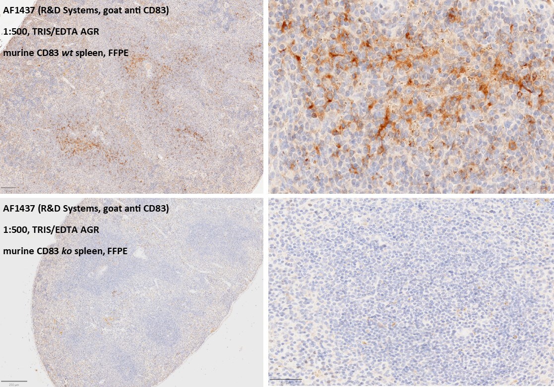
CD83 in Mouse Spleen Tissue.
Dilution: 1:500. AGR with Tris/EDTA pH 9.0 for 10min. Serum blocking with normal rabbit serum (5% for 1hour). anti-CD83 incubation for 16h at 4°C overnight. Secondary antibody: rabbit anti goat IgG Biotin. DAB. Image from a verified customer review.
Detection of CD83 by Western Blot
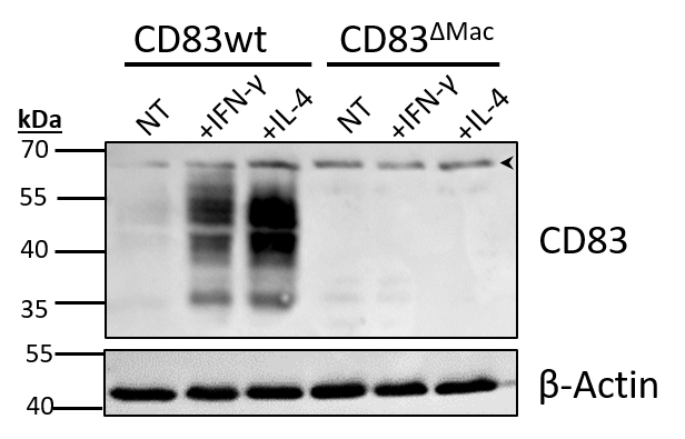
Analyses of CD83 deficient murine Mφ. Mφ were generated from CD83wt or CD83 cKO mice and subsequently stimulated with IFN-gamma or IL-4 for 16h or left untreated. (A)Cd83 expression levels were determined by qPCR and normalized to CD83wt BMDMs (n = 10). (B) Assessment of CD83 expression levels by flow cytometry (n = 20). (C) Assessment of knock-out efficiency in whole cell lysates from mock-, IFN-gamma or IL-4 stimulated Mφ by Western blotting. beta -actin served as a loading control. See full uncut gels in Supplemental Material (S1)(D) Cell viability assessment using flow cytometry (n = 24). (E) Differentiation efficacy assessing the percentage of F4/80+CD11b+ cells by flow cytometry, representing the Mφ population (n ≥ 24). (F) Expression levels of F4/80 and CD11b within the Mφ population (n ≥ 40). The gating strategy for the Mφ population is depicted in Supplementary Figure 2. Statistical analyses were performed by One-way ANOVA or the appropriate corresponding non-parametric test. Data are represented as mean ± SEM. Experiments were performed at least three times. ***p<0.001; **** p< 0.0001. The absence of asterisks indicates that there is no statistical significance. Image collected and cropped by CiteAb from the following open publication (https://pubmed.ncbi.nlm.nih.gov/36875129), licensed under a CC-BY license. Not internally tested by R&D Systems.
Detection of CD83 by Western Blot
Analyses of CD83 deficient murine Mφ. Mφ were generated from CD83wt or CD83 cKO mice and subsequently stimulated with IFN-gamma or IL-4 for 16h or left untreated. (A)Cd83 expression levels were determined by qPCR and normalized to CD83wt BMDMs (n = 10). (B) Assessment of CD83 expression levels by flow cytometry (n = 20). (C) Assessment of knock-out efficiency in whole cell lysates from mock-, IFN-gamma or IL-4 stimulated Mφ by Western blotting. beta -actin served as a loading control. See full uncut gels in Supplemental Material (S1)(D) Cell viability assessment using flow cytometry (n = 24). (E) Differentiation efficacy assessing the percentage of F4/80+CD11b+ cells by flow cytometry, representing the Mφ population (n ≥ 24). (F) Expression levels of F4/80 and CD11b within the Mφ population (n ≥ 40). The gating strategy for the Mφ population is depicted in Supplementary Figure 2. Statistical analyses were performed by One-way ANOVA or the appropriate corresponding non-parametric test. Data are represented as mean ± SEM. Experiments were performed at least three times. ***p<0.001; **** p< 0.0001. The absence of asterisks indicates that there is no statistical significance. Image collected and cropped by CiteAb from the following open publication (https://pubmed.ncbi.nlm.nih.gov/36875129), licensed under a CC-BY license. Not internally tested by R&D Systems.Applications for Mouse CD83 Antibody
Application
Recommended Usage
Adhesion Blockade
The adhesion of human monocyte-derived dendritic cells (5 x 10⁴ cells/well) to immobilized Recombinant Mouse CD83 Fc Chimera (Catalog # 1437-CD, 2.5 µg/mL, 100 µL/well) was maximally inhibited (80-100%) by 40 µg/mL of the antibody.
CyTOF-ready
Ready to be labeled using established conjugation methods. No BSA or other carrier proteins that could interfere with conjugation.
Flow Cytometry
2.5 µg/106 cells
Sample: LPS-treated mature mouse dendritic cells
Sample: LPS-treated mature mouse dendritic cells
Immunocytochemistry
5-15 µg/mL
Sample: Immersion fixed mouse splenocytes and dendritic cells
Sample: Immersion fixed mouse splenocytes and dendritic cells
Western Blot
0.1 µg/mL
Sample: Recombinant Mouse CD83 Fc Chimera (Catalog # 1437-CD)
Sample: Recombinant Mouse CD83 Fc Chimera (Catalog # 1437-CD)
Reviewed Applications
Read 2 reviews rated 5 using AF1437 in the following applications:
Flow Cytometry Panel Builder
Bio-Techne Knows Flow Cytometry
Save time and reduce costly mistakes by quickly finding compatible reagents using the Panel Builder Tool.
Advanced Features
- Spectra Viewer - Custom analysis of spectra from multiple fluorochromes
- Spillover Popups - Visualize the spectra of individual fluorochromes
- Antigen Density Selector - Match fluorochrome brightness with antigen density

Formulation, Preparation, and Storage
Purification
Antigen Affinity-purified
Reconstitution
Reconstitute at 0.2 mg/mL in sterile PBS. For liquid material, refer to CoA for concentration.
Loading...
Formulation
Lyophilized from a 0.2 μm filtered solution in PBS with Trehalose. *Small pack size (SP) is supplied either lyophilized or as a 0.2 µm filtered solution in PBS.
Shipping
Lyophilized product is shipped at ambient temperature. Liquid small pack size (-SP) is shipped with polar packs. Upon receipt, store immediately at the temperature recommended below.
Stability & Storage
Use a manual defrost freezer and avoid repeated freeze-thaw cycles.
- 12 months from date of receipt, -20 to -70 °C as supplied.
- 1 month, 2 to 8 °C under sterile conditions after reconstitution.
- 6 months, -20 to -70 °C under sterile conditions after reconstitution.
Calculators
Background: CD83
References
- Berchtold, S. et. al. (1999) FEBS Lett. 461:211.
- Fujimoto, Y. and T.F. Tedder (2006) J. Med. Dent. Sci. 53:85.
- Scholler, N. et. al. (2001) J. Immunol. 166:3865.
- Lechmann, M. et al. (2005) Biochem. Biophys. Res. Commun. 329:132.
- Zhou, L-J. et. al. (1992) J. Immunol. 149:735.
- Hock, B.D. et al. (2001) Int. Immunol. 13:959.
- Dudziak, D. et al. (2005) J. Immunol. 174:6672.
- Kotzor, N. et al. (2004) Immunobiology 209:129.
- Zinser, E. et al. (2006) Immunobiology 21:449.
- Hirano, N. et al. (2006) Blood 107:1528.
- Cramer, S.O. et al. (2000) Int. Immunol. 12:1347.
- Yamashiro, S. et al. (2000) Blood 96:3958.
- Cao, W. et al. (2005) Biochem. J. 385:85.
- Scholler, N. et al. (2002) J. Immunol. 168:2599.
- Garcia-Martinez, L.F. et al. (2004) J. Immunol. 173:2995.
Alternate Names
BL11, CD83, HB15
Gene Symbol
CD83
UniProt
Additional CD83 Products
Product Documents for Mouse CD83 Antibody
Certificate of Analysis
To download a Certificate of Analysis, please enter a lot or batch number in the search box below.
Note: Certificate of Analysis not available for kit components.
Product Specific Notices for Mouse CD83 Antibody
For research use only
Citations for Mouse CD83 Antibody
 Powered by Bioz
Customer Reviews for Mouse CD83 Antibody (2)
5 out of 5
2 Customer Ratings
Have you used Mouse CD83 Antibody?
Submit a review and receive an Amazon gift card!
$25/€18/£15/$25CAN/¥2500 Yen for a review with an image
$10/€7/£6/$10CAN/¥1110 Yen for a review without an image
Submit a review
Customer Images
Showing
1
-
2 of
2 reviews
Showing All
Filter By:
-
Application: ImmunohistochemistrySample Tested: Spleen tissueSpecies: MouseVerified Customer | Posted 08/18/2025Dilution: 1:500. AGR with Tris/EDTA pH 9.0 for 10min. Serum blocking with normal rabbit serum (5% for 1hour). anti-CD83 incubation for 16h at 4°C overnight. Secondary antibody: rabbit anti goat IgG Biotin. DAB.
 Bio-Techne ResponseThis review reflects a new species or application tested on a primary antibody.
 -
Application: Western BlotSample Tested: Bone marrow-derived macrophagesSpecies: MouseVerified Customer | Posted 09/30/2020Bone marrow-derived macrophages from wt or conditional KO mice were stimulated either with IFN-y or IL-4 for 16 h. Whole cell lysates were prepared and 50 µg of protein was used for SDS-PAGE (16%) and western blotting (primary anti-CD83 1:500; secondary anti-goat-HRP 1:7500). Specific signal only in WT-macrophages, no signal in KO cells. One unspecific band at approx. 65 kDa (see arrow-head).
There are no reviews that match your criteria.
Protocols
Find general support by application which include: protocols, troubleshooting, illustrated assays, videos and webinars.
- 7-Amino Actinomycin D (7-AAD) Cell Viability Flow Cytometry Protocol
- Appropriate Fixation of IHC/ICC Samples
- Cellular Response to Hypoxia Protocols
- ClariTSA™ Fluorophore Kits
- Detection & Visualization of Antibody Binding
- Extracellular Membrane Flow Cytometry Protocol
- Flow Cytometry Protocol for Cell Surface Markers
- Flow Cytometry Protocol for Staining Membrane Associated Proteins
- Flow Cytometry Staining Protocols
- Flow Cytometry Troubleshooting Guide
- ICC Cell Smear Protocol for Suspension Cells
- ICC Immunocytochemistry Protocol Videos
- ICC for Adherent Cells
- Immunocytochemistry (ICC) Protocol
- Immunocytochemistry Troubleshooting
- Immunofluorescence of Organoids Embedded in Cultrex Basement Membrane Extract
- Immunohistochemistry (IHC) and Immunocytochemistry (ICC) Protocols
- Intracellular Flow Cytometry Protocol Using Alcohol (Methanol)
- Intracellular Flow Cytometry Protocol Using Detergents
- Intracellular Nuclear Staining Flow Cytometry Protocol Using Detergents
- Intracellular Staining Flow Cytometry Protocol Using Alcohol Permeabilization
- Intracellular Staining Flow Cytometry Protocol Using Detergents to Permeabilize Cells
- Preparing Samples for IHC/ICC Experiments
- Preventing Non-Specific Staining (Non-Specific Binding)
- Primary Antibody Selection & Optimization
- Propidium Iodide Cell Viability Flow Cytometry Protocol
- Protocol for Liperfluo
- Protocol for VisUCyte™ HRP Polymer Detection Reagent
- Protocol for the Characterization of Human Th22 Cells
- Protocol for the Characterization of Human Th9 Cells
- Protocol for the Fluorescent ICC Staining of Cell Smears - Graphic
- Protocol for the Fluorescent ICC Staining of Cultured Cells on Coverslips - Graphic
- Protocol for the Preparation and Fluorescent ICC Staining of Cells on Coverslips
- Protocol for the Preparation and Fluorescent ICC Staining of Non-adherent Cells
- Protocol for the Preparation and Fluorescent ICC Staining of Stem Cells on Coverslips
- Protocol for the Preparation of a Cell Smear for Non-adherent Cell ICC - Graphic
- Protocol: Annexin V and PI Staining by Flow Cytometry
- Protocol: Annexin V and PI Staining for Apoptosis by Flow Cytometry
- R&D Systems Quality Control Western Blot Protocol
- TUNEL and Active Caspase-3 Detection by IHC/ICC Protocol
- The Importance of IHC/ICC Controls
- Troubleshooting Guide: Fluorokine Flow Cytometry Kits
- Troubleshooting Guide: Western Blot Figures
- Western Blot Conditions
- Western Blot Protocol
- Western Blot Protocol for Cell Lysates
- Western Blot Troubleshooting
- Western Blot Troubleshooting Guide
- View all Protocols, Troubleshooting, Illustrated assays and Webinars
Loading...
Associated Pathways

