CD8, also known as Ly-2, is a heterodimeric glycoprotein consisting of an alpha and beta chain. It is expressed on cytolytic T cells and functions in conjunction with the T cell receptor in the recognition of MHC/peptide complexes. Mouse CD8 (containing an alpha /Ly-2 or alpha ′/Lyt-2 chain) is an antigen co‑receptor on the T cell surface which interacts with MHC I molecules on antigen presenting cells (1). CD8 alpha beta heterodimer is expressed on a subpopulation of mature T cells (2, 3). CD8 alpha, without CD8 beta, has been detected on subsets of gamma delta TCR-bearing T cells (4), intestinal intrathymic lymphocytes (5, 6) and dendritic cells (7, 8).

Key Product Details
Species Reactivity
Validated:
Mouse
Cited:
Mouse, Transgenic Mouse
Applications
Validated:
Flow Cytometry, Immunocytochemistry, Immunoprecipitation, Cell depletion, Inhibition of T Cell Function, CyTOF-ready
Cited:
Immunohistochemistry, Immunohistochemistry-Paraffin, Immunohistochemistry-Frozen, Flow Cytometry, Cell Selection
Label
Unconjugated
Antibody Source
Monoclonal Rat IgG2A Clone # 53-6.7
Loading...
Product Specifications
Immunogen
Mouse thymus or spleen
Specificity
Detects both the alpha and alpha ' chains of mouse CD8 (1).
Clonality
Monoclonal
Host
Rat
Isotype
IgG2A
Endotoxin Level
<0.10 EU per 1 μg of the antibody by the LAL method.
Scientific Data Images for Mouse CD8 Antibody (53-6.7)
CD8 alpha in Mouse Splenocytes.
CD8a was detected in immersion fixed mouse splenocytes using 10 µg/mL Rat Anti-Mouse CD8a Monoclonal Antibody (Catalog # MAB116) for 3 hours at room temperature. Cells were stained with the NorthernLights™ 557-conjugated Anti-Rat IgG Secondary Antibody (red; Catalog # NL013) and counter-stained with DAPI (blue). View our protocol for Fluorescent ICC Staining of Non-adherent Cells.
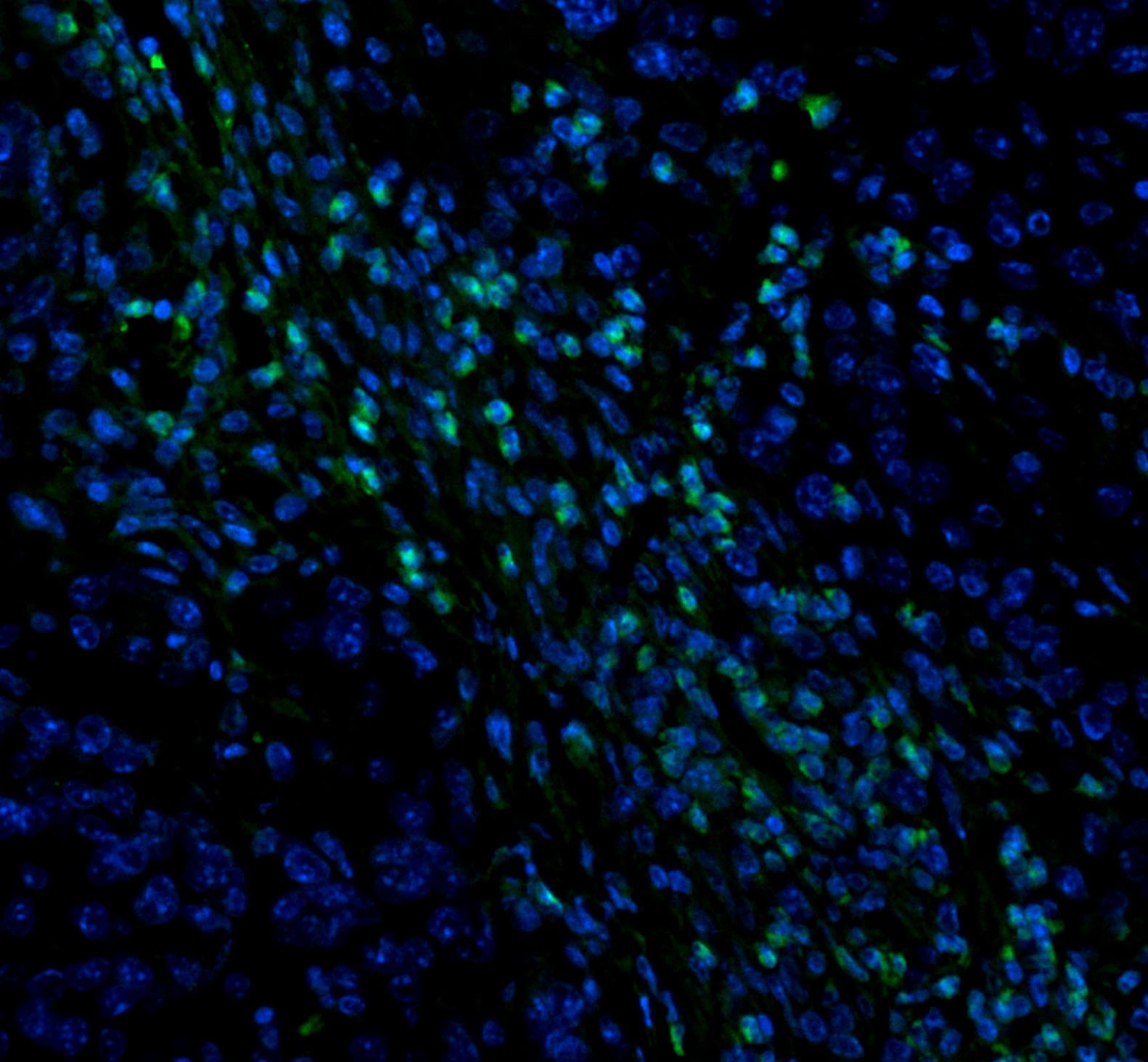
CD8 alpha in Mouse Splenocytes.
CD8a was detected in mouse splenocytes using Rat Anti-Mouse CD8a Monoclonal Antibody (Catalog # MAB116) at 10 µg/mL for 3 hours at room temperature. Cells were stained using the NorthernLights™ 557-conjugated Anti-Rat IgG Secondary Antibody (yellow; Catalog # NL013) and counterstained with DAPI (blue). View our protocol for Fluorescent ICC Staining of Non-adherent Cells.
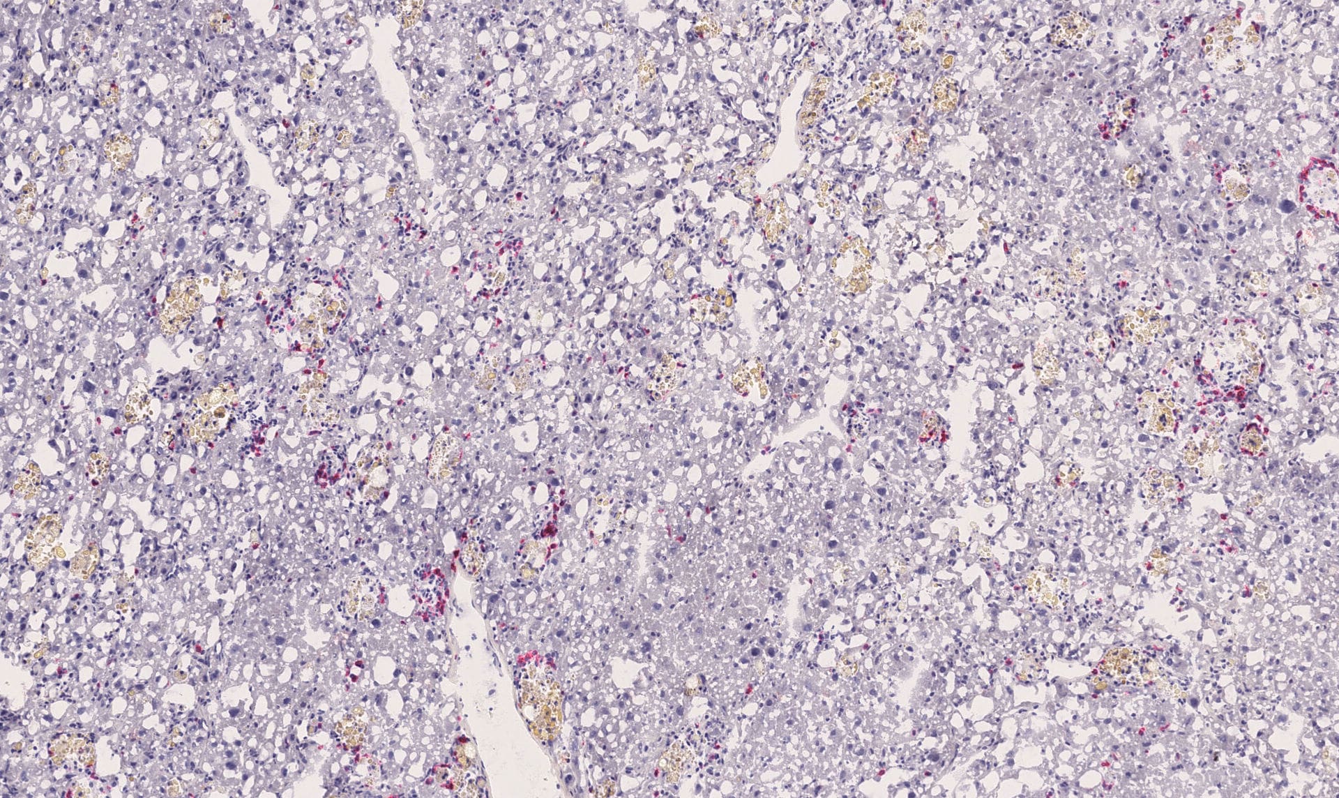
Detection of CD8 alpha by Immunohistochemistry
T cells and Con A-induced hepatitis. Isotype, CD4 and/or CD8 mAbs (100 µg per mouse) was administrated i.p. to Balb/c mice (n = 8 per group) 24h before Con A injection (20 mg kg–1). The effect of anti-CD4 orCD-8 antibodies administration on depleting these cells in the liver of mice prior injecting Con A was determined by FACS. Representative results were shown in (A). Sera were collected 24 h after Con A injection. Serum ALT (B) and AST (C) levels were measured. The results were presented as the mean ± SD of three separate experiments. ***, p<0.001 vs control. (D) The livers were removed 24 h later. Paraffin sections were stained with hematoxylin and eosin (H&E) staining. Representative liver sections were shown for each group, original magnification: ×200. Image collected and cropped by CiteAb from the following open publication (https://pubmed.ncbi.nlm.nih.gov/23118999), licensed under a CC-BY license. Not internally tested by R&D Systems.
Detection of CD8 alpha by Flow Cytometry
Endogenous T cells accumulate in K14E7 skin with an enrichment for CCR6-expressing CD4 T cells.(A) Ear skin tissue was taken from K14E7 mice in addition to control mice expressing the SIY epitope under the K14 promoter. CD4 and CD8 expression was measured on gated CD45+CD3+ cells. Data is representative of at least 4 mice/group (B) The numbers of CD4+ and CD8+ T cells in K14E7 skin or control C57 skin were enumerated per square centimetre of ear skin using flow count beads in flow cytometry. Data represents pooled mice from at least two independent experiments. (C) Gated CD3+ CD4+ T cells from the lymph node or skin of K14E7 mice were analysed for expression of the chemokine receptors, CCR6 and CCR4. The left hand panels show representative plots of isotype and CCR6 antibody staining for CD4+ T cells while the graph summarises chemokine receptor staining representative of 6 mice/group from 3 independent experiments. (D) Both K14E7 mice and C57 mice were analysed for the proliferative marker, Ki67, using intracellular staining of lymphocytes derived from the skin and inguinal lymph nodes (iLN). Naive C57 spleen cells (negative control) or C57 spleen cells treated for 3 days with PMA/Ionomycin (positive control) were also analysed. The K14E7 and C57 data represent 7 mice/group derived from 3 independent experiments while controls are spleen cells from a single mouse in 3 independent experiments. The lower right hand panels are representative plots showing isotype and Ki67 staining in K14E7 or C57 mouse skin. Image collected and cropped by CiteAb from the following open publication (https://pubmed.ncbi.nlm.nih.gov/23469070), licensed under a CC0-1.0 license. Not internally tested by R&D Systems.
Detection of CD8 alpha by Flow Cytometry
T cells and Con A-induced hepatitis. Isotype, CD4 and/or CD8 mAbs (100 µg per mouse) was administrated i.p. to Balb/c mice (n = 8 per group) 24h before Con A injection (20 mg kg–1). The effect of anti-CD4 orCD-8 antibodies administration on depleting these cells in the liver of mice prior injecting Con A was determined by FACS. Representative results were shown in (A). Sera were collected 24 h after Con A injection. Serum ALT (B) and AST (C) levels were measured. The results were presented as the mean ± SD of three separate experiments. ***, p<0.001 vs control. (D) The livers were removed 24 h later. Paraffin sections were stained with hematoxylin and eosin (H&E) staining. Representative liver sections were shown for each group, original magnification: ×200. Image collected and cropped by CiteAb from the following open publication (https://pubmed.ncbi.nlm.nih.gov/23118999), licensed under a CC-BY license. Not internally tested by R&D Systems.Applications for Mouse CD8 Antibody (53-6.7)
Application
Recommended Usage
Cell depletion
Hathcock, K.S. (1991) Current protocols in immunology, pp. 3.4.1. Kruisbeek, A.M. (1991) Current protocols in immunology, pp. 4.1.1.
CyTOF-ready
Ready to be labeled using established conjugation methods. No BSA or other carrier proteins that could interfere with conjugation.
Flow Cytometry
0.25 µg/106 cells
Sample: Mouse splenocytes
Sample: Mouse splenocytes
Immunocytochemistry
8-25 µg/mL
Sample: Immersion fixed mouse splenocytes
Sample: Immersion fixed mouse splenocytes
Immunoprecipitation
Ledbetter, J.A. and L.A. Herzenberg (1979) Immunol. Rev. 47:63.
Inhibition of T Cell Function
This antibody has been used for inhibition of T cell responses to IL-2 (Takahashi, K. et al., 1992, Proc. Natl. Acad. Sci. USA 89:5557 - 5561), MHC class I (Anel, A. et al., 1996, Eur. J. Immunol. 26:2310 - 2319) or antigens (Alexander-Miller, M.A. et al., 1996, J. Exp. Med. 184:485 - 492).
Reviewed Applications
Read 3 reviews rated 4.3 using MAB116 in the following applications:
Flow Cytometry Panel Builder
Bio-Techne Knows Flow Cytometry
Save time and reduce costly mistakes by quickly finding compatible reagents using the Panel Builder Tool.
Advanced Features
- Spectra Viewer - Custom analysis of spectra from multiple fluorochromes
- Spillover Popups - Visualize the spectra of individual fluorochromes
- Antigen Density Selector - Match fluorochrome brightness with antigen density

Formulation, Preparation, and Storage
Purification
Protein A or G purified from hybridoma culture supernatant
Reconstitution
Reconstitute at 0.5 mg/mL in sterile PBS. For liquid material, refer to CoA for concentration.
Loading...
Formulation
Lyophilized from a 0.2 μm filtered solution in PBS with Trehalose. *Small pack size (SP) is supplied either lyophilized or as a 0.2 µm filtered solution in PBS.
Shipping
Lyophilized product is shipped at ambient temperature. Liquid small pack size (-SP) is shipped with polar packs. Upon receipt, store immediately at the temperature recommended below.
Stability & Storage
Use a manual defrost freezer and avoid repeated freeze-thaw cycles.
- 12 months from date of receipt, -20 to -70 °C as supplied.
- 1 month, 2 to 8 °C under sterile conditions after reconstitution.
- 6 months, -20 to -70 °C under sterile conditions after reconstitution.
Calculators
Background: CD8
References
- Bierer, B.E. et al. (1989) Annu. Rev. Immunol. 7:579.
- Ledbetter, J.A. et al. (1980) J. Exp. Med. 152:280.
- Hayakawa, K. et al. (1994) Science 263:1131.
- MacDonald, H.R. et al. (1990) Eur. J. Immunol. 20:927.
- Rocha, B. et al. (1992) Immunol. Today 13:449.
- Wang, J. and J.R. Klein (1994) Science 265:1860.
- Vermec, D. et al. (1992) J. Exp. Med. 176:47.
- Suss, G. and K. Shortman (1996) J. Exp. Med. 183:1789.
Alternate Names
CD8, CD8A
Entrez Gene IDs
Gene Symbol
CD8A
Additional CD8 Products
Product Documents for Mouse CD8 Antibody (53-6.7)
Certificate of Analysis
To download a Certificate of Analysis, please enter a lot or batch number in the search box below.
Note: Certificate of Analysis not available for kit components.
Product Specific Notices for Mouse CD8 Antibody (53-6.7)
For research use only
Citations for Mouse CD8 Antibody (53-6.7)
 Powered by Bioz
Customer Reviews for Mouse CD8 Antibody (53-6.7) (3)
4.3 out of 5
3 Customer Ratings
Have you used Mouse CD8 Antibody (53-6.7)?
Submit a review and receive an Amazon gift card!
$25/€18/£15/$25CAN/¥2500 Yen for a review with an image
$10/€7/£6/$10CAN/¥1110 Yen for a review without an image
Submit a review
Customer Images
Showing
1
-
3 of
3 reviews
Showing All
Filter By:
-
Application: IHC on Frozen tissueSample Tested: Liver tissueSpecies: MouseVerified Customer | Posted 06/05/2018

-
Application: Immunocytochemistry/ImmunofluorescenceSample Tested: Pancreatic cancer tissueSpecies: MouseVerified Customer | Posted 08/08/20171:100 coccentration
-
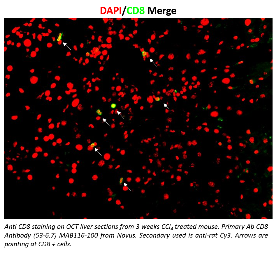
Application: Immunohistochemistry-FrozenSample Tested: Frozen mouse liverSpecies: MouseVerified Customer | Posted 03/03/2017Anti CD8 staining on OCT liver sections from 3 weeks CCl4 treated mouse. Primary Ab CD8 Antibody (53-6.7) MAB116-100 from Novus. Secondary used is anti-rat Cy3. Arrows are pointing at CD8 + cells.The staining is clean, and higly specific. Very low background signal.
There are no reviews that match your criteria.
Protocols
Find general support by application which include: protocols, troubleshooting, illustrated assays, videos and webinars.
- 7-Amino Actinomycin D (7-AAD) Cell Viability Flow Cytometry Protocol
- Appropriate Fixation of IHC/ICC Samples
- Cellular Response to Hypoxia Protocols
- ClariTSA™ Fluorophore Kits
- Detection & Visualization of Antibody Binding
- Extracellular Membrane Flow Cytometry Protocol
- Flow Cytometry Protocol for Cell Surface Markers
- Flow Cytometry Protocol for Staining Membrane Associated Proteins
- Flow Cytometry Staining Protocols
- Flow Cytometry Troubleshooting Guide
- ICC Cell Smear Protocol for Suspension Cells
- ICC Immunocytochemistry Protocol Videos
- ICC for Adherent Cells
- Immunocytochemistry (ICC) Protocol
- Immunocytochemistry Troubleshooting
- Immunofluorescence of Organoids Embedded in Cultrex Basement Membrane Extract
- Immunohistochemistry (IHC) and Immunocytochemistry (ICC) Protocols
- Immunoprecipitation Protocol
- Intracellular Flow Cytometry Protocol Using Alcohol (Methanol)
- Intracellular Flow Cytometry Protocol Using Detergents
- Intracellular Nuclear Staining Flow Cytometry Protocol Using Detergents
- Intracellular Staining Flow Cytometry Protocol Using Alcohol Permeabilization
- Intracellular Staining Flow Cytometry Protocol Using Detergents to Permeabilize Cells
- Preparing Samples for IHC/ICC Experiments
- Preventing Non-Specific Staining (Non-Specific Binding)
- Primary Antibody Selection & Optimization
- Propidium Iodide Cell Viability Flow Cytometry Protocol
- Protocol for Liperfluo
- Protocol for VisUCyte™ HRP Polymer Detection Reagent
- Protocol for the Characterization of Human Th22 Cells
- Protocol for the Characterization of Human Th9 Cells
- Protocol for the Fluorescent ICC Staining of Cell Smears - Graphic
- Protocol for the Fluorescent ICC Staining of Cultured Cells on Coverslips - Graphic
- Protocol for the Preparation and Fluorescent ICC Staining of Cells on Coverslips
- Protocol for the Preparation and Fluorescent ICC Staining of Non-adherent Cells
- Protocol for the Preparation and Fluorescent ICC Staining of Stem Cells on Coverslips
- Protocol for the Preparation of a Cell Smear for Non-adherent Cell ICC - Graphic
- Protocol: Annexin V and PI Staining by Flow Cytometry
- Protocol: Annexin V and PI Staining for Apoptosis by Flow Cytometry
- TUNEL and Active Caspase-3 Detection by IHC/ICC Protocol
- The Importance of IHC/ICC Controls
- Troubleshooting Guide: Fluorokine Flow Cytometry Kits
- View all Protocols, Troubleshooting, Illustrated assays and Webinars
FAQs for Mouse CD8 Antibody (53-6.7)
Showing
1
-
1 of
1 FAQ
Showing All
-
Q: Is Mouse CD8 alpha Antibody, Catalog # MAB116, suitable for IHC with paraffin-embedded tissue?
A: We test this antibody agasint cells and do not have data to show its use against paraffin-embedded tissue. We have citations where customers have used the clone in paraffin-embedded tissue; please see the citation link on the webpage. We are also aware the literature suggests this clone works best with acetone-fixed frozen sections or zinc-fixed paraffin sections. https://www.ncbi.nlm.nih.gov/pmc/articles/PMC4539256/


