
Key Product Details
Validated by
Biological Validation
Species Reactivity
Validated:
Mouse
Cited:
Human, Mouse, Rat, Transgenic Mouse
Applications
Validated:
Immunohistochemistry, Neutralization, Flow Cytometry, Immunocytochemistry, CyTOF-ready
Cited:
Immunohistochemistry, Immunohistochemistry-Paraffin, Immunohistochemistry-Frozen, Western Blot, Neutralization, Flow Cytometry, Immunocytochemistry, Bioassay, In vivo assay
Label
Unconjugated
Antibody Source
Monoclonal Rat IgG2B Clone # 247506
Loading...
Product Specifications
Immunogen
Y3 rat myeloid cell line transfected with mouse CXCR4
Met1-Ser359
Accession # P70658
Met1-Ser359
Accession # P70658
Specificity
Detects mouse CXCR4 transfectants. Does not stain irrelevant transfectants.
Clonality
Monoclonal
Host
Rat
Isotype
IgG2B
Endotoxin Level
<0.10 EU per 1 μg of the antibody by the LAL method.
Scientific Data Images for Mouse CXCR4 Antibody (247506)
Detection of CXCR4 in Mouse Thymocytes by Flow Cytometry.
Mouse thymocytes were stained with (A) Rat Anti-Mouse CXCR4 Monoclonal Antibody (Catalog # MAB21651) or (B) Rat IgG2B isotype control antibody (Catalog # MAB0061) followed by anti-Rat IgG PE-conjugated Secondary Antibody (Catalog # F0105B) and Rat anti-Mouse CD8 alpha APC-conjugated Monoclonal Antibody (Catalog # FAB116A). View our protocol for Staining Membrane-associated Proteins.
CXCR4 in Mouse Neural Progenitor Cells.
CXCR4 was detected in immersion fixed mouse neural progenitor cells using 10 µg/mL Rat Anti-Mouse CXCR4 Monoclonal Antibody (Catalog # MAB21651) for 3 hours at room temperature. Cells were stained with the NorthernLights™ 557-conjugated Anti-Rat IgG Secondary Antibody (red; Catalog # NL013) and counterstained with DAPI (blue). View our protocol for Fluorescent ICC Staining of Cells on Coverslips.
CXCR4 in Mouse Splenocytes.
CXCR4 was detected in immersion fixed mouse splenocytes using 10 µg/mL Rat Anti-Mouse CXCR4 Monoclonal Antibody (Catalog # MAB21651) for 3 hours at room temperature. Cells were stained with the NorthernLights™ 557-conjugated Anti-Rat IgG Secondary Antibody (red; Catalog # NL013) and counterstained with DAPI (blue). View our protocol for Fluorescent ICC Staining of Non-adherent Cells.
CXCR4 in Mouse Spleen.
CXCR4 was detected in immersion fixed frozen sections of mouse spleen using Rat Anti-Mouse CXCR4 Monoclonal Antibody (Catalog # MAB21651) at 15 µg/mL overnight at 4 °C. Tissue was stained using the Anti-Rat HRP-DAB Cell & Tissue Staining Kit (brown; Catalog # CTS017) and counterstained with hematoxylin (blue). Lower panel shows a lack of labeling if primary antibodies are omitted and tissue is stained only with secondary antibody followed by incubation with detection reagents. View our protocol for Chromogenic IHC Staining of Frozen Tissue Sections.
Chemotaxis Induced by CXCL12/SDF‑1 alpha and Neutralization by Mouse CXCR4 Antibody.
Recombinant Mouse CXCL12/SDF-1a (Catalog # 460-SD) chemo-attracts the BaF3 mouse pro-B cell line transfected with mouse CXCR4 in a dose-dependent manner (orange line). The amount of cells that migrated through to the lower chemotaxis chamber was measured by Resazurin (Catalog # AR002). Chemotaxis elicited by Recombinant Mouse CXCL12/ SDF-1a (10 ng/mL) is neutral-ized (green line) by increasing concentrations of Rat Anti-Mouse CXCR4 Monoclonal Antibody (Catalog # MAB21651). The ND50 is typically 8-40 µg/mL.
Detection of Mouse CXCR4 by Western Blot
Effects of SDF-1-CXCR4/CXCR7 pathway on MSC chemotaxis in vitro.(A) The chemotaxis in response to SDF-1 alpha (10 ng/ml for 12 h) was performed in the NP-MSCs and HP-MSCs treated with a neutralizing anti-CXCR4 antibody, an anti-CXCR7 antibody, and the respective isotype-matched control antibodies. *P<0.05, vs NP-MSCs; †P<0.05, vs the respective isotype-matched control antibodies. (B) NP-MSCs were transiently overexpressed with CXCR4 using pORF9-mCXCR4 vector or with CXCR7 using pORF9-mCXCR7 vector (n = 6). A negative control empty (pORF9-MCS) vector was used. (C) The transfected cells were subjected to chemotaxis in response to the indicated concentrations of SDF-1 alpha for 12 h. *P<0.05, vs the empty vector. (D and E) Western blot analysis (D) and FCM (E) were performed to determine the intracellular and extracellular expression of both CXCR4 and CXCR7 in the cells treated with or without SDF-1 alpha (50 ng/ml for 60 min). (F) The chemotaxis in response to SDF-1 alpha was performed in the cells treated with or without SDF-1 alpha. Image collected and cropped by CiteAb from the following publication (https://pubmed.ncbi.nlm.nih.gov/22511954), licensed under a CC-BY license. Not internally tested by R&D Systems.
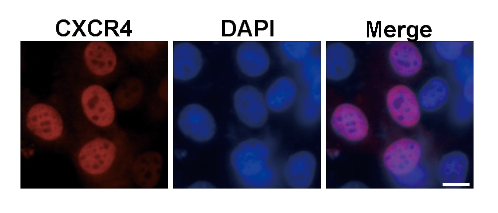
Detection of Mouse CXCR4 by Immunocytochemistry/Immunofluorescence
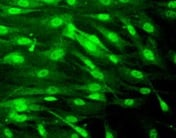
Downregulation of Cxcr4, SSEA1 and expression of TRA98 after release from GoST induction.(A) Immunofluorescence of Cxcr4 (white) and SSEA1 (green) after release from GoST induction. Cxcr4 and SSEA1 were detected simultaneously. DNA was counterstained with DAPI (blue). Cxcr4 and SSEA1 were expressed in ES, cES and GoST cells. Cxcr4 was downregulated at 4, 8 and 12 days after release from GoST induction, while SSEA1 remained expressed until 4 days after release from GoST induction and was downregulated afterwards at 8 and 12 days after release. Scalebar = 50 μm. (B) Immunofluorescence of TRA98 (white) and SSEA1 (green) after release from GoST induction. TRA98 and SSEA1 were detected simultaneously. DNA was counterstained with DAPI (blue). TRA98 and SSEA1 were expressed in ES, cES and GoST cells. Expression of TRA98 slightly decreased but remained detectable in the nucleus at 4, 8 and 12 days after release from GoST induction, while SSEA1 remained expressed until 4 days after release from GoST induction and was downregulated afterwards at 8 and 12 days after release. Scalebar = 50 μm. See Fig. S6. Image collected and cropped by CiteAb from the following publication (https://www.nature.com/articles/srep25104), licensed under a CC-BY license. Not internally tested by R&D Systems.
Detection of Human CXCR4 by Functional
CXCL12 induces CXCR4/CXCR7 and cell polarization in HUVECs. HUVECs were cultured on plastic Petri dishes (pretreated with PDL) with BSA stripes (the control group) and BSA plus CXCL12 stripes (CXCL12 group) for 5 min. HUVECs were fixed and stained with antibodies against CXCR4 (purple), CXCR7 (blue) and F-Actin (red). The micro stripes of fluorescein-conjugated BSA (A) or CXCL12 (H) are shown. CXCR4 (B), CXCR7 (C) and F-Actin (D) in HUVECs cultured on BSA stripes stayed in the resting state, while the polarization of CXCR4 (arrow: I), CXCR7 (arrow: J) and F-Actin (arrow: K) was observed in HUVECs cultured on CXCL12 stripes for 5 min. Moreover, the HUVECs in the BSA control group showed no morphological changes (E–G). The CXCL12 group HUVECs shows polarization towards the CXCL12 stripe (L–N). Scale bar: 10 μm. Image collected and cropped by CiteAb from the following publication (https://pubmed.ncbi.nlm.nih.gov/28811579), licensed under a CC-BY license. Not internally tested by R&D Systems.
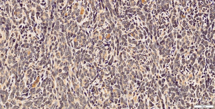
Detection of Mouse CXCR4 by Immunohistochemistry
RhoA modulates the CXCR4-CXCL12 axis in breast tumors. (a) Representative immunohistochemical (IHC) staining images of breast primary tumor sections of mice orthotopically injected with 4T1 GFP-LUC cells carrying indicated lentiviral modifications, stained with antibodies for CD31 (top, left), F4/80 (middle, left) and alpha -SMA (bottom, left). CD31+ area (top, right), F4/80+ cells (middle, right) and alpha -SMA+ area (bottom, right) per tumor field of view (FOV) (mean ± SE) quantifications of IHC images. N = 4. (b) Representative IHC staining images of breast primary tumor sections of above mice stained with CXCL12 antibody (left). CXCL12+ area per tumor FOV (mean ± SE) quantification of IHC images. N = 4. (c) Representative immunofluorescent images of breast primary tumor sections of above mice co-stained with alpha -SMA, CXCL12 and DRAQ5 nuclear stain, showing the co-localization of alpha -SMA and CXCL12 signals. Scale bar: 50 µm. (d) Representative IHC staining images of breast primary tumor sections of above mice stained with CXCR4 antibody (left). CXCR4+ area per tumor FOV (mean ± SE) quantification of IHC images. N = 4. (e) Relative mCXCR4 mRNA levels normalized with mActin (mean ± SE), as quantified by qRT-PCR in primary breast tumors of above mice (top) and in 4T1 GFP-LUC cells carrying indicated lentiviral modifications (bottom). N = 4, ns- not significant *P < 0.05, unpaired Student’s t-test (two-tailed). Image collected and cropped by CiteAb from the following publication (https://pubmed.ncbi.nlm.nih.gov/31705019), licensed under a CC-BY license. Not internally tested by R&D Systems.Applications for Mouse CXCR4 Antibody (247506)
Application
Recommended Usage
CyTOF-ready
Ready to be labeled using established conjugation methods. No BSA or other carrier proteins that could interfere with conjugation.
Flow Cytometry
0.25 µg/106 cells
Sample: Mouse thymocytes
Sample: Mouse thymocytes
Immunocytochemistry
8-25 µg/mL
Sample: Immersion fixed mouse neural progenitor cells and immersion fixed mouse splenocytes
Sample: Immersion fixed mouse neural progenitor cells and immersion fixed mouse splenocytes
Immunohistochemistry
8-25 µg/mL
Sample: Immersion fixed frozen sections of mouse spleen
Sample: Immersion fixed frozen sections of mouse spleen
Neutralization
Measured by its ability to neutralize CXCL12/SDF‑1 alpha -induced chemotaxis in the BaF3 mouse pro‑B cell line transfected with mouse CXCR4. The Neutralization Dose (ND50) is typically 8‑40 µg/mL in the presence of 10 ng/mL Recombinant Mouse CXCL12/SDF‑1 alpha.
Reviewed Applications
Read 5 reviews rated 4.4 using MAB21651 in the following applications:
Flow Cytometry Panel Builder
Bio-Techne Knows Flow Cytometry
Save time and reduce costly mistakes by quickly finding compatible reagents using the Panel Builder Tool.
Advanced Features
- Spectra Viewer - Custom analysis of spectra from multiple fluorochromes
- Spillover Popups - Visualize the spectra of individual fluorochromes
- Antigen Density Selector - Match fluorochrome brightness with antigen density

Formulation, Preparation, and Storage
Purification
Protein A or G purified from hybridoma culture supernatant
Reconstitution
Reconstitute at 0.5 mg/mL in sterile PBS. For liquid material, refer to CoA for concentration.
Loading...
Formulation
Lyophilized from a 0.2 μm filtered solution in PBS with Trehalose. *Small pack size (SP) is supplied either lyophilized or as a 0.2 µm filtered solution in PBS.
Shipping
Lyophilized product is shipped at ambient temperature. Liquid small pack size (-SP) is shipped with polar packs. Upon receipt, store immediately at the temperature recommended below.
Stability & Storage
Use a manual defrost freezer and avoid repeated freeze-thaw cycles.
- 12 months from date of receipt, -20 to -70 °C as supplied.
- 1 month, 2 to 8 °C under sterile conditions after reconstitution.
- 6 months, -20 to -70 °C under sterile conditions after reconstitution.
Calculators
Background: CXCR4
References
- Orsini, M.J. et al. (1999) J. Biol. Chem. 274:31076.
- Zagzag, D. et al. (2005) Cancer Res. 65:6178.
- Speetjens, F.M. et al. (2009) Cancer Microenvironment 2:1.
- Wang, L. et al. (2009) Oncology Reports 22:1333.
- Amara, S. et al. (2015) Cancer Biomark. 15:869.
Long Name
C-X-C Motif Chemokine Receptor 4
Alternate Names
CD184, D2S201E, FB22, Fusin, HM89, HSY3RR, LAP-3, LAP3, LCR1, LESTR, NPY3R, NPYRL, NPYY3R, WHIMS
Gene Symbol
CXCR4
UniProt
Additional CXCR4 Products
Product Documents for Mouse CXCR4 Antibody (247506)
Certificate of Analysis
To download a Certificate of Analysis, please enter a lot or batch number in the search box below.
Note: Certificate of Analysis not available for kit components.
Product Specific Notices for Mouse CXCR4 Antibody (247506)
For research use only
Related Research Areas
Citations for Mouse CXCR4 Antibody (247506)
 Powered by Bioz
Customer Reviews for Mouse CXCR4 Antibody (247506) (5)
4.4 out of 5
5 Customer Ratings
Have you used Mouse CXCR4 Antibody (247506)?
Submit a review and receive an Amazon gift card!
$25/€18/£15/$25CAN/¥2500 Yen for a review with an image
$10/€7/£6/$10CAN/¥1110 Yen for a review without an image
Submit a review
Customer Images
Showing
1
-
5 of
5 reviews
Showing All
Filter By:
-
Application: Immunocytochemistry/ImmunofluorescenceSample Tested: BMM stem cellsSpecies: MouseVerified Customer | Posted 08/20/2021

-
Application: Immunocytochemistry/ImmunofluorescenceSample Tested: Hepa 1-6 mouse hepatoma cell lineSpecies: MouseVerified Customer | Posted 03/07/2020
-
Application: ImmunohistochemistrySample Tested: 4T1 mouse breast cancer cell lineSpecies: MouseVerified Customer | Posted 03/05/20191:75 Citrate buffer antigen retrieval
-
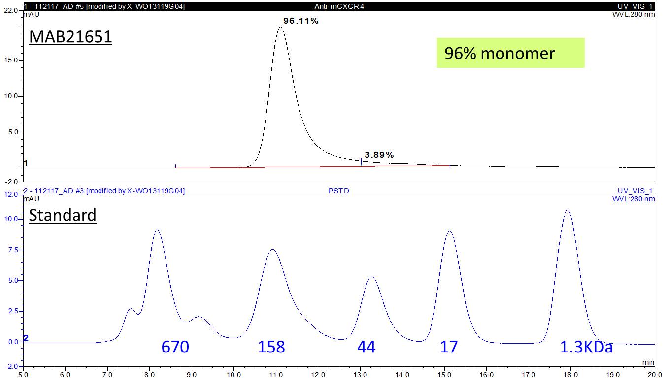
Application: Block/NeutralizeSample Tested: BaF3 mouse pro-B cell lineSpecies: MouseVerified Customer | Posted 12/18/2017> 95% monomer
-
Application: ImmunofluorescenceSample Tested: See PMID 22752632Species: MouseVerified Customer | Posted 02/19/2015
There are no reviews that match your criteria.
Protocols
Find general support by application which include: protocols, troubleshooting, illustrated assays, videos and webinars.
- 7-Amino Actinomycin D (7-AAD) Cell Viability Flow Cytometry Protocol
- Antigen Retrieval Protocol (PIER)
- Antigen Retrieval for Frozen Sections Protocol
- Appropriate Fixation of IHC/ICC Samples
- Cellular Response to Hypoxia Protocols
- Chromogenic IHC Staining of Formalin-Fixed Paraffin-Embedded (FFPE) Tissue Protocol
- Chromogenic Immunohistochemistry Staining of Frozen Tissue
- ClariTSA™ Fluorophore Kits
- Detection & Visualization of Antibody Binding
- Extracellular Membrane Flow Cytometry Protocol
- Flow Cytometry Protocol for Cell Surface Markers
- Flow Cytometry Protocol for Staining Membrane Associated Proteins
- Flow Cytometry Staining Protocols
- Flow Cytometry Troubleshooting Guide
- Fluorescent IHC Staining of Frozen Tissue Protocol
- Graphic Protocol for Heat-induced Epitope Retrieval
- Graphic Protocol for the Preparation and Fluorescent IHC Staining of Frozen Tissue Sections
- Graphic Protocol for the Preparation and Fluorescent IHC Staining of Paraffin-embedded Tissue Sections
- Graphic Protocol for the Preparation of Gelatin-coated Slides for Histological Tissue Sections
- ICC Cell Smear Protocol for Suspension Cells
- ICC Immunocytochemistry Protocol Videos
- ICC for Adherent Cells
- IHC Sample Preparation (Frozen sections vs Paraffin)
- Immunocytochemistry (ICC) Protocol
- Immunocytochemistry Troubleshooting
- Immunofluorescence of Organoids Embedded in Cultrex Basement Membrane Extract
- Immunofluorescent IHC Staining of Formalin-Fixed Paraffin-Embedded (FFPE) Tissue Protocol
- Immunohistochemistry (IHC) and Immunocytochemistry (ICC) Protocols
- Immunohistochemistry Frozen Troubleshooting
- Immunohistochemistry Paraffin Troubleshooting
- Intracellular Flow Cytometry Protocol Using Alcohol (Methanol)
- Intracellular Flow Cytometry Protocol Using Detergents
- Intracellular Nuclear Staining Flow Cytometry Protocol Using Detergents
- Intracellular Staining Flow Cytometry Protocol Using Alcohol Permeabilization
- Intracellular Staining Flow Cytometry Protocol Using Detergents to Permeabilize Cells
- Preparing Samples for IHC/ICC Experiments
- Preventing Non-Specific Staining (Non-Specific Binding)
- Primary Antibody Selection & Optimization
- Propidium Iodide Cell Viability Flow Cytometry Protocol
- Protocol for Heat-Induced Epitope Retrieval (HIER)
- Protocol for Liperfluo
- Protocol for Making a 4% Formaldehyde Solution in PBS
- Protocol for VisUCyte™ HRP Polymer Detection Reagent
- Protocol for the Characterization of Human Th22 Cells
- Protocol for the Characterization of Human Th9 Cells
- Protocol for the Fluorescent ICC Staining of Cell Smears - Graphic
- Protocol for the Fluorescent ICC Staining of Cultured Cells on Coverslips - Graphic
- Protocol for the Preparation & Fixation of Cells on Coverslips
- Protocol for the Preparation and Chromogenic IHC Staining of Frozen Tissue Sections
- Protocol for the Preparation and Chromogenic IHC Staining of Frozen Tissue Sections - Graphic
- Protocol for the Preparation and Chromogenic IHC Staining of Paraffin-embedded Tissue Sections
- Protocol for the Preparation and Chromogenic IHC Staining of Paraffin-embedded Tissue Sections - Graphic
- Protocol for the Preparation and Fluorescent ICC Staining of Cells on Coverslips
- Protocol for the Preparation and Fluorescent ICC Staining of Non-adherent Cells
- Protocol for the Preparation and Fluorescent ICC Staining of Stem Cells on Coverslips
- Protocol for the Preparation and Fluorescent IHC Staining of Frozen Tissue Sections
- Protocol for the Preparation and Fluorescent IHC Staining of Paraffin-embedded Tissue Sections
- Protocol for the Preparation of Gelatin-coated Slides for Histological Tissue Sections
- Protocol for the Preparation of a Cell Smear for Non-adherent Cell ICC - Graphic
- Protocol: Annexin V and PI Staining by Flow Cytometry
- Protocol: Annexin V and PI Staining for Apoptosis by Flow Cytometry
- TUNEL and Active Caspase-3 Detection by IHC/ICC Protocol
- The Importance of IHC/ICC Controls
- Troubleshooting Guide: Fluorokine Flow Cytometry Kits
- Troubleshooting Guide: Immunohistochemistry
- View all Protocols, Troubleshooting, Illustrated assays and Webinars


