Mouse IL-1 beta/IL-1F2 Antibody [Biotin]
R&D Systems | Catalog # BAF401

Scientific images may be resized, cropped, or adjusted for brightness or contrast for presentation purposes. These updates are limited to presentation and do not affect the underlying scientific interpretation of the data. Where available, additional source data may be provided upon request.
Key Product Details
Validated by
Species Reactivity
Validated:
Cited:
Applications
Validated:
Cited:
Label
Antibody Source
Product Specifications
Immunogen
Val118-Ser269
Accession # NP_032387
Specificity
Clonality
Host
Isotype
Scientific Data Images for Mouse IL-1 beta/IL-1F2 Antibody [Biotin]
Detection of Mouse IL-1 beta/IL-1F2 by Western Blot
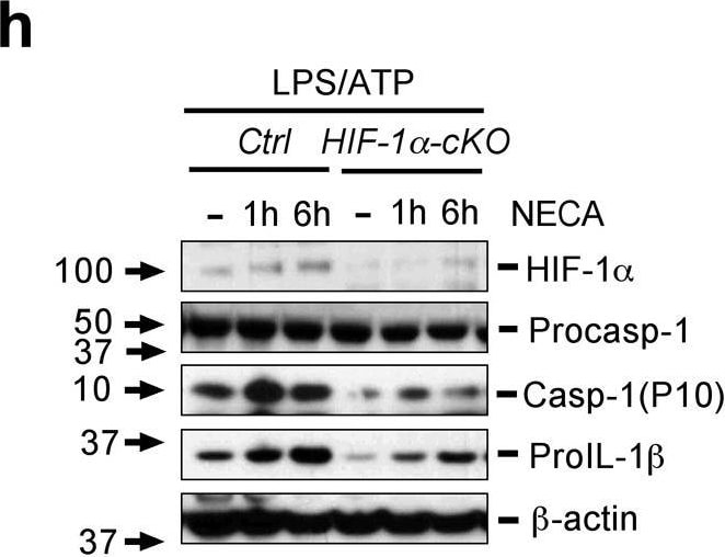
Adenosine mediates increase in pro-IL-1 beta via a HIF-1 alpha -dependent pathway. (a) Consensus NF kappa B and HRE binding sites in the IL-1 beta promoter. (b) LPS primed PECs obtained from A2AR-cKO and controls were stimulated with or without NECA or CGS21680. (c) LPS primed PECs obtained from HIF-1 alpha -cKO and controls were stimulated with or without NECA at time points as indicated. Cells were harvested and RNA isolated after each treatment and gene expression of Il1b and Hif1 alpha quantified by real-time PCR. (d) THP-1 cells were transfected with HRE- promoter luciferase construct and beta -galactosidase plasmid in the presence or absence of CREB dominant negative plasmid (CREB delta ), and then primed with LPS/PMA followed by NECA. (e) THP-1 cells were transfected with human IL-1 beta promoter luciferase construct and beta -galactosidase plasmid in the presence or absence of CREB delta, and then primed with LPS/PMA followed by CGS21680 or NECA. Luciferase activities were measured and normalized to beta -galactosidase activity and normalized with controls. Data are mean± SD of triplicate cultures and are representative of three independent experiments. (f) CD14-MD2-TLR4- HEK 293 cells were transfected with NF kappa B promoter luciferase construct and Renilla luciferase (Rluc) control reporter vector, and then treated with CGS21680, NECA or ZM241395 in the presence of LPS, Luciferase activities were measured and normalized to Rluc activity and the normalized value with controls as indicated. Data are mean± SD of triplicate cultures and are representative of three independent experiments for b-f. (g) LPS primed PECs obtained from HIF-1 alpha -cKO and controls were treated with or without NECA at different time points as indicated followed pulsing with ATP. Cell supernatants were collected in 5 hrs after ATP pulsing. IL-1 beta was measured in cell supernatant by ELISA. Data are expressed as the mean ± SD from three independent experiments. (h) LPS primed PECs obtained from HIF-1 alpha -cKO and control mice were treated with or without NEC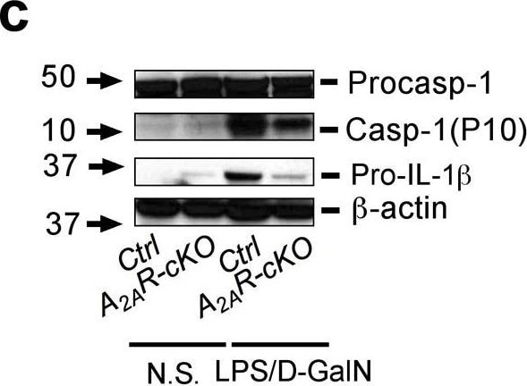
Detection of Mouse IL-1 beta/IL-1F2 by Western Blot
Liver injury and fibrosis is dependent on A2A receptor signaling in macrophages(a)A2AR-cKO and control mice were injected intraperitoneally with LPS (1 mg/kg) and D-galactosamine (500 mg kg−1) for 6 hrs followed by liver tissue and serum collection for H&E staining and ALT assay. (b) Liver RNA samples were collected and Il1b gene assayed by real-time PCR using specific primers. (c) Liver tissue lysates were assayed for pro-caspase-1, cleaved caspase-1 (p10), and beta -actin protein level by immunoblot analysis using specific antibodies. Data are expressed as the mean ± SD from 10–11 mice from each group for a-d. (d) Serum was collected for measurement of IL-1 beta. (e)A2AR-cKO and control mice were injected intraperitoneally with single dose of TAA followed by liver tissue collection as indicated for H&E staining. (f) Liver RNA samples were collected and Il1b gene was assayed by real-time PCR. (g) Liver tissue was also obtained at day 7 after TAA injection and stained for H&E and Sirius red for fibrosis. (h) Sera were collected and the serum ALT assay was performed (Data are expressed as the mean ± SD from 5 mice in each group). * p <0.05 determined by Student’s t-test. Scale bars correspond to 500μm. Image collected and cropped by CiteAb from the following publication (https://pubmed.ncbi.nlm.nih.gov/24352507), licensed under a CC-BY license. Not internally tested by R&D Systems.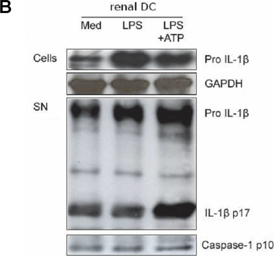
Detection of IL-1 beta/IL-1F2 by Western Blot
IL-1 beta release and caspase-1 activation in renal DC, but not in glomeruli and glomerular cells.A. IL-1 beta release from glomeruli isolated from healthy C57BL/6 wildtype mice, primary mesangial cells (PMC), podocytes, glomerular endothelial cells (GEnC), and renal dendritic cells (DC) stimulated with LPS or LPS followed by ATP was measured by ELISA. Bone marrow dendritic cells (BMDC) served as a positive control. Note that only renal DCs but none of the other glomerular cells released IL-1 beta after LPS/ATP stimulation. B–D. This was confirmed by Western blot for IL-1 beta. The blots illustrate the 37 kDa pro-IL-1 beta in the upper bands and the 17 kDa mature IL-1 beta in the lower bands. Western blot for caspase-1 illustrates the 10 kDa caspase-1 cleaved product which indicates caspase-1 activation. GAPDH is shown as a loading control. Stimulated bone marrow dendritic cells served as a positive control. Image collected and cropped by CiteAb from the following open publication (https://pubmed.ncbi.nlm.nih.gov/22046355), licensed under a CC-BY license. Not internally tested by R&D Systems.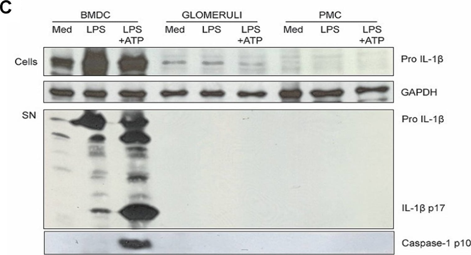
Detection of IL-1 beta/IL-1F2 by Western Blot
IL-1 beta release and caspase-1 activation in renal DC, but not in glomeruli and glomerular cells.A. IL-1 beta release from glomeruli isolated from healthy C57BL/6 wildtype mice, primary mesangial cells (PMC), podocytes, glomerular endothelial cells (GEnC), and renal dendritic cells (DC) stimulated with LPS or LPS followed by ATP was measured by ELISA. Bone marrow dendritic cells (BMDC) served as a positive control. Note that only renal DCs but none of the other glomerular cells released IL-1 beta after LPS/ATP stimulation. B–D. This was confirmed by Western blot for IL-1 beta. The blots illustrate the 37 kDa pro-IL-1 beta in the upper bands and the 17 kDa mature IL-1 beta in the lower bands. Western blot for caspase-1 illustrates the 10 kDa caspase-1 cleaved product which indicates caspase-1 activation. GAPDH is shown as a loading control. Stimulated bone marrow dendritic cells served as a positive control. Image collected and cropped by CiteAb from the following open publication (https://pubmed.ncbi.nlm.nih.gov/22046355), licensed under a CC-BY license. Not internally tested by R&D Systems.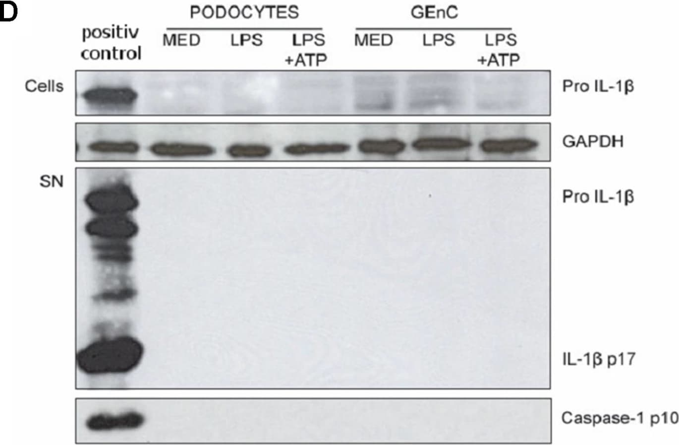
Detection of IL-1 beta/IL-1F2 by Western Blot
IL-1 beta release and caspase-1 activation in renal DC, but not in glomeruli and glomerular cells.A. IL-1 beta release from glomeruli isolated from healthy C57BL/6 wildtype mice, primary mesangial cells (PMC), podocytes, glomerular endothelial cells (GEnC), and renal dendritic cells (DC) stimulated with LPS or LPS followed by ATP was measured by ELISA. Bone marrow dendritic cells (BMDC) served as a positive control. Note that only renal DCs but none of the other glomerular cells released IL-1 beta after LPS/ATP stimulation. B–D. This was confirmed by Western blot for IL-1 beta. The blots illustrate the 37 kDa pro-IL-1 beta in the upper bands and the 17 kDa mature IL-1 beta in the lower bands. Western blot for caspase-1 illustrates the 10 kDa caspase-1 cleaved product which indicates caspase-1 activation. GAPDH is shown as a loading control. Stimulated bone marrow dendritic cells served as a positive control. Image collected and cropped by CiteAb from the following open publication (https://pubmed.ncbi.nlm.nih.gov/22046355), licensed under a CC-BY license. Not internally tested by R&D Systems.
Mouse IL-1 beta / IL-1F2 ELISA Standard Curve
Recombinant Mouse IL‑1 beta /IL‑1F2 (Catalog # 401-ML) was serially diluted and captured by Rat Anti-Mouse IL‑1 beta /IL‑1F2 Monoclonal Antibody (Catalog # MAB401) coated on a Clear Polystyrene Microplate (Catalog # DY990). Goat Anti-Mouse IL‑1 beta /IL‑1F2 Antigen Affinity-purified Polyclonal Antibody (Catalog # AF-401-NA) was biotinylated and incubated with the protein captured on the plate. Detection of the standard curve was achieved by incubating Streptavidin-HRP (Catalog # DY998)
Detection of Mouse IL-1 beta/IL-1F2 by ELISA
Recombinant Mouse IL‑1 beta /IL‑1F2 (Catalog # 401-ML) was serially diluted and captured by Rat Anti-Mouse IL‑1 beta /IL‑1F2 Monoclonal Antibody (Catalog # MAB401) coated on a Clear Polystyrene Microplate (Catalog # DY990). Goat Anti-Mouse IL‑1 beta /IL‑1F2 Antigen Affinity-purified Polyclonal Antibody (Catalog # AF-401-NA) was biotinylated and incubated with the protein captured on the plate. Detection of the standard curve was achieved by incubating Streptavidin-HRP (Catalog # DY998)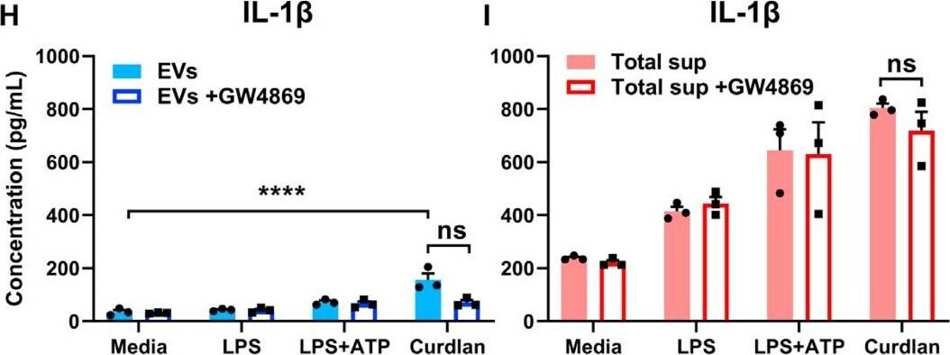
Detection of Mouse IL-1 beta/IL-1F2 by ELISA
Recombinant Mouse IL‑1 beta /IL‑1F2 (Catalog # 401-ML) was serially diluted and captured by Rat Anti-Mouse IL‑1 beta /IL‑1F2 Monoclonal Antibody (Catalog # MAB401) coated on a Clear Polystyrene Microplate (Catalog # DY990). Goat Anti-Mouse IL‑1 beta /IL‑1F2 Antigen Affinity-purified Polyclonal Antibody (Catalog # AF-401-NA) was biotinylated and incubated with the protein captured on the plate. Detection of the standard curve was achieved by incubating Streptavidin-HRP (Catalog # DY998)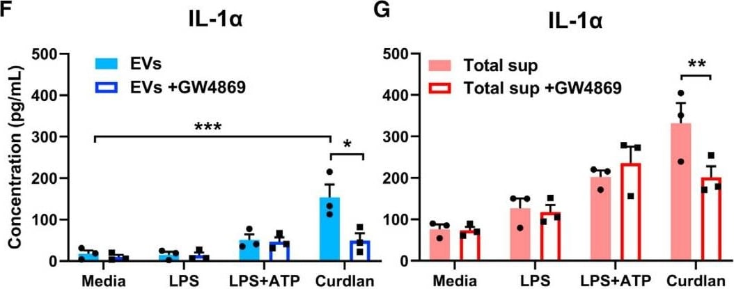
Detection of Mouse IL-1 beta/IL-1F2 by ELISA
Recombinant Mouse IL‑1 beta /IL‑1F2 (Catalog # 401-ML) was serially diluted and captured by Rat Anti-Mouse IL‑1 beta /IL‑1F2 Monoclonal Antibody (Catalog # MAB401) coated on a Clear Polystyrene Microplate (Catalog # DY990). Goat Anti-Mouse IL‑1 beta /IL‑1F2 Antigen Affinity-purified Polyclonal Antibody (Catalog # AF-401-NA) was biotinylated and incubated with the protein captured on the plate. Detection of the standard curve was achieved by incubating Streptavidin-HRP (Catalog # DY998)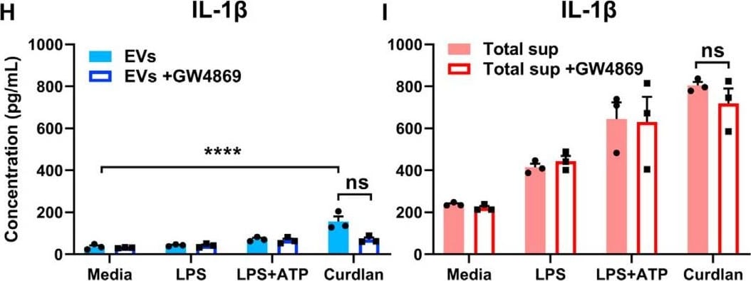
Detection of Mouse IL-1 beta/IL-1F2 by ELISA
Recombinant Mouse IL‑1 beta /IL‑1F2 (Catalog # 401-ML) was serially diluted and captured by Rat Anti-Mouse IL‑1 beta /IL‑1F2 Monoclonal Antibody (Catalog # MAB401) coated on a Clear Polystyrene Microplate (Catalog # DY990). Goat Anti-Mouse IL‑1 beta /IL‑1F2 Antigen Affinity-purified Polyclonal Antibody (Catalog # AF-401-NA) was biotinylated and incubated with the protein captured on the plate. Detection of the standard curve was achieved by incubating Streptavidin-HRP (Catalog # DY998)Applications for Mouse IL-1 beta/IL-1F2 Antibody [Biotin]
Western Blot
Sample: Recombinant Mouse IL‑1 beta /IL‑1F2 (Catalog # 401-ML)
Mouse IL-1 beta /IL-1F2 Sandwich Immunoassay
Reviewed Applications
Read 1 review rated 5 using BAF401 in the following applications:
Formulation, Preparation, and Storage
Purification
Reconstitution
Reconstitute at 0.2 mg/mL in sterile PBS.
Formulation
Shipping
Stability & Storage
- 12 months from date of receipt, -20 to -70 °C as supplied.
- 1 month, 2 to 8 °C under sterile conditions after reconstitution.
- 6 months, -20 to -70 °C under sterile conditions after reconstitution.
Calculators
Background: IL-1 beta/IL-1F2
Long Name
Alternate Names
Entrez Gene IDs
Gene Symbol
UniProt
Additional IL-1 beta/IL-1F2 Products
Product Documents for Mouse IL-1 beta/IL-1F2 Antibody [Biotin]
Certificate of Analysis
To download a Certificate of Analysis, please enter a lot or batch number in the search box below.
Note: Certificate of Analysis not available for kit components.
Product Specific Notices for Mouse IL-1 beta/IL-1F2 Antibody [Biotin]
For research use only
Related Research Areas
Citations for Mouse IL-1 beta/IL-1F2 Antibody [Biotin]
 Powered by Bioz
Customer Reviews for Mouse IL-1 beta/IL-1F2 Antibody [Biotin] (1)
Have you used Mouse IL-1 beta/IL-1F2 Antibody [Biotin]?
Submit a review and receive an Amazon gift card!
$25/€18/£15/$25CAN/¥2500 for a review with an image
$10/€7/£6/$10CAN/¥1110 for a review without an image
Submit a review
Customer Images
![Mouse IL-1 beta/IL-1F2 Antibody [Biotin] BAF401](https://resources.rndsystems.com/images/reviews/review_baf401_16661.png)
-
Application: ELISASample Tested: Serum and Plasma and Tissue Culture MediaSpecies: MouseVerified Customer | Posted 11/21/2017This antibody was paired with MAB4012 to create an ELISA to measure IL-1b in mouse samples.
![Mouse IL-1 beta/IL-1F2 Antibody [Biotin] BAF401](data:image/png;base64,R0lGODlhAQABAAD/ACwAAAAAAQABAAACADs=)
There are no reviews that match your criteria.
Protocols
Find general support by application which include: protocols, troubleshooting, illustrated assays, videos and webinars.
- Cellular Response to Hypoxia Protocols
- R&D Systems Quality Control Western Blot Protocol
- Troubleshooting Guide: Western Blot Figures
- Western Blot Conditions
- Western Blot Protocol
- Western Blot Protocol for Cell Lysates
- Western Blot Troubleshooting
- Western Blot Troubleshooting Guide
- View all Protocols, Troubleshooting, Illustrated assays and Webinars
Associated Pathways