Mouse Integrin beta 1/CD29 Antibody (265917)
R&D Systems | Catalog # MAB2405

Key Product Details
Species Reactivity
Validated:
Mouse
Cited:
Mouse, Rat
Applications
Validated:
Immunohistochemistry, Western Blot, Flow Cytometry, Immunocytochemistry, CyTOF-ready
Cited:
Immunohistochemistry, Immunohistochemistry-Paraffin, Immunohistochemistry-Frozen, Western Blot, Neutralization, Flow Cytometry, Immunofluorescence, Immunocytochemistry
Label
Unconjugated
Antibody Source
Monoclonal Rat IgG2A Clone # 265917
Loading...
Product Specifications
Immunogen
Chinese hamster ovary cell line CHO-derived recombinant mouse Integrin beta 1/CD29
Gln21-Asp728
Accession # P09055
Gln21-Asp728
Accession # P09055
Specificity
Detects mouse Integrin beta 1/CD29 in direct ELISAs and Western blots. In Western blots, no cross-reactivity with recombinant human Integrin beta 1, recombinant mouse (rm) Integrin beta 2, rmIntegrin beta 3, rmIntegrin beta 4, or rmIntegrin beta 6 is observed.
Clonality
Monoclonal
Host
Rat
Isotype
IgG2A
Scientific Data Images for Mouse Integrin beta 1/CD29 Antibody (265917)
Detection of Integrin beta 1/CD29 in B16‑F1 Mouse Cell Line by Flow Cytometry.
B16-F1 mouse melanoma cell line was stained with Rat Anti-Mouse Integrin beta 1/CD29 Monoclonal Antibody (Catalog # MAB2405, filled histogram) or isotype control antibody (Catalog # MAB006, open histogram), followed by Phycoerythrin-conjugated Anti-Rat IgG Secondary Antibody (Catalog # F0105B).
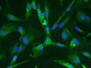
Integrin beta 1/CD29 in L‑929 Mouse Cell Line.
Integrin beta 1/CD29 was detected in immersion fixed L-929 mouse fibroblast cell line using Rat Anti-Mouse Integrin beta 1/CD29 Monoclonal Antibody (Catalog # MAB2405) at 10 µg/mL for 3 hours at room temperature. Cells were stained using the NorthernLights™ 557-conjugated Anti-Rat IgG Secondary Antibody (yellow; Catalog # NL013) and counterstained with DAPI (blue). View our protocol for Fluorescent ICC Staining of Cells on Coverslips.Applications for Mouse Integrin beta 1/CD29 Antibody (265917)
Application
Recommended Usage
CyTOF-ready
Ready to be labeled using established conjugation methods. No BSA or other carrier proteins that could interfere with conjugation.
Flow Cytometry
0.25 µg/106 cells
Sample: B16‑F10 mouse melanoma cell line
Sample: B16‑F10 mouse melanoma cell line
Immunocytochemistry
8-25 µg/mL
Sample: Immersion fixed L-929 mouse fibroblast cell line
Sample: Immersion fixed L-929 mouse fibroblast cell line
Immunohistochemistry
8-25 µg/mL
Sample: Perfusion fixed frozen sections of mouse spinal cord
Sample: Perfusion fixed frozen sections of mouse spinal cord
Western Blot
1 µg/mL
Sample: Recombinant Mouse Integrin beta 1/CD29 under non-reducing conditions only
Sample: Recombinant Mouse Integrin beta 1/CD29 under non-reducing conditions only
Reviewed Applications
Read 3 reviews rated 2.3 using MAB2405 in the following applications:
Flow Cytometry Panel Builder
Bio-Techne Knows Flow Cytometry
Save time and reduce costly mistakes by quickly finding compatible reagents using the Panel Builder Tool.
Advanced Features
- Spectra Viewer - Custom analysis of spectra from multiple fluorochromes
- Spillover Popups - Visualize the spectra of individual fluorochromes
- Antigen Density Selector - Match fluorochrome brightness with antigen density

Formulation, Preparation, and Storage
Purification
Protein A or G purified from hybridoma culture supernatant
Reconstitution
Reconstitute at 0.5 mg/mL in sterile PBS. For liquid material, refer to CoA for concentration.
Loading...
Formulation
Lyophilized from a 0.2 μm filtered solution in PBS with Trehalose. *Small pack size (SP) is supplied either lyophilized or as a 0.2 µm filtered solution in PBS.
Shipping
Lyophilized product is shipped at ambient temperature. Liquid small pack size (-SP) is shipped with polar packs. Upon receipt, store immediately at the temperature recommended below.
Stability & Storage
Use a manual defrost freezer and avoid repeated freeze-thaw cycles.
- 12 months from date of receipt, -20 to -70 °C as supplied.
- 1 month, 2 to 8 °C under sterile conditions after reconstitution.
- 6 months, -20 to -70 °C under sterile conditions after reconstitution.
Calculators
Background: Integrin beta 1/CD29
Alternate Names
CD29, ITGB1
Gene Symbol
ITGB1
UniProt
Additional Integrin beta 1/CD29 Products
Product Documents for Mouse Integrin beta 1/CD29 Antibody (265917)
Certificate of Analysis
To download a Certificate of Analysis, please enter a lot or batch number in the search box below.
Note: Certificate of Analysis not available for kit components.
Product Specific Notices for Mouse Integrin beta 1/CD29 Antibody (265917)
For research use only
Citations for Mouse Integrin beta 1/CD29 Antibody (265917)
 Powered by Bioz
Customer Reviews for Mouse Integrin beta 1/CD29 Antibody (265917) (3)
2.3 out of 5
3 Customer Ratings
Have you used Mouse Integrin beta 1/CD29 Antibody (265917)?
Submit a review and receive an Amazon gift card!
$25/€18/£15/$25CAN/¥2500 Yen for a review with an image
$10/€7/£6/$10CAN/¥1110 Yen for a review without an image
Submit a review
Customer Images
Showing
1
-
3 of
3 reviews
Showing All
Filter By:
-
Application: Immunocytochemistry/ImmunofluorescenceSample Tested: Endothelial cellsSpecies: MouseVerified Customer | Posted 01/25/2022

-
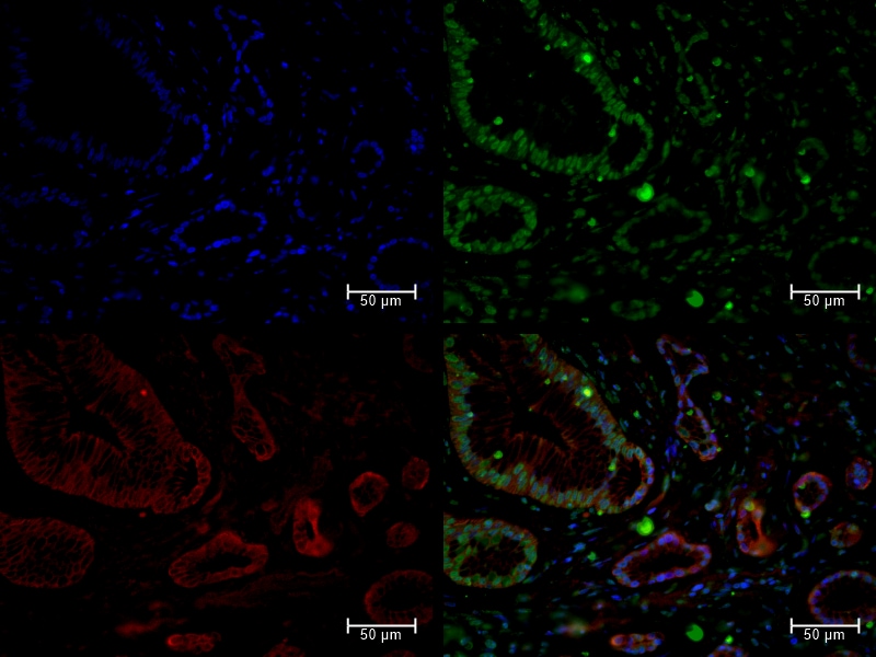
Application: Immunocytochemistry/ImmunofluorescenceSample Tested: Pancreatic cancer tissueSpecies: HumanVerified Customer | Posted 10/12/2021Not-so-negative control IF staining of rat anti-mouse integrin beta 1/CD29 at 15ug/mL on FFPE human tissue section, incubated at 4C overnight, and followed by Invitrogen Goat anti-Rat Alexa 488 (green) for 1 hour. Counterstained for mitochondria (red) and DAPI (blue).
Bio-Techne ResponseThank you for reviewing our product. We are sorry to hear that this product did not perform as expected. We have been in touch with the customer to resolve this issue according to our Product Guarantee and to the customer’s satisfaction. This customer needed an anti-mouse specific antibody that could be used in an immunohistochemistry (IHC) application as a negative control to differentiate transplanted human cells from surrounding mouse tissue. The MAB2405 datasheet makes no statement regarding potential cross-reactivity between human and mouse Integrin beta 1 when analyzing natural samples by IHC. Cross-reactivity has only been examined using a recombinant human Integrin beta 1 protein in Western Blotting. Because the anti-mouse Integrin beta 1 antibody displayed some putative cross-reactivity with human Integrin beta 1 protein in IHC samples, other suitable antibodies were discussed and offered as substitutes. None met the customer's dual needs of specific localized expression and zero guaranteed cross-reactivity with the human version of the protein in natural samples studied by the IHC application.
-
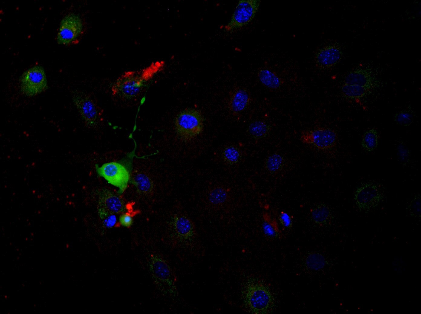
Application: Immunocytochemistry/ImmunofluorescenceSample Tested: Primary enteric nervous system (ENS) cellsSpecies: MouseVerified Customer | Posted 03/06/2019Integrin B1 antibody was used at the highest concentration (25 µg/ml) with O/N incubation at 4 degrees Celsius, using a secondary antibody chicken anti-rat Alexa 594 (Thermofisher). Dapi was used to stain the cell nuclei. A TUBB3 antibody with green labeling was used to visualize enteric neurons. Signal from Integrin B1 could not be detected where it was expected.
Bio-Techne ResponseThank you for reviewing our product. We are sorry to hear that this antibody did not perform as expected. We have been in touch with the customer to resolve this issue according to our Product Guarantee and to the customer’s satisfaction.
There are no reviews that match your criteria.
Protocols
Find general support by application which include: protocols, troubleshooting, illustrated assays, videos and webinars.
- 7-Amino Actinomycin D (7-AAD) Cell Viability Flow Cytometry Protocol
- Antigen Retrieval Protocol (PIER)
- Antigen Retrieval for Frozen Sections Protocol
- Appropriate Fixation of IHC/ICC Samples
- Cellular Response to Hypoxia Protocols
- Chromogenic IHC Staining of Formalin-Fixed Paraffin-Embedded (FFPE) Tissue Protocol
- Chromogenic Immunohistochemistry Staining of Frozen Tissue
- ClariTSA™ Fluorophore Kits
- Detection & Visualization of Antibody Binding
- Extracellular Membrane Flow Cytometry Protocol
- Flow Cytometry Protocol for Cell Surface Markers
- Flow Cytometry Protocol for Staining Membrane Associated Proteins
- Flow Cytometry Staining Protocols
- Flow Cytometry Troubleshooting Guide
- Fluorescent IHC Staining of Frozen Tissue Protocol
- Graphic Protocol for Heat-induced Epitope Retrieval
- Graphic Protocol for the Preparation and Fluorescent IHC Staining of Frozen Tissue Sections
- Graphic Protocol for the Preparation and Fluorescent IHC Staining of Paraffin-embedded Tissue Sections
- Graphic Protocol for the Preparation of Gelatin-coated Slides for Histological Tissue Sections
- ICC Cell Smear Protocol for Suspension Cells
- ICC Immunocytochemistry Protocol Videos
- ICC for Adherent Cells
- IHC Sample Preparation (Frozen sections vs Paraffin)
- Immunocytochemistry (ICC) Protocol
- Immunocytochemistry Troubleshooting
- Immunofluorescence of Organoids Embedded in Cultrex Basement Membrane Extract
- Immunofluorescent IHC Staining of Formalin-Fixed Paraffin-Embedded (FFPE) Tissue Protocol
- Immunohistochemistry (IHC) and Immunocytochemistry (ICC) Protocols
- Immunohistochemistry Frozen Troubleshooting
- Immunohistochemistry Paraffin Troubleshooting
- Intracellular Flow Cytometry Protocol Using Alcohol (Methanol)
- Intracellular Flow Cytometry Protocol Using Detergents
- Intracellular Nuclear Staining Flow Cytometry Protocol Using Detergents
- Intracellular Staining Flow Cytometry Protocol Using Alcohol Permeabilization
- Intracellular Staining Flow Cytometry Protocol Using Detergents to Permeabilize Cells
- Preparing Samples for IHC/ICC Experiments
- Preventing Non-Specific Staining (Non-Specific Binding)
- Primary Antibody Selection & Optimization
- Propidium Iodide Cell Viability Flow Cytometry Protocol
- Protocol for Heat-Induced Epitope Retrieval (HIER)
- Protocol for Liperfluo
- Protocol for Making a 4% Formaldehyde Solution in PBS
- Protocol for VisUCyte™ HRP Polymer Detection Reagent
- Protocol for the Characterization of Human Th22 Cells
- Protocol for the Characterization of Human Th9 Cells
- Protocol for the Fluorescent ICC Staining of Cell Smears - Graphic
- Protocol for the Fluorescent ICC Staining of Cultured Cells on Coverslips - Graphic
- Protocol for the Preparation & Fixation of Cells on Coverslips
- Protocol for the Preparation and Chromogenic IHC Staining of Frozen Tissue Sections
- Protocol for the Preparation and Chromogenic IHC Staining of Frozen Tissue Sections - Graphic
- Protocol for the Preparation and Chromogenic IHC Staining of Paraffin-embedded Tissue Sections
- Protocol for the Preparation and Chromogenic IHC Staining of Paraffin-embedded Tissue Sections - Graphic
- Protocol for the Preparation and Fluorescent ICC Staining of Cells on Coverslips
- Protocol for the Preparation and Fluorescent ICC Staining of Non-adherent Cells
- Protocol for the Preparation and Fluorescent ICC Staining of Stem Cells on Coverslips
- Protocol for the Preparation and Fluorescent IHC Staining of Frozen Tissue Sections
- Protocol for the Preparation and Fluorescent IHC Staining of Paraffin-embedded Tissue Sections
- Protocol for the Preparation of Gelatin-coated Slides for Histological Tissue Sections
- Protocol for the Preparation of a Cell Smear for Non-adherent Cell ICC - Graphic
- Protocol: Annexin V and PI Staining by Flow Cytometry
- Protocol: Annexin V and PI Staining for Apoptosis by Flow Cytometry
- R&D Systems Quality Control Western Blot Protocol
- TUNEL and Active Caspase-3 Detection by IHC/ICC Protocol
- The Importance of IHC/ICC Controls
- Troubleshooting Guide: Fluorokine Flow Cytometry Kits
- Troubleshooting Guide: Immunohistochemistry
- Troubleshooting Guide: Western Blot Figures
- Western Blot Conditions
- Western Blot Protocol
- Western Blot Protocol for Cell Lysates
- Western Blot Troubleshooting
- Western Blot Troubleshooting Guide
- View all Protocols, Troubleshooting, Illustrated assays and Webinars
Loading...
Associated Pathways


Embryonic and Induced Pluripotent Stem Cell Differentiation Pathways & Lineage-specific Markers
 Mesenchymal Stem Cell Differentiation Pathways & Lineage-specific Markers
