The low density lipoprotein receptor (LDL R) is the founding member of the LDL R family of scavenger receptors (1, 2, 3, 4). This family contains type I transmembrane molecules that are characterized by the presence of EGF repeats, complement-like repeats, and YWTD motifs that form beta -propellers. Although members of the family were originally thought to be endocytic receptors, it is now clear that some members interact with adjacent cell-surface molecules, expanding their range of activities (2, 4). Mouse LDL R is synthesized as a 864 amino acid (aa) precursor that contains a 21 aa signal sequence, a 769 aa extracellular region, a 22 aa transmembrane segment and a 52 aa cytoplasmic tail (5). The extracellular region is complex. It consists of seven N-terminal complement-like cysteine-rich repeats (class A LDL domains) that bind LDL. Cysteines in this region participate in intrachain disulfide bonds. This region is followed by two EGF-like domains and six class B LDL repeats that generate a beta -propeller whose blades each contain a YWTD motif. This area is likely responsible for ligand dissociation (6). Finally, there is a 50 aa membrane proximal Ser/Thr-rich region that shows extensive O-linked glycosylation, generating a native molecular weight for LDL R of 135 kDa (5). Within the 52 aa cytoplasmic region, there is an NPxY motif that links the receptor to clathrin pits and binds to select adaptor proteins (1, 7, 8). The extracellular region of mouse LDL R shares 78% and 87% aa identity with the extracellular region of human and rat LDL R, respectively. LDL R is constitutively expressed and binds apoB of LDL and apoE of VLDL (9). It is responsible for clearing 70% of plasma LDL in liver (9).

Key Product Details
Validated by
Biological Validation
Species Reactivity
Validated:
Mouse
Cited:
Human, Mouse, Rat, Transgenic Mouse
Applications
Validated:
Immunohistochemistry, Western Blot, Blockade of Receptor-ligand Interaction, Flow Cytometry, CyTOF-ready
Cited:
Immunohistochemistry, Immunohistochemistry-Frozen, Western Blot, Neutralization, Flow Cytometry, Immunocytochemistry, Blocking, ELISA Development
Label
Unconjugated
Antibody Source
Polyclonal Goat IgG
Loading...
Product Specifications
Immunogen
Mouse myeloma cell line NS0-derived recombinant mouse LDL R
Ala22-Arg790 (Ala23Val, Cys27Gly)
Accession # Q6GTJ9
Ala22-Arg790 (Ala23Val, Cys27Gly)
Accession # Q6GTJ9
Specificity
Detects mouse LDL R in direct ELISAs and Western blots. In direct ELISAs, approximately 20% cross-reactivity with recombinant human LDL R is observed.
Clonality
Polyclonal
Host
Goat
Isotype
IgG
Endotoxin Level
<0.10 EU per 1 μg of the antibody by the LAL method.
Scientific Data Images for Mouse LDLR Antibody
Detection of Mouse LDLR by Western Blot.
Western blot shows lysates of mouse liver tissue. PVDF membrane was probed with 0.1 µg/mL of Goat Anti-Mouse LDLR Antigen Affinity-purified Polyclonal Antibody (Catalog # AF2255) followed by HRP-conjugated Anti-Goat IgG Secondary Antibody (Catalog # HAF017). A specific band was detected for LDLR at approximately 145 kDa (as indicated). This experiment was conducted under reducing conditions and using Immunoblot Buffer Group 1.
LDL R in Mouse Liver.
LDL R was detected in perfusion fixed frozen sections of mouse liver using Mouse LDL R Antigen Affinity-purified Polyclonal Antibody (Catalog # AF2255) at 15 µg/mL overnight at 4 °C. Tissue was stained using the Anti-Goat HRP-DAB Cell & Tissue Staining Kit (brown; Catalog # CTS008) and counterstained with hematoxylin (blue). Specific labeling was localized to the bile canaliculi. View our protocol for Chromogenic IHC Staining of Frozen Tissue Sections.
Detection of Mouse LDLR by Western Blot
Effect of Pcsk9 deletion on LDLR and CD81 levels in age- and sex-matched Pcsk9-/- mice (n = 5) and their wild type littermates (n = 4).Shown are (A) mean ± SE serum LDL-C levels and (B) Western blot analysis of CD81 and LDLR levels in liver extracts with TfR as loading control, in animals sacrificed after a 4-hour fast. Lanes in panel B represent samples from individual mice. LDL-C, low-density lipoprotein cholesterol; LDLR, low-density lipoprotein receptor; PCSK9, proprotein convertase subtilisin/kexin type 9; SE, standard error; TfR, transferrin receptor; wt, wild type. Image collected and cropped by CiteAb from the following publication (https://dx.plos.org/10.1371/journal.pone.0154498), licensed under a CC-BY license. Not internally tested by R&D Systems.
Detection of Mouse LDLR by Western Blot
Effect of administration of alirocumab (monoclonal antibody to PCSK9) or control antibody (10 mg/kg) on LDLR and CD81 levels in hyperlipidemic Pcsk9hum/humLdlr+/- mice.Shown are mean ± SE serum LDL-C levels (A) and Western blot analysis of CD81 and LDLR levels in liver extracts using GAPDH as loading control (B). Western blot in B was quantified using Image J. The intensities of CD81 and LDLR bands were adjusted to respective loading control for each lane and presented as a fold change from control antibody treated group. Means ± SE are shown. Serum and livers were collected on Day 4 after antibody administration. In panel B, the three columns represent three livers collected for each of the two treatments. Ab, antibody; GAPDH, glyceraldehyde 3-phosphate dehydrogenase; LDL-C, low-density lipoprotein cholesterol; LDLR, low-density lipoprotein receptor; mAb, monoclonal Ab. Image collected and cropped by CiteAb from the following publication (https://dx.plos.org/10.1371/journal.pone.0154498), licensed under a CC-BY license. Not internally tested by R&D Systems.
Detection of Mouse LDLR by Western Blot
Hypercholesterolemia induced by recombinant Adeno-Associated Virus (rAAV)8-D377Y-murine Proprotein Convertase Subtilisin/Kexin type 9 (mPCSK9) in chow diet-fed mice.A. Serum levels of murine PCSK9 protein in mice injected with AAVmPCSK9 virus, AAVnull–injected or saline-injected control mice. **P<0.01. B. Liver LDL receptor protein levels analyzed by western blot and normalized to alpha -tubulin (n = 3). C. Plasma total cholesterol of chow diet—fed mice. **P<0.01 compared with control, ***P<0.01 compared with control and AAVmPCSK9. D. Serum alanine aminotransferase (ALT) and aspartate aminotransferase (AST) were measured at the same time point and determined as described in Materials and Methods. Values are mean ± SEM; n = 3. Image collected and cropped by CiteAb from the following publication (https://pubmed.ncbi.nlm.nih.gov/28291840), licensed under a CC-BY license. Not internally tested by R&D Systems.
Detection of Mouse LDLR by Western Blot
Lymphatic function assessment in mice following a specific knockdown of LDLR in murine endothelial cells. Two weeks after the intraperitoneal injection of an adeno-associated virus type 1 (AAV1) containing a shRNA targeting LDLR, skin draining lymph nodes were harvested, digested and analyzed by flow cytometry to assess lymphatic endothelial cell (LEC)-specific knockdown of LDLR expression in mice. Membrane-bound LDLR expression was measured on (A) CD45-CD31+Podoplanin+ and (B) CD45+ cells. Fluorescence minus one (FMO) control was used for LDLR expression, as depicted by the dotted line in the first histogram. White histogram, shSCR and dark histogram, shLldlr. LDLR expression was also determine by immunoblotting in (C) aorta isolated from female mice and (D) in liver from wild-type and Pcsk9-/- male (full dot) and female (empty dot) mice. ShSCR in black and ShLdlr in grey. Total plasmatic cholesterol was measured following FPLC in each liproprotein subfraction in (E) wild-type and (F)Pcsk9-/-female (dotted lines) and male (solid lines) mice treated with shSCR (black lines) and shLdlr (grey lines). Lymphatic contraction capacity was assessed by fluorescent in vivo imaging in (G) wild-type and (H)Pcsk9-/- mice. Black histogram, ShSCR and grey histogram, ShLdlr. n=3-9. Statistics **p < 0,01. Image collected and cropped by CiteAb from the following open publication (https://pubmed.ncbi.nlm.nih.gov/35154499), licensed under a CC-BY license. Not internally tested by R&D Systems.
Detection of Mouse LDLR by Western Blot
Lymphatic function assessment in mice following a specific knockdown of LDLR in murine endothelial cells. Two weeks after the intraperitoneal injection of an adeno-associated virus type 1 (AAV1) containing a shRNA targeting LDLR, skin draining lymph nodes were harvested, digested and analyzed by flow cytometry to assess lymphatic endothelial cell (LEC)-specific knockdown of LDLR expression in mice. Membrane-bound LDLR expression was measured on (A) CD45-CD31+Podoplanin+ and (B) CD45+ cells. Fluorescence minus one (FMO) control was used for LDLR expression, as depicted by the dotted line in the first histogram. White histogram, shSCR and dark histogram, shLldlr. LDLR expression was also determine by immunoblotting in (C) aorta isolated from female mice and (D) in liver from wild-type and Pcsk9-/- male (full dot) and female (empty dot) mice. ShSCR in black and ShLdlr in grey. Total plasmatic cholesterol was measured following FPLC in each liproprotein subfraction in (E) wild-type and (F)Pcsk9-/-female (dotted lines) and male (solid lines) mice treated with shSCR (black lines) and shLdlr (grey lines). Lymphatic contraction capacity was assessed by fluorescent in vivo imaging in (G) wild-type and (H)Pcsk9-/- mice. Black histogram, ShSCR and grey histogram, ShLdlr. n=3-9. Statistics **p < 0,01. Image collected and cropped by CiteAb from the following open publication (https://pubmed.ncbi.nlm.nih.gov/35154499), licensed under a CC-BY license. Not internally tested by R&D Systems.Applications for Mouse LDLR Antibody
Application
Recommended Usage
Blockade of Receptor-ligand Interaction
In a functional ELISA, 0.04‑0.2 µg/mL of this antibody will block 50% of the binding of 200 ng/mL of Recombinant Mouse LDL R (Catalog # 2255-LD) to immobilized human Low Density Lipoprotein coated at 2 µg/mL (100 µL/well). At 3 µg/mL, this antibody will block >95% of the binding.
CyTOF-ready
Ready to be labeled using established conjugation methods. No BSA or other carrier proteins that could interfere with conjugation.
Flow Cytometry
2.5 µg/106 cells
Sample: Serum‑deprived RAW 264.7 mouse monocyte/macrophage cell line
Sample: Serum‑deprived RAW 264.7 mouse monocyte/macrophage cell line
Immunohistochemistry
5-15 µg/mL
Sample: Perfusion fixed frozen sections of mouse liver
Sample: Perfusion fixed frozen sections of mouse liver
Western Blot
0.1 µg/mL
Sample: Mouse Liver Tissue
Sample: Mouse Liver Tissue
Reviewed Applications
Read 1 review rated 5 using AF2255 in the following applications:
Flow Cytometry Panel Builder
Bio-Techne Knows Flow Cytometry
Save time and reduce costly mistakes by quickly finding compatible reagents using the Panel Builder Tool.
Advanced Features
- Spectra Viewer - Custom analysis of spectra from multiple fluorochromes
- Spillover Popups - Visualize the spectra of individual fluorochromes
- Antigen Density Selector - Match fluorochrome brightness with antigen density

Formulation, Preparation, and Storage
Purification
Antigen Affinity-purified
Reconstitution
Reconstitute at 0.2 mg/mL in sterile PBS. For liquid material, refer to CoA for concentration.
Loading...
Formulation
Lyophilized from a 0.2 μm filtered solution in PBS with Trehalose. *Small pack size (SP) is supplied either lyophilized or as a 0.2 µm filtered solution in PBS.
Shipping
Lyophilized product is shipped at ambient temperature. Liquid small pack size (-SP) is shipped with polar packs. Upon receipt, store immediately at the temperature recommended below.
Stability & Storage
Use a manual defrost freezer and avoid repeated freeze-thaw cycles.
- 12 months from date of receipt, -20 to -70 °C as supplied.
- 1 month, 2 to 8 °C under sterile conditions after reconstitution.
- 6 months, -20 to -70 °C under sterile conditions after reconstitution.
Calculators
Background: LDLR
References
- Strickland, D.K. et al. (2002) Trends Endocrinol. Metab. 13:66.
- Nykjaer, A. and T.E. Willnow (2002) Trends Cell Biol. 12:273.
- Gent, J. and I. Braakman (2004) Cell. Mol. Life Sci. 61:2461.
- Bujo, H. and Y. Saito (2006) Arterioscler. Thromb. Vasc. Biol. 26:1246.
- Hoffer, M.J. V. et al. (1993) Biochem. Biophys. Res. Commun. 191:880.
- Rudenko, G. and J. Deisenhofer (2003) Curr. Opin. Struct. Biol. 13:683.
- Trommsdorff, M. et al. (1998) J. Biol. Chem. 273:33556.
- Stolt, P.C. and H.H. Bock (2006) Cell. Signal. 18:1560
- Defesche, J.C. (2004) Semin. Vasc. Med. 4:5.
Long Name
Low Density Lipoprotein Receptor
Alternate Names
LDL R
Entrez Gene IDs
Gene Symbol
LDLR
UniProt
Additional LDLR Products
Product Documents for Mouse LDLR Antibody
Certificate of Analysis
To download a Certificate of Analysis, please enter a lot or batch number in the search box below.
Note: Certificate of Analysis not available for kit components.
Product Specific Notices for Mouse LDLR Antibody
For research use only
Related Research Areas
Citations for Mouse LDLR Antibody
 Powered by Bioz
Customer Reviews for Mouse LDLR Antibody (1)
5 out of 5
1 Customer Rating
Have you used Mouse LDLR Antibody?
Submit a review and receive an Amazon gift card!
$25/€18/£15/$25CAN/¥2500 Yen for a review with an image
$10/€7/£6/$10CAN/¥1110 Yen for a review without an image
Submit a review
Customer Images
Showing
1
-
1 of
1 review
Showing All
Filter By:
-
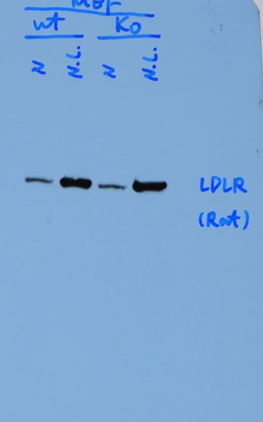
Application: Western BlotSample Tested: MEFSpecies: MouseVerified Customer | Posted 01/30/2019When the cells were lipid-depleted (N.L.), the protein level of endogenous LDLR was significantly increased (shown in the figure).

There are no reviews that match your criteria.
Protocols
Find general support by application which include: protocols, troubleshooting, illustrated assays, videos and webinars.
- 7-Amino Actinomycin D (7-AAD) Cell Viability Flow Cytometry Protocol
- Antigen Retrieval Protocol (PIER)
- Antigen Retrieval for Frozen Sections Protocol
- Appropriate Fixation of IHC/ICC Samples
- Cellular Response to Hypoxia Protocols
- Chromogenic IHC Staining of Formalin-Fixed Paraffin-Embedded (FFPE) Tissue Protocol
- Chromogenic Immunohistochemistry Staining of Frozen Tissue
- ClariTSA™ Fluorophore Kits
- Detection & Visualization of Antibody Binding
- Extracellular Membrane Flow Cytometry Protocol
- Flow Cytometry Protocol for Cell Surface Markers
- Flow Cytometry Protocol for Staining Membrane Associated Proteins
- Flow Cytometry Staining Protocols
- Flow Cytometry Troubleshooting Guide
- Fluorescent IHC Staining of Frozen Tissue Protocol
- Graphic Protocol for Heat-induced Epitope Retrieval
- Graphic Protocol for the Preparation and Fluorescent IHC Staining of Frozen Tissue Sections
- Graphic Protocol for the Preparation and Fluorescent IHC Staining of Paraffin-embedded Tissue Sections
- Graphic Protocol for the Preparation of Gelatin-coated Slides for Histological Tissue Sections
- IHC Sample Preparation (Frozen sections vs Paraffin)
- Immunofluorescent IHC Staining of Formalin-Fixed Paraffin-Embedded (FFPE) Tissue Protocol
- Immunohistochemistry (IHC) and Immunocytochemistry (ICC) Protocols
- Immunohistochemistry Frozen Troubleshooting
- Immunohistochemistry Paraffin Troubleshooting
- Intracellular Flow Cytometry Protocol Using Alcohol (Methanol)
- Intracellular Flow Cytometry Protocol Using Detergents
- Intracellular Nuclear Staining Flow Cytometry Protocol Using Detergents
- Intracellular Staining Flow Cytometry Protocol Using Alcohol Permeabilization
- Intracellular Staining Flow Cytometry Protocol Using Detergents to Permeabilize Cells
- Preparing Samples for IHC/ICC Experiments
- Preventing Non-Specific Staining (Non-Specific Binding)
- Primary Antibody Selection & Optimization
- Propidium Iodide Cell Viability Flow Cytometry Protocol
- Protocol for Heat-Induced Epitope Retrieval (HIER)
- Protocol for Liperfluo
- Protocol for Making a 4% Formaldehyde Solution in PBS
- Protocol for VisUCyte™ HRP Polymer Detection Reagent
- Protocol for the Characterization of Human Th22 Cells
- Protocol for the Characterization of Human Th9 Cells
- Protocol for the Preparation & Fixation of Cells on Coverslips
- Protocol for the Preparation and Chromogenic IHC Staining of Frozen Tissue Sections
- Protocol for the Preparation and Chromogenic IHC Staining of Frozen Tissue Sections - Graphic
- Protocol for the Preparation and Chromogenic IHC Staining of Paraffin-embedded Tissue Sections
- Protocol for the Preparation and Chromogenic IHC Staining of Paraffin-embedded Tissue Sections - Graphic
- Protocol for the Preparation and Fluorescent IHC Staining of Frozen Tissue Sections
- Protocol for the Preparation and Fluorescent IHC Staining of Paraffin-embedded Tissue Sections
- Protocol for the Preparation of Gelatin-coated Slides for Histological Tissue Sections
- Protocol: Annexin V and PI Staining by Flow Cytometry
- Protocol: Annexin V and PI Staining for Apoptosis by Flow Cytometry
- R&D Systems Quality Control Western Blot Protocol
- TUNEL and Active Caspase-3 Detection by IHC/ICC Protocol
- The Importance of IHC/ICC Controls
- Troubleshooting Guide: Fluorokine Flow Cytometry Kits
- Troubleshooting Guide: Immunohistochemistry
- Troubleshooting Guide: Western Blot Figures
- Western Blot Conditions
- Western Blot Protocol
- Western Blot Protocol for Cell Lysates
- Western Blot Troubleshooting
- Western Blot Troubleshooting Guide
- View all Protocols, Troubleshooting, Illustrated assays and Webinars
Loading...
Associated Pathways
