Axl (Ufo, Ark), Dtk (Sky, Tyro3, Rse, Brt) and Mer (human and mouse homologues of chicken c-Eyk) constitute a receptor tyrosine kinase subfamily. The extracellular domains of these proteins contain two Ig-like motifs and two fibronectin type III motifs. This characteristic topology is also found in neural cell adhesion molecules and in receptor tyrosine phosphatases. These receptors bind the vitamin K-dependent protein growth-arrest-specific gene 6 (Gas6) which is structurally related to the anticoagulation factor protein S. Binding of Gas6 induces receptor autophosphorylation and downstream signaling pathways that can lead to cell proliferation, migration or the prevention of apoptosis. Studies suggest that this family of tyrosine kinase receptors may be involved in hematopoiesis, embryonic development, tumorigenesis and regulation of testicular functions (1, 2).

Key Product Details
Validated by
Orthogonal Validation
Species Reactivity
Validated:
Mouse
Cited:
Human, Mouse, Rat, Transgenic Mouse
Applications
Validated:
Immunohistochemistry, Flow Cytometry, Dual RNAscope ISH-IHC Compatible, CyTOF-ready
Cited:
Immunohistochemistry, Immunohistochemistry-Paraffin, Immunohistochemistry-Frozen, Western Blot, Neutralization, Flow Cytometry, Immunofluorescence, Immunocytochemistry, Immunoprecipitation, Activation, Cell Culture, ELISA Capture, Proximity Ligation Assay, In vivo assay, Functional Assay, IF/IHC, CyTOF-reported
Label
Unconjugated
Antibody Source
Polyclonal Goat IgG
Loading...
Product Specifications
Immunogen
S. frugiperda insect ovarian cell line Sf 21-derived recombinant mouse Mer
Glu23-Phe498
Accession # Q60805
Glu23-Phe498
Accession # Q60805
Specificity
Detects mouse Mer in direct ELISAs and Western blots. In direct ELISAs, approximately 20% cross‑reactivity with recombinant human Mer is observed and less than 1% cross-reactivity with recombinant mouse (rm) Axl and rmDtk is observed.
Clonality
Polyclonal
Host
Goat
Isotype
IgG
Scientific Data Images for Mouse Mer Antibody
Detection of Mer in J774A.1 Mouse Cell Line by Flow Cytometry.
J774A.1 mouse reticulum cell sarcoma macrophage cell line was stained with Goat Anti-Mouse Mer Antigen Affinity-purified Polyclonal Antibody (Catalog # AF591, filled histogram) or control antibody (Catalog # AB-108-C, open histogram), followed by Allophycocyanin-conjugated Anti-Goat IgG Secondary Antibody (Catalog # F0108).
Mer in Mouse Testis.
Mer was detected in perfusion fixed frozen sections of mouse testis using Goat Anti-Mouse Mer Antigen Affinity-purified Polyclonal Antibody (Catalog # AF591) at 1 µg/mL for 1 hour at room temperature followed by incubation with the Anti-Goat IgG VisUCyte™ HRP Polymer Antibody (Catalog # VC004). Tissue was stained using DAB (brown) and counterstained with hematoxylin (blue). Specific staining was localized to sperm cells. View our protocol for IHC Staining with VisUCyte HRP Polymer Detection Reagents.
Mer in Mouse Kidney Using Dual RNAscope®ISH and IHC.
Mer mRNA was detected in formalin-fixed paraffin-embedded tissue sections of mouse kidney probed with ACD RNAScope®Probe (Catalog # 441241) and stained using ACD RNAscope®2.5 HD Detection Reagents-Red (top image, Catalog # 32260). Adjacent tissue section was processed for immunohistochemistry using R&D Systems Goat Anti-Mouse Mer Antigen Affinity-purified Polyclonal Antibody (Catalog # AF591) at 1 ug/mL with overnight incubation at 4 °C followed by incubation with the Anti-Goat IgG VisUCyte HRP Polymer Antibody (R&D Systems, Catalog # VC004) and DAB chromogen (lower image, yellow-brown). Tissues were counterstained with hematoxylin (blue).
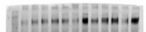
Detection of Mouse Mer by Western Blot
RPE express components of BAI1-signaling pathway and Bai1Tg. (A) Expression of the BAI1 signaling pathway was analyzed by quantitative RT-PCR in RPE isolated from P14 Mertk+/− and Mertk−/− mice (n = 2) Error bars represent SEM. (B) Immunoblot analysis of BAI1 signaling pathway in RPE whole cell lysates isolated from P14 mice. Left panel shows representative immunoblots for Mer, Dock180, Elmo2 and Rac. Right panel shows combined densitometry analysis of immunoblots from n = 3 mice per genotype. For densitometry analysis, band volumes were normalized to an actin loading control and band densities in Mertk−/− were then normalized to Mertk+/− for comparison across multiple blots. (C) Schematic of Bai1Tg insertion in the Rosa26 locus indicating the N-terminal HA-tag in red and IRES-GFP in green. Mouse image licensed from Motifolio Inc. (D) GFP expression was analyzed in RPE flat mount preparations at 20x magnification. Inset image in the left panel shows a representative flat mount at 1.2x magnification. White scale bars in images are 50 μm. Images are representative of n = 2 mice per genotype. (E) qRT-PCR analysis of Bai1Tg expression in Mertk+/−Bai1Tg (n = 9) and Mertk−/−Bai1Tg (n = 3) RPE isolated on P14. n.d. = not detected. Error bars represent SEM. (F) Immunoblot analysis of HA-tag (Bai1Tg) in RPE whole cell lysates isolated from P13-P14 mice. Immunoblot shown is representative of n = 2 experiments. (G) HA staining and localization was analyzed in eyecups at 40x magnification. Neural retinas were removed prior to fixation and staining. Black lines next to the figure align with apical microvilli of RPE. Scale bars are 20 μm. Images shown are representative of n = 2 mice per genotype. Image collected and cropped by CiteAb from the following publication (https://www.nature.com/articles/s41598-017-15191-1), licensed under a CC-BY license. Not internally tested by R&D Systems.Applications for Mouse Mer Antibody
Application
Recommended Usage
CyTOF-ready
Ready to be labeled using established conjugation methods. No BSA or other carrier proteins that could interfere with conjugation.
Dual RNAscope ISH-IHC Compatible
1-15 µg/mL
Sample: Immersion fixed paraffin-embedded tissue sections of mouse kidney
Sample: Immersion fixed paraffin-embedded tissue sections of mouse kidney
Flow Cytometry
2.5 µg/106 cells
Sample:
Sample:
| J774A.1 mouse reticulum cell sarcoma macrophage cell line |
Immunohistochemistry
1-15 µg/mL
Sample: Perfusion fixed frozen sections of mouse testis
Sample: Perfusion fixed frozen sections of mouse testis
Reviewed Applications
Read 4 reviews rated 4.5 using AF591 in the following applications:
Flow Cytometry Panel Builder
Bio-Techne Knows Flow Cytometry
Save time and reduce costly mistakes by quickly finding compatible reagents using the Panel Builder Tool.
Advanced Features
- Spectra Viewer - Custom analysis of spectra from multiple fluorochromes
- Spillover Popups - Visualize the spectra of individual fluorochromes
- Antigen Density Selector - Match fluorochrome brightness with antigen density

Formulation, Preparation, and Storage
Purification
Antigen Affinity-purified
Reconstitution
Reconstitute at 0.2 mg/mL in sterile PBS. For liquid material, refer to CoA for concentration.
Loading...
Formulation
Lyophilized from a 0.2 μm filtered solution in PBS with Trehalose. *Small pack size (SP) is supplied either lyophilized or as a 0.2 µm filtered solution in PBS.
Shipping
Lyophilized product is shipped at ambient temperature. Liquid small pack size (-SP) is shipped with polar packs. Upon receipt, store immediately at the temperature recommended below.
Stability & Storage
Use a manual defrost freezer and avoid repeated freeze-thaw cycles.
- 12 months from date of receipt, -20 to -70 °C as supplied.
- 1 month, 2 to 8 °C under sterile conditions after reconstitution.
- 6 months, -20 to -70 °C under sterile conditions after reconstitution.
Calculators
Background: Mer
References
- Nagata, K. et al. (1996) J. Biol. Chem. 22:30022.
- Crosier, K.E. and P.S Crosier (1997) Pathology 29:131.
Long Name
Receptor Tyrosine Protein Kinase Mer
Alternate Names
c-Eyk, C-mer, MerTK
Gene Symbol
MERTK
UniProt
Additional Mer Products
Product Documents for Mouse Mer Antibody
Certificate of Analysis
To download a Certificate of Analysis, please enter a lot or batch number in the search box below.
Note: Certificate of Analysis not available for kit components.
Product Specific Notices for Mouse Mer Antibody
For research use only
Citations for Mouse Mer Antibody
 Powered by Bioz
Customer Reviews for Mouse Mer Antibody (4)
4.5 out of 5
4 Customer Ratings
Have you used Mouse Mer Antibody?
Submit a review and receive an Amazon gift card!
$25/€18/£15/$25CAN/¥2500 Yen for a review with an image
$10/€7/£6/$10CAN/¥1110 Yen for a review without an image
Submit a review
Customer Images
Showing
1
-
4 of
4 reviews
Showing All
Filter By:
-
Application: Western BlotSample Tested: lysates of primary bone marrow derived macrophageSpecies: miceVerified Customer | Posted 11/08/2022The band comes around 170Kda

-
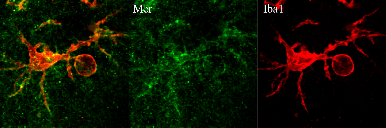
Application: immunohistochemistry free floating sectionSample Tested: Adult mouse brainSpecies: MouseVerified Customer | Posted 03/16/2018Mer is in green and Iba1 is in red.antigen retrieval for 15 mins. 1:250 dilution.
-
Application: Flow CytometrySample Tested: Peritoneal macrophagesSpecies: MouseVerified Customer | Posted 06/17/2017
-
Application: in vivoSample Tested: Adult mouse brain and brain and spinal cordSpecies: MouseVerified Customer | Posted 04/03/2017Used in vivo to inhibit Mer activation.
There are no reviews that match your criteria.
Protocols
Find general support by application which include: protocols, troubleshooting, illustrated assays, videos and webinars.
- 7-Amino Actinomycin D (7-AAD) Cell Viability Flow Cytometry Protocol
- Antigen Retrieval Protocol (PIER)
- Antigen Retrieval for Frozen Sections Protocol
- Appropriate Fixation of IHC/ICC Samples
- Cellular Response to Hypoxia Protocols
- Chromogenic IHC Staining of Formalin-Fixed Paraffin-Embedded (FFPE) Tissue Protocol
- Chromogenic Immunohistochemistry Staining of Frozen Tissue
- ClariTSA™ Fluorophore Kits
- Detection & Visualization of Antibody Binding
- Extracellular Membrane Flow Cytometry Protocol
- Flow Cytometry Protocol for Cell Surface Markers
- Flow Cytometry Protocol for Staining Membrane Associated Proteins
- Flow Cytometry Staining Protocols
- Flow Cytometry Troubleshooting Guide
- Fluorescent IHC Staining of Frozen Tissue Protocol
- Graphic Protocol for Heat-induced Epitope Retrieval
- Graphic Protocol for the Preparation and Fluorescent IHC Staining of Frozen Tissue Sections
- Graphic Protocol for the Preparation and Fluorescent IHC Staining of Paraffin-embedded Tissue Sections
- Graphic Protocol for the Preparation of Gelatin-coated Slides for Histological Tissue Sections
- IHC Sample Preparation (Frozen sections vs Paraffin)
- ISH-IHC Protocol for Chromogenic Detection on Formalin Fixed Paraffin Embedded (FFPE) Tissue
- Immunofluorescent IHC Staining of Formalin-Fixed Paraffin-Embedded (FFPE) Tissue Protocol
- Immunohistochemistry (IHC) and Immunocytochemistry (ICC) Protocols
- Immunohistochemistry Frozen Troubleshooting
- Immunohistochemistry Paraffin Troubleshooting
- Intracellular Flow Cytometry Protocol Using Alcohol (Methanol)
- Intracellular Flow Cytometry Protocol Using Detergents
- Intracellular Nuclear Staining Flow Cytometry Protocol Using Detergents
- Intracellular Staining Flow Cytometry Protocol Using Alcohol Permeabilization
- Intracellular Staining Flow Cytometry Protocol Using Detergents to Permeabilize Cells
- Preparing Samples for IHC/ICC Experiments
- Preventing Non-Specific Staining (Non-Specific Binding)
- Primary Antibody Selection & Optimization
- Propidium Iodide Cell Viability Flow Cytometry Protocol
- Protocol for Heat-Induced Epitope Retrieval (HIER)
- Protocol for Liperfluo
- Protocol for Making a 4% Formaldehyde Solution in PBS
- Protocol for VisUCyte™ HRP Polymer Detection Reagent
- Protocol for the Characterization of Human Th22 Cells
- Protocol for the Characterization of Human Th9 Cells
- Protocol for the Preparation & Fixation of Cells on Coverslips
- Protocol for the Preparation and Chromogenic IHC Staining of Frozen Tissue Sections
- Protocol for the Preparation and Chromogenic IHC Staining of Frozen Tissue Sections - Graphic
- Protocol for the Preparation and Chromogenic IHC Staining of Paraffin-embedded Tissue Sections
- Protocol for the Preparation and Chromogenic IHC Staining of Paraffin-embedded Tissue Sections - Graphic
- Protocol for the Preparation and Fluorescent IHC Staining of Frozen Tissue Sections
- Protocol for the Preparation and Fluorescent IHC Staining of Paraffin-embedded Tissue Sections
- Protocol for the Preparation of Gelatin-coated Slides for Histological Tissue Sections
- Protocol: Annexin V and PI Staining by Flow Cytometry
- Protocol: Annexin V and PI Staining for Apoptosis by Flow Cytometry
- TUNEL and Active Caspase-3 Detection by IHC/ICC Protocol
- The Importance of IHC/ICC Controls
- Troubleshooting Guide: Fluorokine Flow Cytometry Kits
- Troubleshooting Guide: Immunohistochemistry
- View all Protocols, Troubleshooting, Illustrated assays and Webinars
Loading...