Encoded by the THBD gene, Thrombomodulin is also known as CD141 antigen. The deduced amino acid sequence of mouse THBD predicts a signal peptide (aa 1 to 16) and a mature chain (aa 17 to 577) that consists of the following domains: C-type lectin (aa 31 to 167), EGF-like (aa 240 to 280, aa 283 to 323, aa 324 to 362, aa 364 to 404, aa 405 to 439, and aa 440 to 480), transmembrane (aa 518 to 541) and cytoplasmic (aa 542 to 577) (1). The R&D Systems rmTHBD consists of aa 17 to 517, corresponding to the extracellular portion of the type I membrane protein. Predominantly synthesized by vascular endothelial cells, THBD inhibits coagulation and fibrinolysis (2‑4). It functions as a cell surface receptor and an essential cofactor for active thrombin, which in turn activates protein C and thrombin-activatable fibrinolysis inhibitor (TAFI), also known as carboxypeptidase B2 (CPB2). Activated protein C (APC), facilitated by protein S, degrades coagulation factors Va and VIIIa, which are required for thrombin activation. Activated CPB2 cleaves basic C-terminal amino acid residues of its substrates, including fibrin, preventing the conversion of plasminogen to plasmin. In addition, THBD gene polymorphisims are associated with human disease and THBD plays a role in thrombosis, stroke, arteriosclerosis, and cancer (5). For example, increased serum levels of THBD, due to protease cleavage, have been associated with smoking, cardiac surgery, atherosclerosis, liver cirrhosis, diabetes mellitus, cerebral and myocardial infarction, and multiple sclerosis (6).
Mouse Thrombomodulin/BDCA-3 Antibody (461714)
R&D Systems | Catalog # MAB3894

Key Product Details
Species Reactivity
Validated:
Mouse
Cited:
Mouse, Transgenic Mouse
Applications
Validated:
Immunohistochemistry, Western Blot, Flow Cytometry, Immunocytochemistry, CyTOF-ready
Cited:
Immunohistochemistry, Immunohistochemistry-Paraffin, Western Blot, Bioassay
Label
Unconjugated
Antibody Source
Monoclonal Rat IgG2B Clone # 461714
Loading...
Product Specifications
Immunogen
Mouse myeloma cell line NS0-derived recombinant mouse Thrombomodulin/BDCA-3
Leu17-Ser517
Accession # P15306
Leu17-Ser517
Accession # P15306
Specificity
Detects mouse Thrombomodulin/BDCA-3 in direct ELISAs and Western blots. In direct ELISAs and Western blots, no cross-reactivity with recombinant human (rh) Thrombomodulin is observed.
Clonality
Monoclonal
Host
Rat
Isotype
IgG2B
Scientific Data Images for Mouse Thrombomodulin/BDCA-3 Antibody (461714)
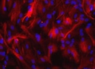
Thrombomodulin/BDCA-3 in bEnd.3 Mouse Cell Line.
Thrombomodulin/BDCA-3 was detected in immersion fixed bEnd.3 mouse endothelioma cell line using Mouse Thrombomodulin/BDCA-3 Monoclonal Antibody (Catalog # MAB3894) at 10 µg/mL for 3 hours at room temperature. Cells were stained using the NorthernLights™ 557-conjugated Anti-Rat IgG Secondary Antibody (red; Catalog # NL013) and counterstained with DAPI (blue). View our protocol for Fluorescent ICC Staining of Cells on Coverslips.
Thrombomodulin/BDCA-3 in Mouse Heart.
Thrombomodulin/BDCA-3 was detected in perfusion fixed frozen sections of mouse heart using 25 µg/mL Mouse Thrombomodulin/BDCA-3 Monoclonal Antibody (Catalog # MAB3894) overnight at 4 °C. Tissue was stained with the Anti-Rat HRP-DAB Cell & Tissue Staining Kit (brown; Catalog # CTS017) and counterstained with hematoxylin (blue). View our protocol for Chromogenic IHC Staining of Paraffin-embedded Tissue Sections.
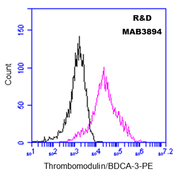
Detection of Thrombomodulin/BDCA‑3 in bEnd.3 Mouse Cell Line by Flow Cytometry.
bEnd.3 mouse endothelioma cell line was stained with Mouse Thrombomodulin/BDCA‑3 Monoclonal Antibody (Catalog # MAB3894, filled histogram) or isotype control antibody (Catalog # MAB0061, open histogram), followed by Phycoerythrin-conjugated Anti-Rat IgG F(ab')2Secondary Antibody (Catalog # F0105B).Applications for Mouse Thrombomodulin/BDCA-3 Antibody (461714)
Application
Recommended Usage
CyTOF-ready
Ready to be labeled using established conjugation methods. No BSA or other carrier proteins that could interfere with conjugation.
Flow Cytometry
2.5 µg/106 cells
Sample: bEnd.3 mouse endothelioma cell line
Sample: bEnd.3 mouse endothelioma cell line
Immunocytochemistry
8-25 µg/mL
Sample: Immersion fixed bEND.3 mouse endothelioma cell line
Sample: Immersion fixed bEND.3 mouse endothelioma cell line
Immunohistochemistry
8-25 µg/mL
Sample: Perfusion fixed frozen sections of mouse kidney and heart
Sample: Perfusion fixed frozen sections of mouse kidney and heart
Western Blot
1 µg/mL
Sample: Recombinant Mouse Thrombomodulin/THBD/CD141 (Catalog # 3894-PA)
under non-reducing conditions only
Sample: Recombinant Mouse Thrombomodulin/THBD/CD141 (Catalog # 3894-PA)
under non-reducing conditions only
Reviewed Applications
Read 2 reviews rated 5 using MAB3894 in the following applications:
Flow Cytometry Panel Builder
Bio-Techne Knows Flow Cytometry
Save time and reduce costly mistakes by quickly finding compatible reagents using the Panel Builder Tool.
Advanced Features
- Spectra Viewer - Custom analysis of spectra from multiple fluorochromes
- Spillover Popups - Visualize the spectra of individual fluorochromes
- Antigen Density Selector - Match fluorochrome brightness with antigen density

Formulation, Preparation, and Storage
Purification
Protein A or G purified from hybridoma culture supernatant
Reconstitution
Reconstitute at 0.5 mg/mL in sterile PBS. For liquid material, refer to CoA for concentration.
Loading...
Formulation
Lyophilized from a 0.2 μm filtered solution in PBS with Trehalose. *Small pack size (SP) is supplied either lyophilized or as a 0.2 µm filtered solution in PBS.
Shipping
Lyophilized product is shipped at ambient temperature. Liquid small pack size (-SP) is shipped with polar packs. Upon receipt, store immediately at the temperature recommended below.
Stability & Storage
Use a manual defrost freezer and avoid repeated freeze-thaw cycles.
- 12 months from date of receipt, -20 to -70 °C as supplied.
- 1 month, 2 to 8 °C under sterile conditions after reconstitution.
- 6 months, -20 to -70 °C under sterile conditions after reconstitution.
Calculators
Background: Thrombomodulin/BDCA-3
References
- Dittman, W.A. and P.W. Majerus (1989) Nucleic Acids Res. 17:802.
- Van de Wouwer, M. et al. (2004) Arterioscler. Thromb. Vasc. 24:1374.
- Wu, K.K. et al. (2000) Ann Med. 32:73.
- Li, Y.H. et al. (2006) Cardiovasc. Hematol. Agents Med. Chem. 4:183.
- Weiler, H. and B.H. Isermann (2003) J. Thromb. Haemost. 1:1515.
- Califano, F. et al. (2000) Eur. Rev. Med. Pharmacol. Sci. 4:59.
Alternate Names
BDCA-3, BDCA3, CD141, Fetomodulin, THBD, THRM
Gene Symbol
THBD
UniProt
Additional Thrombomodulin/BDCA-3 Products
Product Documents for Mouse Thrombomodulin/BDCA-3 Antibody (461714)
Certificate of Analysis
To download a Certificate of Analysis, please enter a lot or batch number in the search box below.
Note: Certificate of Analysis not available for kit components.
Product Specific Notices for Mouse Thrombomodulin/BDCA-3 Antibody (461714)
For research use only
Citations for Mouse Thrombomodulin/BDCA-3 Antibody (461714)
 Powered by Bioz
Customer Reviews for Mouse Thrombomodulin/BDCA-3 Antibody (461714) (2)
5 out of 5
2 Customer Ratings
Have you used Mouse Thrombomodulin/BDCA-3 Antibody (461714)?
Submit a review and receive an Amazon gift card!
$25/€18/£15/$25CAN/¥2500 Yen for a review with an image
$10/€7/£6/$10CAN/¥1110 Yen for a review without an image
Submit a review
Customer Images
Showing
1
-
2 of
2 reviews
Showing All
Filter By:
-
Application: Immunocytochemistry/ImmunofluorescenceSample Tested: AstrocytesSpecies: MouseVerified Customer | Posted 02/10/2022

-
Application: Flow CytometrySample Tested: Peripheral blood mononuclear cells (PBMCs)Species: MouseVerified Customer | Posted 03/22/2017106 Mouse PBMCs were incubated with 2.5 ug of Rat anti-Mouse Thrombomodulin/BDCA-3 antibody (pink) or Isotype Control IgG (black), followed by PE-conjugated Anti-Rat secondary antibody.
There are no reviews that match your criteria.
Protocols
Find general support by application which include: protocols, troubleshooting, illustrated assays, videos and webinars.
- 7-Amino Actinomycin D (7-AAD) Cell Viability Flow Cytometry Protocol
- Antigen Retrieval Protocol (PIER)
- Antigen Retrieval for Frozen Sections Protocol
- Appropriate Fixation of IHC/ICC Samples
- Cellular Response to Hypoxia Protocols
- Chromogenic IHC Staining of Formalin-Fixed Paraffin-Embedded (FFPE) Tissue Protocol
- Chromogenic Immunohistochemistry Staining of Frozen Tissue
- ClariTSA™ Fluorophore Kits
- Detection & Visualization of Antibody Binding
- Extracellular Membrane Flow Cytometry Protocol
- Flow Cytometry Protocol for Cell Surface Markers
- Flow Cytometry Protocol for Staining Membrane Associated Proteins
- Flow Cytometry Staining Protocols
- Flow Cytometry Troubleshooting Guide
- Fluorescent IHC Staining of Frozen Tissue Protocol
- Graphic Protocol for Heat-induced Epitope Retrieval
- Graphic Protocol for the Preparation and Fluorescent IHC Staining of Frozen Tissue Sections
- Graphic Protocol for the Preparation and Fluorescent IHC Staining of Paraffin-embedded Tissue Sections
- Graphic Protocol for the Preparation of Gelatin-coated Slides for Histological Tissue Sections
- ICC Cell Smear Protocol for Suspension Cells
- ICC Immunocytochemistry Protocol Videos
- ICC for Adherent Cells
- IHC Sample Preparation (Frozen sections vs Paraffin)
- Immunocytochemistry (ICC) Protocol
- Immunocytochemistry Troubleshooting
- Immunofluorescence of Organoids Embedded in Cultrex Basement Membrane Extract
- Immunofluorescent IHC Staining of Formalin-Fixed Paraffin-Embedded (FFPE) Tissue Protocol
- Immunohistochemistry (IHC) and Immunocytochemistry (ICC) Protocols
- Immunohistochemistry Frozen Troubleshooting
- Immunohistochemistry Paraffin Troubleshooting
- Intracellular Flow Cytometry Protocol Using Alcohol (Methanol)
- Intracellular Flow Cytometry Protocol Using Detergents
- Intracellular Nuclear Staining Flow Cytometry Protocol Using Detergents
- Intracellular Staining Flow Cytometry Protocol Using Alcohol Permeabilization
- Intracellular Staining Flow Cytometry Protocol Using Detergents to Permeabilize Cells
- Preparing Samples for IHC/ICC Experiments
- Preventing Non-Specific Staining (Non-Specific Binding)
- Primary Antibody Selection & Optimization
- Propidium Iodide Cell Viability Flow Cytometry Protocol
- Protocol for Heat-Induced Epitope Retrieval (HIER)
- Protocol for Liperfluo
- Protocol for Making a 4% Formaldehyde Solution in PBS
- Protocol for VisUCyte™ HRP Polymer Detection Reagent
- Protocol for the Characterization of Human Th22 Cells
- Protocol for the Characterization of Human Th9 Cells
- Protocol for the Fluorescent ICC Staining of Cell Smears - Graphic
- Protocol for the Fluorescent ICC Staining of Cultured Cells on Coverslips - Graphic
- Protocol for the Preparation & Fixation of Cells on Coverslips
- Protocol for the Preparation and Chromogenic IHC Staining of Frozen Tissue Sections
- Protocol for the Preparation and Chromogenic IHC Staining of Frozen Tissue Sections - Graphic
- Protocol for the Preparation and Chromogenic IHC Staining of Paraffin-embedded Tissue Sections
- Protocol for the Preparation and Chromogenic IHC Staining of Paraffin-embedded Tissue Sections - Graphic
- Protocol for the Preparation and Fluorescent ICC Staining of Cells on Coverslips
- Protocol for the Preparation and Fluorescent ICC Staining of Non-adherent Cells
- Protocol for the Preparation and Fluorescent ICC Staining of Stem Cells on Coverslips
- Protocol for the Preparation and Fluorescent IHC Staining of Frozen Tissue Sections
- Protocol for the Preparation and Fluorescent IHC Staining of Paraffin-embedded Tissue Sections
- Protocol for the Preparation of Gelatin-coated Slides for Histological Tissue Sections
- Protocol for the Preparation of a Cell Smear for Non-adherent Cell ICC - Graphic
- Protocol: Annexin V and PI Staining by Flow Cytometry
- Protocol: Annexin V and PI Staining for Apoptosis by Flow Cytometry
- R&D Systems Quality Control Western Blot Protocol
- TUNEL and Active Caspase-3 Detection by IHC/ICC Protocol
- The Importance of IHC/ICC Controls
- Troubleshooting Guide: Fluorokine Flow Cytometry Kits
- Troubleshooting Guide: Immunohistochemistry
- Troubleshooting Guide: Western Blot Figures
- Western Blot Conditions
- Western Blot Protocol
- Western Blot Protocol for Cell Lysates
- Western Blot Troubleshooting
- Western Blot Troubleshooting Guide
- View all Protocols, Troubleshooting, Illustrated assays and Webinars
Loading...
Associated Pathways

