TIGIT (T cell Immunoreceptor with Ig and ITIM domains; also Vstm3 and Vsig9) is a 30-34 kDa (in human) member of the CD28 family, Ig superfamily of molecules. It is expressed by NK cells and multiple subsets of mature T cells, and binds to PVR/CD155 and PVR2/CD112 that appear on dendritic cells (DC) and endothelium. Along with CD226, the TIGIT:CD226/DNAM1 and PVR:PVR2 pairings appear to form a network that parallels the well-characterized B7-1:B7-2 and CD28:CTLA4 system. Binding of TIGIT by DC induces DC IL-10 release and inhibits IL-12 production. Ligation of TIGIT on T cells dampens TCR-mediated activation, while NK cell TIGIT ligation blocks NK cell cytotoxicity. Mature mouse TIGIT is a type I transmembrane protein 215 amino acids (aa) in length. It contains a 116 aa extracellular region (aa 26-141) with a V-type Ig-like domain (aa 27-125), and a 79 aa cytoplasmic domain with one ITIM motif. There is one isoform variant that is quite unusual and shows an addition of nine amino acids spread over three insertion sites (SwissProt #:P86176). Mouse and human TIGIT are highly divergent, and over aa 26-143, mouse TIGIT shares only 68% aa sequence identity with human TIGIT.
Mouse TIGIT Antibody (2190A)
R&D Systems | Catalog # MAB72671
Recombinant Monoclonal Antibody.

Key Product Details
Species Reactivity
Mouse
Applications
Flow Cytometry, CyTOF-ready
Label
Unconjugated
Antibody Source
Recombinant Monoclonal Rabbit IgG Clone # 2190A
Loading...
Product Specifications
Immunogen
Mouse myeloma cell line NS0-derived recombinant mouse TIGIT
Met1-Thr143
Accession # P86176
Met1-Thr143
Accession # P86176
Specificity
Detects mouse TIGIT in direct ELISAs.
Clonality
Monoclonal
Host
Rabbit
Isotype
IgG
Scientific Data Images for Mouse TIGIT Antibody (2190A)
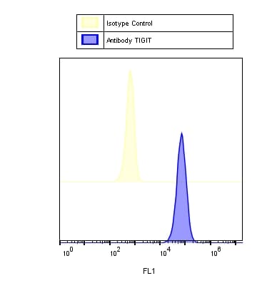
Detection of TIGIT on CD4+ Mouse Splenocytes by Flow Cytometry.
Mouse splenocytes were surface stained with Rat anti-Mouse CD4 FITC-conjugated Monoclonal Antibody (Catalog # FAB554F) and either (A) Rabbit Anti-Mouse TIGIT Monoclonal Antibody (Catalog # MAB72671) or (B) Rabbit IgG control antibody (Catalog # MAB1050) followed by APC-conjugated Anti-Rabbit IgG Secondary Antibody (Catalog # F0111). To facilitate intracellular staining, cells were fixed and permeabilized with FlowX FoxP3 Fixation & Permeabilization Buffer Kit (Catalog # FC012). Then stained with Rabbit anti-Human/Mouse FoxP3 PE-conjugated Monoclonal Antibody (Catalog # IC8214P).Applications for Mouse TIGIT Antibody (2190A)
Application
Recommended Usage
CyTOF-ready
Ready to be labeled using established conjugation methods. No BSA or other carrier proteins that could interfere with conjugation.
Flow Cytometry
0.25 µg/106 cells
Sample: Mouse CD4+ splenocytes
Sample: Mouse CD4+ splenocytes
Reviewed Applications
Read 4 reviews rated 5 using MAB72671 in the following applications:
Flow Cytometry Panel Builder
Bio-Techne Knows Flow Cytometry
Save time and reduce costly mistakes by quickly finding compatible reagents using the Panel Builder Tool.
Advanced Features
- Spectra Viewer - Custom analysis of spectra from multiple fluorochromes
- Spillover Popups - Visualize the spectra of individual fluorochromes
- Antigen Density Selector - Match fluorochrome brightness with antigen density

Formulation, Preparation, and Storage
Purification
Protein A or G purified from cell culture supernatant
Reconstitution
Reconstitute at 0.5 mg/mL in sterile PBS. For liquid material, refer to CoA for concentration.
Loading...
Formulation
Lyophilized from a 0.2 μm filtered solution in PBS with Trehalose. *Small pack size (SP) is supplied either lyophilized or as a 0.2 µm filtered solution in PBS.
Shipping
Lyophilized product is shipped at ambient temperature. Liquid small pack size (-SP) is shipped with polar packs. Upon receipt, store immediately at the temperature recommended below.
Stability & Storage
Use a manual defrost freezer and avoid repeated freeze-thaw cycles.
- 12 months from date of receipt, -20 to -70 °C as supplied.
- 1 month, 2 to 8 °C under sterile conditions after reconstitution.
- 6 months, -20 to -70 °C under sterile conditions after reconstitution.
Calculators
Background: TIGIT
Long Name
T Cell Immunoreceptor with Ig and ITIM Domains
Alternate Names
VSIG9, VSTM3, WUCAM
Gene Symbol
TIGIT
UniProt
Additional TIGIT Products
Product Documents for Mouse TIGIT Antibody (2190A)
Certificate of Analysis
To download a Certificate of Analysis, please enter a lot or batch number in the search box below.
Note: Certificate of Analysis not available for kit components.
Product Specific Notices for Mouse TIGIT Antibody (2190A)
For research use only
Customer Reviews for Mouse TIGIT Antibody (2190A) (4)
5 out of 5
4 Customer Ratings
Have you used Mouse TIGIT Antibody (2190A)?
Submit a review and receive an Amazon gift card!
$25/€18/£15/$25CAN/¥2500 Yen for a review with an image
$10/€7/£6/$10CAN/¥1110 Yen for a review without an image
Submit a review
Customer Images


Showing
1
-
4 of
4 reviews
Showing All
Filter By:
-
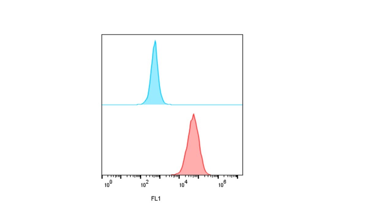
Application: Flow CytometrySample Tested: 4T1 mouse breast cancer cell line and 4T1.2 mouse mammary carcinoma cellsSpecies: MouseVerified Customer | Posted 01/31/2019Antibodies primary and secondary incubated for 60 min at 4 C. The antibody was tested along rabbit isotype control. The analysis was done in FL1 channel using flow cytometry

-
Application: Flow CytometrySample Tested: CT-26 cell lineSpecies: MouseVerified Customer | Posted 12/16/2018
-
Application: Flow CytometrySample Tested: 4T1 mouse breast cancer cell lineSpecies: MouseVerified Customer | Posted 11/16/2018Antibodies primary and secondary incubated for 60 min at 4 C. The antibody was tested along rabbit isotype control
-
Application: Flow CytometrySample Tested: MOuse cell line CT26Species: MouseVerified Customer | Posted 10/24/2018
There are no reviews that match your criteria.
Protocols
Find general support by application which include: protocols, troubleshooting, illustrated assays, videos and webinars.
- 7-Amino Actinomycin D (7-AAD) Cell Viability Flow Cytometry Protocol
- Extracellular Membrane Flow Cytometry Protocol
- Flow Cytometry Protocol for Cell Surface Markers
- Flow Cytometry Protocol for Staining Membrane Associated Proteins
- Flow Cytometry Staining Protocols
- Flow Cytometry Troubleshooting Guide
- Intracellular Flow Cytometry Protocol Using Alcohol (Methanol)
- Intracellular Flow Cytometry Protocol Using Detergents
- Intracellular Nuclear Staining Flow Cytometry Protocol Using Detergents
- Intracellular Staining Flow Cytometry Protocol Using Alcohol Permeabilization
- Intracellular Staining Flow Cytometry Protocol Using Detergents to Permeabilize Cells
- Propidium Iodide Cell Viability Flow Cytometry Protocol
- Protocol for Liperfluo
- Protocol for the Characterization of Human Th22 Cells
- Protocol for the Characterization of Human Th9 Cells
- Protocol: Annexin V and PI Staining by Flow Cytometry
- Protocol: Annexin V and PI Staining for Apoptosis by Flow Cytometry
- Troubleshooting Guide: Fluorokine Flow Cytometry Kits
- View all Protocols, Troubleshooting, Illustrated assays and Webinars
Loading...
Associated Pathways
