Notch-1 Antibody (mN1A) - BSA Free
Novus Biologicals | Catalog # NB100-78486
Key Product Details
Validated by
Species Reactivity
Validated:
Cited:
Applications
Validated:
Cited:
Label
Antibody Source
Format
Product Specifications
Immunogen
Reactivity Notes
Specificity
Clonality
Host
Isotype
Theoretical MW
Disclaimer note: The observed molecular weight of the protein may vary from the listed predicted molecular weight due to post translational modifications, post translation cleavages, relative charges, and other experimental factors.
Scientific Data Images for Notch-1 Antibody (mN1A) - BSA Free
![Knockout Validated: Notch-1 Antibody (mN1A) - BSA Free [NB100-78486]](https://resources.rndsystems.com/images/products/Notch-1-Antibody-mN1A-Knockout-Validated-NB100-78486-img0011.jpg "Western Blot: Notch-1 Antibody (mN1A) - BSA Free [NB100-78486]")
Western Blot: Notch-1 Antibody (mN1A) - BSA Free [NB100-78486]
Western Blot: Notch-1 Antibody (mN1A) [NB100-78486] - Lysates of HeLa human cervical epithelial carcinoma parental cell line and Notch-1 knockout (KO) HeLa cell line. PVDF membrane was probed with 2.0 ug/mL of Mouse Anti-Human Notch-1 Monoclonal Antibody (Catalog # NB100-78486) followed by HRP-conjugated Anti-Mouse IgG Secondary Antibody (Catalog #HAF018). Specific band was detected for Notch-1 at approximately 110 kDa (as indicated) in the parental HeLa cell line, but is not detectable in the knockout HeLa cell line. This experiment was conducted under reducing conditions.![Western Blot: Notch-1 Antibody (mN1A)BSA Free [NB100-78486]](https://resources.rndsystems.com/images/products/Notch-1-Antibody-mN1A-Western-Blot-NB100-78486-img0009.jpg "Western Blot: Notch-1 Antibody (mN1A)BSA Free [NB100-78486]")
Western Blot: Notch-1 Antibody (mN1A)BSA Free [NB100-78486]
Western Blot: Notch-1 Antibody (mN1A) [NB100-78486] - Cell extracts from Jurkat (Lane 1) or mouse thymocytes (Lane 2) were analyzed with monoclonal anti-NOTCH1 antibody. The mN1A antibody recognizes both mouse and human 270 kDa full-length NOTCH1 and 110-120 kDa cleaved NOTCH 1 (NICD).![Immunocytochemistry/ Immunofluorescence: Notch-1 Antibody (mN1A) - BSA Free [NB100-78486]](https://resources.rndsystems.com/images/products/Notch-1-Antibody-mN1A-Immunocytochemistry-Immunofluorescence-NB100-78486-img0008.jpg "Immunocytochemistry/ Immunofluorescence: Notch-1 Antibody (mN1A) - BSA Free [NB100-78486]")
Immunocytochemistry/ Immunofluorescence: Notch-1 Antibody (mN1A) - BSA Free [NB100-78486]
Immunocytochemistry/Immunofluorescence: Notch-1 Antibody (mN1A) [NB100-78486] - The Notch1 Antibody was tested in HEK293 cells at a 1:50 dilution with DyLight 488 (Green). Actin was counterstained with Phalloidin 568 (Red) and cells were mounted in DAPI Flouromount (Blue).![Immunohistochemistry-Paraffin: Notch-1 Antibody (mN1A) - BSA Free [NB100-78486]](https://resources.rndsystems.com/images/products/Notch-1-Antibody-mN1A-Immunohistochemistry-Paraffin-NB100-78486-img0010.jpg "Immunohistochemistry-Paraffin: Notch-1 Antibody (mN1A) - BSA Free [NB100-78486]")
Immunohistochemistry-Paraffin: Notch-1 Antibody (mN1A) - BSA Free [NB100-78486]
Immunohistochemistry-Paraffin: Notch-1 Antibody (mN1A) [NB100-78486] - Analysis of FFPE human pancreatic cancer using 1:10 dilution of Notch-1 antibody on a Bond Rx autostainer (Leica Biosystems). The assay involved 20 minutes of heat induced antigen retrieval (HIER) using 10mM sodium citrate buffer (pH 6.0) and endogenous peroxidase quenching with peroxide block. The sections were incubated with primary antibody for 30 minutes and Bond Polymer Refine Detection (Leica Biosystems) with DAB was used for signal development followed by counterstaining with hematoxylin. Whole slide scanning and capturing of representative images (20X) was performed using Aperio AT2 (Leica Biosystems). Cytoplasmic staining in epithelial cells was observed. Staining was performed by Histowiz.![Flow Cytometry: Notch-1 Antibody (mN1A) - BSA Free [NB100-78486]](https://resources.rndsystems.com/images/products/Notch-1-Antibody-mN1A-Flow-Cytometry-NB100-78486-img0007.jpg "Flow Cytometry: Notch-1 Antibody (mN1A) - BSA Free [NB100-78486]")
Flow Cytometry: Notch-1 Antibody (mN1A) - BSA Free [NB100-78486]
Flow Cytometry: Notch-1 Antibody (mN1A) [NB100-78486] - Intracellular flow cytometric staining of 1 x 10^6 CHO (A) and MCF-7 (B) cells using Notch1 antibody (dark blue). Isotype control shown in orange. An antibody concentration of 1 ug/1x10^6 cells was used. - BSA Free [NB100-78486] -")
Western Blot: Notch-1 Antibody (mN1A) - BSA Free [NB100-78486] -
The DA-MeHA hydrogel regulated Notch signaling.A–E DA-MeHA hydrogel-encapsulated spheroids or suspensions of ADSCs upregulated the expression of Notch1 and Notch2 while downregulating the expression of Notch3, Jagged1 and Jagged2. F No significant difference in Hes1 expression was observed among the groups. n = 4 samples/group (*p < 0.05 and **p < 0.01). Image collected and cropped by CiteAb from the following open publication (https://pubmed.ncbi.nlm.nih.gov/36030275), licensed under a CC-BY license. Not internally tested by Novus Biologicals. - BSA Free [NB100-78486] -")
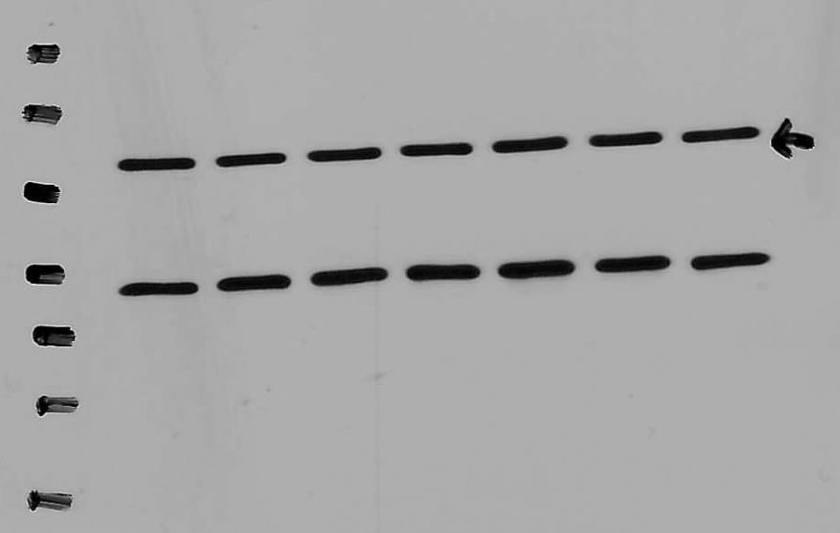
Western Blot: Notch-1 Antibody (mN1A) - BSA Free [NB100-78486] -
Interleukin-33 (IL-33) decreases bone marrow-derived monocytes (BMMs) osteoclastogenesis by inhibiting Notch1 activity. (A) Western blot analysis of Notch1 and Hes-1 expression in osteoclast progenitors incubated with CM-CRE in the presence of miR-34a-5p antagomir. (B–D) Quantification of Notch1, Jag1, and Hes-1 expression in osteoclast progenitors incubated with CM-CRE in the presence of miR-34a-5p antagomir. (E) Western blot analysis of Notch1 and Hes-1 expression in osteoclast progenitors induced by IL-33 in the presence of miR-34a-5p antagomir. (F–H) Quantification of Notch1, Jag1, and Hes-1 expression in osteoclast progenitors treated with IL-33 in the presence of miR-34a-5p antagomir. (I–K) BMMs osteoclastic formation and tartrate-resistant acid phosphatase (TRAP) activity assay after treatment with 20 ng/ml IL-33 in the presence of Jag-1. Red arrows indicated multinucleated osteoclasts. Scale bars represent 100 um. (L–O) Quantification of Trap, Cathepsin K, Nfatc1, and C-fos expression in osteoclast progenitors treated with 20 ng/ml IL-33 in the presence of Jag-1. *P < 0.05, **P < 0.01, ***P < 0.001. P values were analyzed by one-way ANOVA. Image collected and cropped by CiteAb from the following open publication (https://pubmed.ncbi.nlm.nih.gov/29085370), licensed under a CC-BY license. Not internally tested by Novus Biologicals. - BSA Free [NB100-78486] -")
Western Blot: Notch-1 Antibody (mN1A) - BSA Free [NB100-78486] -
Interleukin-33 (IL-33) decreases bone marrow-derived monocytes (BMMs) osteoclastogenesis by inhibiting Notch1 activity. (A) Western blot analysis of Notch1 and Hes-1 expression in osteoclast progenitors incubated with CM-CRE in the presence of miR-34a-5p antagomir. (B–D) Quantification of Notch1, Jag1, and Hes-1 expression in osteoclast progenitors incubated with CM-CRE in the presence of miR-34a-5p antagomir. (E) Western blot analysis of Notch1 and Hes-1 expression in osteoclast progenitors induced by IL-33 in the presence of miR-34a-5p antagomir. (F–H) Quantification of Notch1, Jag1, and Hes-1 expression in osteoclast progenitors treated with IL-33 in the presence of miR-34a-5p antagomir. (I–K) BMMs osteoclastic formation and tartrate-resistant acid phosphatase (TRAP) activity assay after treatment with 20 ng/ml IL-33 in the presence of Jag-1. Red arrows indicated multinucleated osteoclasts. Scale bars represent 100 um. (L–O) Quantification of Trap, Cathepsin K, Nfatc1, and C-fos expression in osteoclast progenitors treated with 20 ng/ml IL-33 in the presence of Jag-1. *P < 0.05, **P < 0.01, ***P < 0.001. P values were analyzed by one-way ANOVA. Image collected and cropped by CiteAb from the following open publication (https://pubmed.ncbi.nlm.nih.gov/29085370), licensed under a CC-BY license. Not internally tested by Novus Biologicals.Applications for Notch-1 Antibody (mN1A) - BSA Free
Flow Cytometry
Immunocytochemistry/ Immunofluorescence
Immunohistochemistry
Immunohistochemistry-Paraffin
Immunoprecipitation
Western Blot
This antibody is CyTOF ready.
Reviewed Applications
Read 2 reviews rated 5 using NB100-78486 in the following applications:
Flow Cytometry Panel Builder
Bio-Techne Knows Flow Cytometry
Save time and reduce costly mistakes by quickly finding compatible reagents using the Panel Builder Tool.
Advanced Features
- Spectra Viewer - Custom analysis of spectra from multiple fluorochromes
- Spillover Popups - Visualize the spectra of individual fluorochromes
- Antigen Density Selector - Match fluorochrome brightness with antigen density

Formulation, Preparation, and Storage
Purification
Formulation
Format
Preservative
Concentration
Shipping
Stability & Storage
Background: Notch-1
Alternate Names
Gene Symbol
UniProt
Additional Notch-1 Products
Product Documents for Notch-1 Antibody (mN1A) - BSA Free
Certificate of Analysis
To download a Certificate of Analysis, please enter a lot or batch number in the search box below.
Product Specific Notices for Notch-1 Antibody (mN1A) - BSA Free
This product is for research use only and is not approved for use in humans or in clinical diagnosis. Primary Antibodies are guaranteed for 1 year from date of receipt.
Related Research Areas
Citations for Notch-1 Antibody (mN1A) - BSA Free
 Powered by Bioz
Customer Reviews for Notch-1 Antibody (mN1A) - BSA Free (2)
Have you used Notch-1 Antibody (mN1A) - BSA Free?
Submit a review and receive an Amazon gift card!
$25/€18/£15/$25CAN/¥2500 Yen for a review with an image
$10/€7/£6/$10CAN/¥1110 Yen for a review without an image
Submit a review
Customer Images
-
Application: Western BlotSample Tested: Vascular Smooth Muscle CellsSpecies: MouseVerified Customer | Posted 12/07/2021Excellent Notch-1 antibody

-
Application: Western BlotSample Tested:Species: OtherVerified Customer | Posted 01/10/2014
There are no reviews that match your criteria.
Protocols
Find general support by application which include: protocols, troubleshooting, illustrated assays, videos and webinars.
- 7-Amino Actinomycin D (7-AAD) Cell Viability Flow Cytometry Protocol
- Antigen Retrieval Protocol (PIER)
- Antigen Retrieval for Frozen Sections Protocol
- Appropriate Fixation of IHC/ICC Samples
- Cellular Response to Hypoxia Protocols
- Chromogenic IHC Staining of Formalin-Fixed Paraffin-Embedded (FFPE) Tissue Protocol
- Chromogenic Immunohistochemistry Staining of Frozen Tissue
- ClariTSA™ Fluorophore Kits
- Detection & Visualization of Antibody Binding
- Extracellular Membrane Flow Cytometry Protocol
- Flow Cytometry Protocol for Cell Surface Markers
- Flow Cytometry Protocol for Staining Membrane Associated Proteins
- Flow Cytometry Staining Protocols
- Flow Cytometry Troubleshooting Guide
- Fluorescent IHC Staining of Frozen Tissue Protocol
- Graphic Protocol for Heat-induced Epitope Retrieval
- Graphic Protocol for the Preparation and Fluorescent IHC Staining of Frozen Tissue Sections
- Graphic Protocol for the Preparation and Fluorescent IHC Staining of Paraffin-embedded Tissue Sections
- Graphic Protocol for the Preparation of Gelatin-coated Slides for Histological Tissue Sections
- ICC Cell Smear Protocol for Suspension Cells
- ICC Immunocytochemistry Protocol Videos
- ICC for Adherent Cells
- IHC Sample Preparation (Frozen sections vs Paraffin)
- Immunocytochemistry (ICC) Protocol
- Immunocytochemistry Troubleshooting
- Immunofluorescence of Organoids Embedded in Cultrex Basement Membrane Extract
- Immunofluorescent IHC Staining of Formalin-Fixed Paraffin-Embedded (FFPE) Tissue Protocol
- Immunohistochemistry (IHC) and Immunocytochemistry (ICC) Protocols
- Immunohistochemistry Frozen Troubleshooting
- Immunohistochemistry Paraffin Troubleshooting
- Immunoprecipitation Protocol
- Intracellular Flow Cytometry Protocol Using Alcohol (Methanol)
- Intracellular Flow Cytometry Protocol Using Detergents
- Intracellular Nuclear Staining Flow Cytometry Protocol Using Detergents
- Intracellular Staining Flow Cytometry Protocol Using Alcohol Permeabilization
- Intracellular Staining Flow Cytometry Protocol Using Detergents to Permeabilize Cells
- Preparing Samples for IHC/ICC Experiments
- Preventing Non-Specific Staining (Non-Specific Binding)
- Primary Antibody Selection & Optimization
- Propidium Iodide Cell Viability Flow Cytometry Protocol
- Protocol for Heat-Induced Epitope Retrieval (HIER)
- Protocol for Liperfluo
- Protocol for Making a 4% Formaldehyde Solution in PBS
- Protocol for VisUCyte™ HRP Polymer Detection Reagent
- Protocol for the Characterization of Human Th22 Cells
- Protocol for the Characterization of Human Th9 Cells
- Protocol for the Fluorescent ICC Staining of Cell Smears - Graphic
- Protocol for the Fluorescent ICC Staining of Cultured Cells on Coverslips - Graphic
- Protocol for the Preparation & Fixation of Cells on Coverslips
- Protocol for the Preparation and Chromogenic IHC Staining of Frozen Tissue Sections
- Protocol for the Preparation and Chromogenic IHC Staining of Frozen Tissue Sections - Graphic
- Protocol for the Preparation and Chromogenic IHC Staining of Paraffin-embedded Tissue Sections
- Protocol for the Preparation and Chromogenic IHC Staining of Paraffin-embedded Tissue Sections - Graphic
- Protocol for the Preparation and Fluorescent ICC Staining of Cells on Coverslips
- Protocol for the Preparation and Fluorescent ICC Staining of Non-adherent Cells
- Protocol for the Preparation and Fluorescent ICC Staining of Stem Cells on Coverslips
- Protocol for the Preparation and Fluorescent IHC Staining of Frozen Tissue Sections
- Protocol for the Preparation and Fluorescent IHC Staining of Paraffin-embedded Tissue Sections
- Protocol for the Preparation of Gelatin-coated Slides for Histological Tissue Sections
- Protocol for the Preparation of a Cell Smear for Non-adherent Cell ICC - Graphic
- Protocol: Annexin V and PI Staining by Flow Cytometry
- Protocol: Annexin V and PI Staining for Apoptosis by Flow Cytometry
- R&D Systems Quality Control Western Blot Protocol
- TUNEL and Active Caspase-3 Detection by IHC/ICC Protocol
- The Importance of IHC/ICC Controls
- Troubleshooting Guide: Fluorokine Flow Cytometry Kits
- Troubleshooting Guide: Immunohistochemistry
- Troubleshooting Guide: Western Blot Figures
- Western Blot Conditions
- Western Blot Protocol
- Western Blot Protocol for Cell Lysates
- Western Blot Troubleshooting
- Western Blot Troubleshooting Guide
- View all Protocols, Troubleshooting, Illustrated assays and Webinars
FAQs for Notch-1 Antibody (mN1A) - BSA Free
-
Q: Hello, does this product (NB300-251) only recognize the cleavage Notch1?
A: This antibody with recognize full length Notch1 if it is in the mix, as well as the cleaved product.
Associated Pathways


