![Western Blot: STAT3 Antibody (9D8) [NBP2-22471]](https://resources.rndsystems.com/images/products/STAT3-Antibody-9D8-Western-Blot-NBP2-22471-img0023.jpg "Western Blot: STAT3 Antibody (9D8) [NBP2-22471]")
Key Product Details
Validated by
Species Reactivity
Validated:
Cited:
Applications
Validated:
Cited:
Label
Antibody Source
Product Specifications
Immunogen
Reactivity Notes
Clonality
Host
Isotype
Scientific Data Images for STAT3 Antibody (9D8)
![Immunocytochemistry/ Immunofluorescence: STAT3 Antibody (9D8) [NBP2-22471]](https://resources.rndsystems.com/images/products/STAT3-Antibody-9D8-Immunocytochemistry-Immunofluorescence-NBP2-22471-img0016.jpg "Immunocytochemistry/ Immunofluorescence: STAT3 Antibody (9D8) [NBP2-22471]")
Immunocytochemistry/ Immunofluorescence: STAT3 Antibody (9D8) [NBP2-22471]
Immunocytochemistry/Immunofluorescence: STAT3 Antibody (9D8) [NBP2-22471] - Analysis of STAT3 using anti-STAT3 (9D8) monoclonal antibody (shown in green) in HeLa cells. Formalin fixed cells were permeabilized with 0.1% Triton X-100 in TBS for 10 minutes at room temperature. Cells were then blocked with 1% Blocker BSA for 15 minutes at room temperature. Cells were probed with a mouse monoclonal antibody recognizing STAT3, at a dilution of 1:100 for at least 1 hour at room temperature. Cells were then washed with PBS and incubated with DyLight 488 goat-anti-mouse secondary antibody at a dilution of 1:400 for 30 minutes at room temperature. Nuclei (blue) were stained with Hoechst 33342 dye.![Immunohistochemistry-Paraffin: STAT3 Antibody (9D8) [NBP2-22471]](https://resources.rndsystems.com/images/products/STAT3-Antibody-9D8-Immunohistochemistry-Paraffin-NBP2-22471-img0017.jpg "Immunohistochemistry-Paraffin: STAT3 Antibody (9D8) [NBP2-22471]")
Immunohistochemistry-Paraffin: STAT3 Antibody (9D8) [NBP2-22471]
Immunohistochemistry-Paraffin: STAT3 Antibody (9D8) [NBP2-22471] - Biopsies of normal and cancer tissues.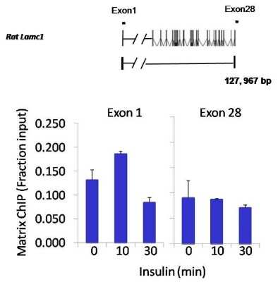
![Western Blot: STAT3 Antibody (9D8) [NBP2-22471]](https://resources.rndsystems.com/images/products/STAT3-Antibody-9D8-Western-Blot-NBP2-22471-img0010.jpg "Western Blot: STAT3 Antibody (9D8) [NBP2-22471]")
Western Blot: STAT3 Antibody (9D8) [NBP2-22471]
Western Blot: STAT3 Antibody (9D8) [NBP2-22471] - Analysis of 25ug HepG2 total lysate.![Western Blot: STAT3 Antibody (9D8) [NBP2-22471]](https://resources.rndsystems.com/images/products/STAT3-Antibody-9D8-Western-Blot-NBP2-22471-img0019.jpg "Western Blot: STAT3 Antibody (9D8) [NBP2-22471]")
Western Blot: STAT3 Antibody (9D8) [NBP2-22471]
Western Blot: STAT3 Antibody (9D8) [NBP2-22471] - Analysis of 25ug of various whole cell lysates.![Western Blot: STAT3 Antibody (9D8) [NBP2-22471]](https://resources.rndsystems.com/images/products/STAT3-Antibody-9D8-Western-Blot-NBP2-22471-img0022.jpg "Western Blot: STAT3 Antibody (9D8) [NBP2-22471]")
Western Blot: STAT3 Antibody (9D8) [NBP2-22471]
Western Blot: STAT3 Antibody (9D8) [NBP2-22471] - Analysis of 25ug of U2-OS lysate from STAT3 SMART pool siRNA transfected or non-targeting control transfected U2-OS cells onto a 4-20% Tris-HCl polyacrylamide gel.![Immunoprecipitation: STAT3 Antibody (9D8) [NBP2-22471]](https://resources.rndsystems.com/images/products/STAT3-Antibody-9D8-Immunoprecipitation-NBP2-22471-img0021.jpg "Immunoprecipitation: STAT3 Antibody (9D8) [NBP2-22471]")
Immunoprecipitation: STAT3 Antibody (9D8) [NBP2-22471]
Immunoprecipitation: STAT3 Antibody (9D8) [NBP2-22471] - Analysis of STAT3 was performed on HepG2 cells. The antigen: antibody complex was formed by incubating 750ug whole cell lysate with 2ug of mouse monoclonal antibody recognizing STAT3 overnight on a rocking platform at 4C. The immune-complex was then captured on 50ul Protein A/G Plus Agarose. Captured immune-complexes were then washed extensively and proteins eluted with 5X Reducing Sample Loading Dye. Samples were then resolved on a 4-20% Tris-HCl polyacrylamide gel. Proteins were transferred to PVDF membrane and blocked with 5% Milk/TBS-0.1%Tween for at least 1 hour. Membranes were then probed with a mouse monoclonal antibody recognizing STAT3 at a dilution of 1:5000 overnight rotating at 4C. Membranes were washed in TBST and probed with Clean-blot IP detection reagent at a dilution of 1:2000 for at least one hour. Membranes were washed and chemiluminescent detection was performed using Super Signal West Dura. [NBP2-22471] -")
Western Blot: STAT3 Antibody (9D8) [NBP2-22471] -
N-EV & H-EV treatment promote macrophage M2 polarization by delivering miR-21-5p that targets PTEN. a, western blot analysis of PTEN protein expression level in induced macrophages. H/i-miR-EV, monocytes were induced with the presence of EV secreted by miR-21-5p-inhibited, hypoxia pre-challenged MSCs; H-EV + i-miR, monocytes were transfected with miR-21-5p inhibitor-expressing vector before induction with the presence of H-EV. Macrophages induced without MSC-EV were used as negative control (NC). b, c, flow cytometry determining the percentage of CD163+CD206+ cells among total CD68+ cells after induction. N-EV + O/E PTEN or H-EV + O/E PTEN, monocytes were transfected with PTEN overexpressing vector before N-EV or H-EV treatment, respectively. d–f, western blot detecting Akt & STAT3 protein expression as well as their activating phosphorylation (p-Ser473 for Akt & p-tyr705 for STAT3) in macrophages after induction. g–i, ELISA evaluating IL-10, TGF-beta & VEGF-alpha in macrophage culture medium after induction. Macrophages induced with the presence of N-EV were used as negative control in b–i. Tukey’s test was used for statistical analysis. *, p < 0.05; **, p < 0.01; ***, p < 0.001; ****, p < 0.0001 Image collected & cropped by CiteAb from the following publication (https://pubmed.ncbi.nlm.nih.gov/30736829), licensed under a CC-BY license. Not internally tested by Novus Biologicals.Applications for STAT3 Antibody (9D8)
Chromatin Immunoprecipitation (ChIP)
Immunocytochemistry/ Immunofluorescence
Immunohistochemistry
Immunohistochemistry-Paraffin
Immunoprecipitation
Western Blot
Reviewed Applications
Read 1 review rated 4 using NBP2-22471 in the following applications:
Formulation, Preparation, and Storage
Purification
Formulation
Preservative
Concentration
Shipping
Stability & Storage
Background: STAT3
Long Name
Alternate Names
Entrez Gene IDs
Gene Symbol
UniProt
Additional STAT3 Products
Product Documents for STAT3 Antibody (9D8)
Certificate of Analysis
To download a Certificate of Analysis, please enter a lot or batch number in the search box below.
Product Specific Notices for STAT3 Antibody (9D8)
This product is for research use only and is not approved for use in humans or in clinical diagnosis. Primary Antibodies are guaranteed for 1 year from date of receipt.
Citations for STAT3 Antibody (9D8)
 Powered by Bioz
Customer Reviews for STAT3 Antibody (9D8) (1)
Have you used STAT3 Antibody (9D8)?
Submit a review and receive an Amazon gift card!
$25/€18/£15/$25CAN/¥2500 Yen for a review with an image
$10/€7/£6/$10CAN/¥1110 Yen for a review without an image
Submit a review
Customer Images
-
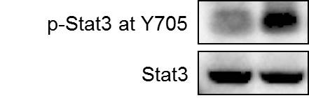
Application: Western BlotSample Tested: Breast cancer cellsSpecies: HumanVerified Customer | Posted 10/11/2018Human breast cancer cell MDA-MB-231 was treated with carboplatin for 72 hours and the expression of p-Stat3 at Y705 and total Stat3 were detected by western blot.

There are no reviews that match your criteria.
Protocols
Find general support by application which include: protocols, troubleshooting, illustrated assays, videos and webinars.
- Antigen Retrieval Protocol (PIER)
- Antigen Retrieval for Frozen Sections Protocol
- Appropriate Fixation of IHC/ICC Samples
- Cellular Response to Hypoxia Protocols
- ChIP Protocol Video
- Chromatin Immunoprecipitation (ChIP) Protocol
- Chromatin Immunoprecipitation Protocol
- Chromogenic IHC Staining of Formalin-Fixed Paraffin-Embedded (FFPE) Tissue Protocol
- Chromogenic Immunohistochemistry Staining of Frozen Tissue
- ClariTSA™ Fluorophore Kits
- Detection & Visualization of Antibody Binding
- Fluorescent IHC Staining of Frozen Tissue Protocol
- Graphic Protocol for Heat-induced Epitope Retrieval
- Graphic Protocol for the Preparation and Fluorescent IHC Staining of Frozen Tissue Sections
- Graphic Protocol for the Preparation and Fluorescent IHC Staining of Paraffin-embedded Tissue Sections
- Graphic Protocol for the Preparation of Gelatin-coated Slides for Histological Tissue Sections
- ICC Cell Smear Protocol for Suspension Cells
- ICC Immunocytochemistry Protocol Videos
- ICC for Adherent Cells
- IHC Sample Preparation (Frozen sections vs Paraffin)
- Immunocytochemistry (ICC) Protocol
- Immunocytochemistry Troubleshooting
- Immunofluorescence of Organoids Embedded in Cultrex Basement Membrane Extract
- Immunofluorescent IHC Staining of Formalin-Fixed Paraffin-Embedded (FFPE) Tissue Protocol
- Immunohistochemistry (IHC) and Immunocytochemistry (ICC) Protocols
- Immunohistochemistry Frozen Troubleshooting
- Immunohistochemistry Paraffin Troubleshooting
- Immunoprecipitation Protocol
- Preparing Samples for IHC/ICC Experiments
- Preventing Non-Specific Staining (Non-Specific Binding)
- Primary Antibody Selection & Optimization
- Protocol for Heat-Induced Epitope Retrieval (HIER)
- Protocol for Making a 4% Formaldehyde Solution in PBS
- Protocol for VisUCyte™ HRP Polymer Detection Reagent
- Protocol for the Fluorescent ICC Staining of Cell Smears - Graphic
- Protocol for the Fluorescent ICC Staining of Cultured Cells on Coverslips - Graphic
- Protocol for the Preparation & Fixation of Cells on Coverslips
- Protocol for the Preparation and Chromogenic IHC Staining of Frozen Tissue Sections
- Protocol for the Preparation and Chromogenic IHC Staining of Frozen Tissue Sections - Graphic
- Protocol for the Preparation and Chromogenic IHC Staining of Paraffin-embedded Tissue Sections
- Protocol for the Preparation and Chromogenic IHC Staining of Paraffin-embedded Tissue Sections - Graphic
- Protocol for the Preparation and Fluorescent ICC Staining of Cells on Coverslips
- Protocol for the Preparation and Fluorescent ICC Staining of Non-adherent Cells
- Protocol for the Preparation and Fluorescent ICC Staining of Stem Cells on Coverslips
- Protocol for the Preparation and Fluorescent IHC Staining of Frozen Tissue Sections
- Protocol for the Preparation and Fluorescent IHC Staining of Paraffin-embedded Tissue Sections
- Protocol for the Preparation of Gelatin-coated Slides for Histological Tissue Sections
- Protocol for the Preparation of a Cell Smear for Non-adherent Cell ICC - Graphic
- R&D Systems Quality Control Western Blot Protocol
- TUNEL and Active Caspase-3 Detection by IHC/ICC Protocol
- The Importance of IHC/ICC Controls
- Troubleshooting Guide: Immunohistochemistry
- Troubleshooting Guide: Western Blot Figures
- Western Blot Conditions
- Western Blot Protocol
- Western Blot Protocol for Cell Lysates
- Western Blot Troubleshooting
- Western Blot Troubleshooting Guide
- View all Protocols, Troubleshooting, Illustrated assays and Webinars
FAQs for STAT3 Antibody (9D8)
-
Q: For antibodies : STAT5b H00006777-M03; STAT 5a NBR2-00622; STAT3 NBP2-22471; STAT 6 H00006778-M01 - do these detect phosphorylated, unphosphorylated or both forms of STATs?
A: Antibodies H00006777-M03, NBR2-00622, NBP2-22471, and H00006778-M01 were raised to recognize total STAT proteins, they do not differentiate between any posttranslational modifications and will recognize both phosphorylated and non-phosphorylated protein.
Associated Pathways


