Green fluorescent protein (GFP) is a 27 kDa protein originally isolated from the jellyfish Aequorea victoria. In the presence of UV light (490-520 nm), it emits a green fluorescent color that can be used to pinpoint locations of various intracellular proteins. GFP is 238 amino acids (aa) in length. It is a globular monomer that has a tendency to dimerize. The monomer has the shape of a beta -barrel with a chromophore (aa 65-67) containing alpha -helix running up its center. GFPuv is the Aequorea sequence with three aa substitutions; Phe to Ser at # 99, Met to Thr at # 153, and Val to Ala at # 163. This form expresses faster and is 18-fold brighter than native GFP; excitation peaks at 395 nm and emission at 508 nm.

Key Product Details
Species Reactivity
Validated:
Multi-Species
Cited:
Human, Mouse, Rat
Applications
Validated:
Western Blot, Intracellular Staining by Flow Cytometry, Immunocytochemistry, Simple Western, Immunoprecipitation, CyTOF-ready
Cited:
Immunohistochemistry, Western Blot, Immunocytochemistry, Simple Western, Immunoprecipitation, Bioassay, Purification
Label
Unconjugated
Antibody Source
Polyclonal Goat IgG
Loading...
Product Specifications
Immunogen
E. coli-derived recombinant GFPuv
Ser2-Lys238
Accession # P42212
Ser2-Lys238
Accession # P42212
Specificity
Detects GFP in direct ELISAs and Western blots.
Clonality
Polyclonal
Host
Goat
Isotype
IgG
Scientific Data Images for GFP Antibody
Detection of GFP by Western Blot.
Western blot shows lysates of NS0 mouse myeloma cell line either mock transfected or transfected with eGFP-tagged EDG6. PVDF membrane was probed with 1 µg/mL of Goat Anti-GFP Antigen Affinity-purified Polyclonal Antibody (Catalog # AF4240) followed by HRP-conjugated Anti-Goat IgG Secondary Antibody (Catalog # HAF017). A specific band was detected for GFP at approximately 80 kDa (as indicated). This experiment was conducted under reducing conditions and using Immunoblot Buffer Group 1.
GFP in HEK293 human embryonic kidney cells transfected with GFP.
GFP was detected in immersion fixed HEK293 human embryonic kidney cells transfected with GFP (green) using Goat Anti-GFP Antigen Affinity-purified Polyclonal Antibody (Catalog # AF4240) at 1.7 µg/mL for 3 hours at room temperature. Cells were stained using the NorthernLights™ 557-conjugated Anti-Goat IgG Secondary Antibody (red, middle panel; Catalog # NL001) and counterstained with DAPI (blue). Specific staining was localized to the cytoplasm of GFP-positive cells. View our protocol for Fluorescent ICC Staining of Cells on Coverslips.
Detection of GFP in HEK293 human embryonic kidney cells transfected with GFP.
HEK293 human embryonic kidney cells transfected with GFP was stained with Allophycocyanin-conjugated Anti-Goat IgG Secondary Antibody only (A, Catalog # F0108) or with Goat Anti-GFP Antigen Affinity-purified Polyclonal Antibody (Catalog # AF4240) followed by Secondary Antibody (B). To facilitate intracellular staining, cells were fixed with paraformaldehyde and permeabilized with saponin. Quadrant markers were set based on control antibody staining (Catalog # AB-108-C).
Detection of GFP by Simple WesternTM.
Simple Western lane view shows lysates of HEK293T human embryonic kidney cell line either mock transfected (-) or transfected with eGFP-tagged EDG6 (+), loaded at 0.2 mg/mL. A specific band was detected for GFP at approximately 114 kDa (as indicated) using 2.5 µg/mL of Goat Anti-GFP Antigen Affinity-purified Polyclonal Antibody (Catalog # AF4240) followed by 1:50 dilution of HRP-conjugated Anti-Goat IgG Secondary Antibody (Catalog # HAF109). This experiment was conducted under reducing conditions and using the 12-230 kDa separation system.
Simple Western: GFP Antibody [Unconjugated] [AF4240] -
Simple Western: GFP Antibody [Unconjugated] [AF4240] - Design for a membrane-localized ACE2 expression system. (A) Our ACE2 construct is driven by a CMV promoter followed by the first 25 residues of ACE2 containing the leader sequence that direct ACE2 to the plasma membrane. This is followed by a 3xHA tag linked to the remainder of ACE2 (20-805) and a C-terminal sfGFP. Both 3xHA and sfGFP fusions are separated from ACE2 by flexible 3xGGGGS linkers. (B) The ACE2 fusion protein is designed to be embedded in the plasma membrane where it can perform extracellular carboxypeptidase-mediated metabolism and its levels can be detected by cell staining with antibodies to HA. (C) Lysates from untransfected or 3xHA-ACE2-sfGFP-transfected HEK293 cells were analyzed by automated Jess capillary immunoassay using antibodies to HA, GFP, and two ACE2 antibodies. (D) Confocal fluorescence microscopy of HEK cells transfected with 3xHA-ACE2-sfGFP and stained with HA and the nuclear stain DAPI. Image collected and cropped by CiteAb from the following publication (https://pubmed.ncbi.nlm.nih.gov/37644110), licensed under a CC-BY license. Not internally tested by R&D Systems.
Detection of GFP by Western Blot
GFP-rMSCs following in vitro stress assays. (A) Trypsinized and re-plated GFP-rMSCs at 24 h following hydrogen peroxide treatment. PSF-treated cells were much more abundant and had begun dividing, but GFP-rMSCs not treated with PSF appeared to have lasting negative effects of H2O2 exposure. (B) Lactate dehydrogenase released into medium of the hydrogen peroxide-treated cells was a means to measure cell death. (C) Changes in expression of stress-related proteins Hif-1a, HSP70, and SOD1 in GFP-rMSCs following hydrogen peroxide treatment. * Indicates p < 0.5. Experiments were repeated twice. Bar =100 µm. Image collected and cropped by CiteAb from the following open publication (https://pubmed.ncbi.nlm.nih.gov/35054878), licensed under a CC-BY license. Not internally tested by R&D Systems.Applications for GFP Antibody
Application
Recommended Usage
CyTOF-ready
Ready to be labeled using established conjugation methods. No BSA or other carrier proteins that could interfere with conjugation.
Immunocytochemistry
1-15 µg/mL
Sample: Immersion fixed HEK293 human embryonic kidney cells transfected with wild type GFP
Sample: Immersion fixed HEK293 human embryonic kidney cells transfected with wild type GFP
Immunoprecipitation
25 µg/mL
Sample: Conditioned cell culture medium spiked with Recombinant GFP, see our available Western blot detection antibodies
Sample: Conditioned cell culture medium spiked with Recombinant GFP, see our available Western blot detection antibodies
Intracellular Staining by Flow Cytometry
0.25 µg/106 cells
Sample: Immersion fixed HEK293 human embryonic kidney cells transfected with wild type GFP
Sample: Immersion fixed HEK293 human embryonic kidney cells transfected with wild type GFP
Simple Western
2.5 µg/mL
Sample: HEK293T human embryonic kidney cell line transfected with eGFP-tagged proteins
Sample: HEK293T human embryonic kidney cell line transfected with eGFP-tagged proteins
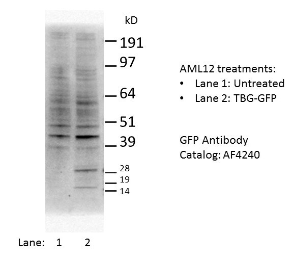
Western Blot
1 µg/mL
Sample: NS0 mouse myeloma cell line either transfected with eGFP-tagged proteins
Sample: NS0 mouse myeloma cell line either transfected with eGFP-tagged proteins
Reviewed Applications
Read 4 reviews rated 4.3 using AF4240 in the following applications:
Flow Cytometry Panel Builder
Bio-Techne Knows Flow Cytometry
Save time and reduce costly mistakes by quickly finding compatible reagents using the Panel Builder Tool.
Advanced Features
- Spectra Viewer - Custom analysis of spectra from multiple fluorochromes
- Spillover Popups - Visualize the spectra of individual fluorochromes
- Antigen Density Selector - Match fluorochrome brightness with antigen density

Formulation, Preparation, and Storage
Purification
Antigen Affinity-purified
Reconstitution
Reconstitute at 0.2 mg/mL in sterile PBS. For liquid material, refer to CoA for concentration.
Loading...
Formulation
Lyophilized from a 0.2 μm filtered solution in PBS with Trehalose. *Small pack size (SP) is supplied either lyophilized or as a 0.2 µm filtered solution in PBS.
Shipping
Lyophilized product is shipped at ambient temperature. Liquid small pack size (-SP) is shipped with polar packs. Upon receipt, store immediately at the temperature recommended below.
Stability & Storage
Use a manual defrost freezer and avoid repeated freeze-thaw cycles.
- 12 months from date of receipt, -20 to -70 °C as supplied.
- 1 month, 2 to 8 °C under sterile conditions after reconstitution.
- 6 months, -20 to -70 °C under sterile conditions after reconstitution.
Calculators
Background: GFP
Additional GFP Products
Product Documents for GFP Antibody
Certificate of Analysis
To download a Certificate of Analysis, please enter a lot or batch number in the search box below.
Note: Certificate of Analysis not available for kit components.
Product Specific Notices for GFP Antibody
For research use only
Citations for GFP Antibody
 Powered by Bioz
Customer Reviews for GFP Antibody (4)
4.3 out of 5
4 Customer Ratings
Have you used GFP Antibody?
Submit a review and receive an Amazon gift card!
$25/€18/£15/$25CAN/¥2500 Yen for a review with an image
$10/€7/£6/$10CAN/¥1110 Yen for a review without an image
Submit a review
Customer Images
Showing
1
-
4 of
4 reviews
Showing All
Filter By:
-
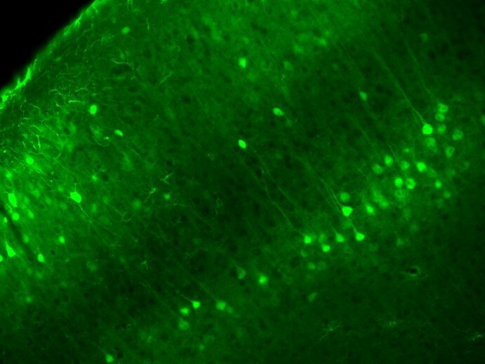
Application: Immunocytochemistry/ImmunofluorescenceSample Tested: Brain (cross-section through blood vessel)Species: MouseVerified Customer | Posted 09/17/2018

-
Application: ImmunoprecipitationSample Tested: HEK293 human embryonic kidney cell lineSpecies: HumanVerified Customer | Posted 01/17/2018
-
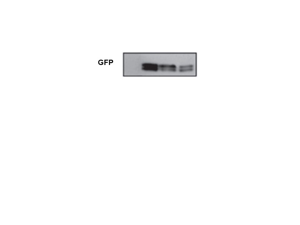
Application: Western BlotSample Tested: Liver cellsSpecies: MouseVerified Customer | Posted 10/27/2017
-
Application: Western BlotSample Tested: See PMID 20484048Species: OtherVerified Customer | Posted 01/08/2015
There are no reviews that match your criteria.
Protocols
Find general support by application which include: protocols, troubleshooting, illustrated assays, videos and webinars.
- 7-Amino Actinomycin D (7-AAD) Cell Viability Flow Cytometry Protocol
- Appropriate Fixation of IHC/ICC Samples
- Cellular Response to Hypoxia Protocols
- ClariTSA™ Fluorophore Kits
- Detection & Visualization of Antibody Binding
- Extracellular Membrane Flow Cytometry Protocol
- Flow Cytometry Protocol for Cell Surface Markers
- Flow Cytometry Protocol for Staining Membrane Associated Proteins
- Flow Cytometry Staining Protocols
- Flow Cytometry Troubleshooting Guide
- ICC Cell Smear Protocol for Suspension Cells
- ICC Immunocytochemistry Protocol Videos
- ICC for Adherent Cells
- Immunocytochemistry (ICC) Protocol
- Immunocytochemistry Troubleshooting
- Immunofluorescence of Organoids Embedded in Cultrex Basement Membrane Extract
- Immunohistochemistry (IHC) and Immunocytochemistry (ICC) Protocols
- Immunoprecipitation Protocol
- Intracellular Flow Cytometry Protocol Using Alcohol (Methanol)
- Intracellular Flow Cytometry Protocol Using Detergents
- Intracellular Nuclear Staining Flow Cytometry Protocol Using Detergents
- Intracellular Staining Flow Cytometry Protocol Using Alcohol Permeabilization
- Intracellular Staining Flow Cytometry Protocol Using Detergents to Permeabilize Cells
- Preparing Samples for IHC/ICC Experiments
- Preventing Non-Specific Staining (Non-Specific Binding)
- Primary Antibody Selection & Optimization
- Propidium Iodide Cell Viability Flow Cytometry Protocol
- Protocol for Liperfluo
- Protocol for VisUCyte™ HRP Polymer Detection Reagent
- Protocol for the Characterization of Human Th22 Cells
- Protocol for the Characterization of Human Th9 Cells
- Protocol for the Fluorescent ICC Staining of Cell Smears - Graphic
- Protocol for the Fluorescent ICC Staining of Cultured Cells on Coverslips - Graphic
- Protocol for the Preparation and Fluorescent ICC Staining of Cells on Coverslips
- Protocol for the Preparation and Fluorescent ICC Staining of Non-adherent Cells
- Protocol for the Preparation and Fluorescent ICC Staining of Stem Cells on Coverslips
- Protocol for the Preparation of a Cell Smear for Non-adherent Cell ICC - Graphic
- Protocol: Annexin V and PI Staining by Flow Cytometry
- Protocol: Annexin V and PI Staining for Apoptosis by Flow Cytometry
- R&D Systems Quality Control Western Blot Protocol
- TUNEL and Active Caspase-3 Detection by IHC/ICC Protocol
- The Importance of IHC/ICC Controls
- Troubleshooting Guide: Fluorokine Flow Cytometry Kits
- Troubleshooting Guide: Western Blot Figures
- Western Blot Conditions
- Western Blot Protocol
- Western Blot Protocol for Cell Lysates
- Western Blot Troubleshooting
- Western Blot Troubleshooting Guide
- View all Protocols, Troubleshooting, Illustrated assays and Webinars
Loading...