
Best Seller
HA Tag Antibody (543851)
R&D Systems | Catalog # MAB060


Scientific images may be resized, cropped, or adjusted for brightness or contrast for presentation purposes. These updates are limited to presentation and do not affect the underlying scientific interpretation of the data. Where available, additional source data may be provided upon request.
Key Product Details
Species Reactivity
Validated:
Multi-Species
Cited:
Human, Mouse
Applications
Validated:
Western Blot
Cited:
Neutralization, Bioassay, Cross-linking, Functional Assay
Label
Unconjugated
Antibody Source
Monoclonal Mouse IgG2B Clone # 543851
Loading...
Product Specifications
Immunogen
HA Tag Peptide
Accession # ABB51961
Accession # ABB51961
Specificity
Specifically detects N-terminal HA-tagged proteins with a free N-terminus.
Clonality
Monoclonal
Host
Mouse
Isotype
IgG2B
Scientific Data Images for HA Tag Antibody (543851)
Detection of N‑terminal HA-tag by Western Blot.
Western blot shows Recombinant Human APRIL/TNFSF13 (Catalog # 5860-AP, 5 ng/lane). PVDF membrane was probed with 1 µg/mL of Mouse Anti-HA Tag Peptide Antibody Monoclonal Antibody (Catalog # MAB060) followed by HRP-conjugated Anti-Mouse IgG Secondary Antibody (Catalog # HAF018). A specific band was detected for N-terminal HA-tagged Recombinant Human APRIL/ TNFSF13 at approximately 26 kDa (as indicated). This experiment was conducted under reducing conditions and using Immunoblot Buffer Group 1.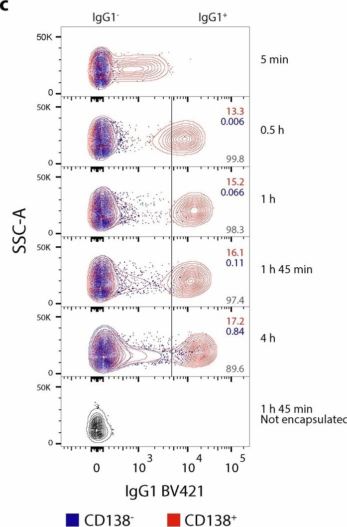
Detection of HA Tag by Flow Cytometry
Cell survival and IgG secretion of encapsulated mouse bone marrow plasma cells over time.(a) Cell viability of encapsulated and non-encapsulated cells after incubation in culture medium for different time periods. Viability was analyzed by flow cytometry (gated on single cells or single beads/CD138+/live; gating strategy in Supplementary Fig. 2). (b) Detection of IgG in the culture supernatant of encapsulated (red) and nonencapsulated cells (dark gray) by ELISA at different time points. The dashed line indicates the sensitivity of the assay (absorbance corresponding to 1.56 ng/mL IgG). The solid lines denote the mean of two technical replicates. (c) IgG1 secretion of encapsulated and non-encapsulated cells over time was analyzed by flow cytometry. To determine the extent of cross-contamination between beads, the IgG1 signal of plasma cells (CD138+) was compared to the signal of empty beads or cells that lack the plasma cell marker (CD138−; gating strategy in Supplementary Fig. 3). The numbers in the top right corner indicate the percentage of IgG+ events in the CD138+ (red) and CD138− (blue) populations (IgG+ gate indicated by a gray line). The number in the bottom right corner indicates the percentage of ‘true positives’ (percentage of IgG+ events originating from CD138+ based on the sum of IgG+ events from both CD138+ and CD138− populations). The plots show a minimum of 10,019 (CD138−) and 2,533 (CD138+) events at 2% contour level. (d) Time course of ASC IgG secretion and background based on c. Left axis: percentage of IgG1-positive events in the CD138+ (red diamonds) and CD138− (blue diamonds) populations for each analyzed time point. Right axis (gray diamonds): percentage of events in the IgG1 gate that originate from CD138+ and not from CD138−.Source data Image collected and cropped by CiteAb from the following open publication (https://www.nature.com/articles/s41587-024-02346-5), licensed under a CC-BY license. Not internally tested by R&D Systems.Applications for HA Tag Antibody (543851)
Application
Recommended Usage
Western Blot
1 µg/mL
Sample: N-terminal HA-tagged Recombinant Human APRIL/TNFSF13 (Catalog # 5860-AP)
Sample: N-terminal HA-tagged Recombinant Human APRIL/TNFSF13 (Catalog # 5860-AP)
Reviewed Applications
Read 8 reviews rated 4.8 using MAB060 in the following applications:
Formulation, Preparation, and Storage
Purification
Protein A or G purified from hybridoma culture supernatant
Reconstitution
Sterile PBS to a final concentration of 0.5 mg/mL. For liquid material, refer to CoA for concentration.
Loading...
Formulation
Lyophilized from a 0.2 μm filtered solution in PBS with Trehalose. *Small pack size (SP) is supplied either lyophilized or as a 0.2 µm filtered solution in PBS.
Shipping
Lyophilized product is shipped at ambient temperature. Liquid small pack size (-SP) is shipped with polar packs. Upon receipt, store immediately at the temperature recommended below.
Stability & Storage
Use a manual defrost freezer and avoid repeated freeze-thaw cycles.
- 12 months from date of receipt, -20 to -70 °C as supplied.
- 1 month, 2 to 8 °C under sterile conditions after reconstitution.
- 6 months, -20 to -70 °C under sterile conditions after reconstitution.
Calculators
Background: HA Tag
References
1. Wilks, S., Graaf, M. D., Smith, D. J., & Burke, D. F. (2012). A review of influenza haemagglutinin receptor binding as it relates to pandemic properties. Vaccine, 30(29), 4369-4376. doi:10.1016/j.vaccine.2012.02.076
2. Wu, N. C., & Wilson, I. A. (2019). Influenza hemagglutinin structures and antibody recognition. Cold Spring Harbor Perspectives in Medicine, 10(8). doi:10.1101/cshperspect.a038778
3. Zhao, X., Li, G., & Liang, S. (2013). Several affinity tags commonly used in chromatographic purification. Journal of Analytical Methods in Chemistry, 2013, 1-8. doi:10.1155/2013/581093
4. Kimple, M. E., Brill, A. L., & Pasker, R. L. (2013). Overview of affinity tags for protein purification. Current Protocols in Protein Science, 73(1). doi:10.1002/0471140864.ps0909s73
5. Schembri, L., Dalibart, R., Tomasello, F., Legembre, P., Ichas, F., & Giorgi, F. D. (2007). The HA tag is cleaved and loses immunoreactivity during apoptosis. Nature Methods, 4(2), 107-108. doi:10.1038/nmeth0207-107
Alternate Names
HA Epitope Tag
Gene Symbol
HA
UniProt
Additional HA Tag Products
Product Documents for HA Tag Antibody (543851)
Certificate of Analysis
To download a Certificate of Analysis, please enter a lot or batch number in the search box below.
Note: Certificate of Analysis not available for kit components.
Product Specific Notices for HA Tag Antibody (543851)
For research use only
Related Research Areas
Citations for HA Tag Antibody (543851)
 Powered by Bioz
Customer Reviews for HA Tag Antibody (543851) (8)
4.8 out of 5
8 Customer Ratings
Have you used HA Tag Antibody (543851)?
Submit a review and receive an Amazon gift card!
$25/€18/£15/$25CAN/¥2500 for a review with an image
$10/€7/£6/$10CAN/¥1110 for a review without an image
Submit a review
Customer Images


Showing
1
-
5 of
8 reviews
Showing All
Filter By:
-
Application: Western BlotSample Tested: 293T human embryonic kidney cell lineSpecies: HumanVerified Customer | Posted 11/04/2021

-
Application: Cell ProliferationSample Tested: B cellsSpecies: MouseVerified Customer | Posted 01/05/2021
-
Application: Cell CultureSample Tested: B cellsSpecies: MouseVerified Customer | Posted 10/22/2020This product has been used in combination with murine CD40L-HaTag protein to culture primary murine B cells.
-
Application: Functional AssaySample Tested: Recombinant proteinSpecies: HumanVerified Customer | Posted 12/06/2017
-
Application: Flow CytometrySample Tested: Jurkat human acute T cell leukemia cell lineSpecies: HumanVerified Customer | Posted 08/23/2017
-
Application: Western BlotSample Tested: 293-EBNA human EBV-expressing embryonic kidney cell lineSpecies: MouseVerified Customer | Posted 07/23/2017
-
Application: Western BlotSample Tested: 293T human embryonic kidney cell lineSpecies: HumanVerified Customer | Posted 07/21/2017
-
Application: Western BlotSample Tested: 293-EBNA human EBV-expressing embryonic kidney cell lineSpecies: MouseVerified Customer | Posted 07/20/2017
There are no reviews that match your criteria.
Protocols
Find general support by application which include: protocols, troubleshooting, illustrated assays, videos and webinars.
- Cellular Response to Hypoxia Protocols
- R&D Systems Quality Control Western Blot Protocol
- Troubleshooting Guide: Western Blot Figures
- Western Blot Conditions
- Western Blot Protocol
- Western Blot Protocol for Cell Lysates
- Western Blot Troubleshooting
- Western Blot Troubleshooting Guide
- View all Protocols, Troubleshooting, Illustrated assays and Webinars
Loading...