
Human Angiopoietin-like Protein 8/Betatrophin Antibody (910248)
R&D Systems | Catalog # MAB8548

Key Product Details
Species Reactivity
Validated:
Human
Cited:
Mouse
Applications
Validated:
Immunohistochemistry, Western Blot, Immunocytochemistry
Cited:
Immunohistochemistry
Label
Unconjugated
Antibody Source
Monoclonal Mouse IgG1 Clone # 910248
Loading...
Product Specifications
Immunogen
Human Angiopoietin-like Protein 8/Betatrophin synthetic peptide with sequences corresponding to the N-term region
Accession # Q6UXH0
Accession # Q6UXH0
Specificity
Detects human Angiopoietin-like Protein 8/Betatrophin in direct ELISA and Western Blots.
Clonality
Monoclonal
Host
Mouse
Isotype
IgG1
Scientific Data Images for Human Angiopoietin-like Protein 8/Betatrophin Antibody (910248)
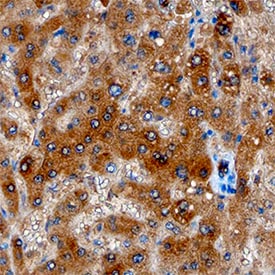
Angiopoietin-like Protein 8/Betatrophin in Human Liver.
Angiopoietin-like Protein 8/Betatrophin was detected in immersion fixed paraffin-embedded sections of human liver using Mouse Anti-Human Angiopoietin-like Protein 8/Betatrophin Antigen Affinity-purified Monoclonal Antibody (Catalog # MAB8548) at 15 µg/mL overnight at 4 °C. Tissue was stained using the Anti-Mouse HRP-DAB Cell & Tissue Staining Kit (brown; Catalog # CTS002) and counterstained with hematoxylin (blue). Specific staining was localized to hepatocytes. View our protocol for Chromogenic IHC Staining of Paraffin-embedded Tissue Sections.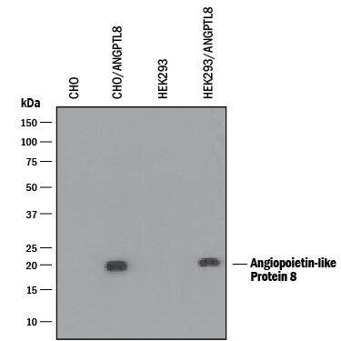
Detection of Human Angiopoietin-like Protein 8/Betatrophin by Western Blot.
Western blot shows lysates of CHO Chinese hamster ovary cell line and HEK293 human embryonic kidney cell line either mock transfected or transfected with human Angiopoietin-like Protein 8 (ANGPTL8)/Betatrophin. PVDF membrane was probed with 1 µg/mL of Mouse Anti-Human Angiopoietin-like Protein 8/Betatrophin Antigen Affinity-purified Polyclonal Antibody (Catalog # MAB8548) followed by HRP-conjugated Anti-Mouse IgG Secondary Antibody (Catalog # HAF018). A specific band was detected for Angiopoietin-like Protein 8/Betatrophin at approximately 23 kDa (as indicated). This experiment was conducted under reducing conditions and using Immunoblot Buffer Group 1.
Angiopoietin-like Protein 8/Betatrophin in HepG2 Human Cell Line.
Angiopoietin-like Protein 8/Betatrophin was detected in immersion fixed HepG2 human hepatocellular carcinoma cell line using Mouse Anti-Human Angiopoietin-like Protein 8/Betatrophin Monoclonal Antibody (Catalog # MAB8548) at 10 µg/mL for 3 hours at room temperature. Cells were stained using the NorthernLights™ 557-conjugated Anti-Mouse IgG Secondary Antibody (red; Catalog # NL007) and counterstained with DAPI (blue). View our protocol for Fluorescent ICC Staining of Cells on Coverslips.Applications for Human Angiopoietin-like Protein 8/Betatrophin Antibody (910248)
Application
Recommended Usage
Immunocytochemistry
8-25 µg/mL
Sample: Immersion fixed HepG2 human hepatocellular carcinoma cell line
Sample: Immersion fixed HepG2 human hepatocellular carcinoma cell line
Immunohistochemistry
8-25 µg/mL
Sample: Immersion fixed paraffin-embedded sections of human liver
Sample: Immersion fixed paraffin-embedded sections of human liver
Western Blot
1 µg/mL
Sample: CHO Chinese hamster ovary cell line and HEK293 human embryonic kidney cell line transfected with human Angiopoietin-like Protein 8 (ANGPTL8)/Betatrophin
Sample: CHO Chinese hamster ovary cell line and HEK293 human embryonic kidney cell line transfected with human Angiopoietin-like Protein 8 (ANGPTL8)/Betatrophin
Formulation, Preparation, and Storage
Purification
Protein A or G purified from hybridoma culture supernatant
Reconstitution
Reconstitute at 0.5 mg/mL in sterile PBS.
Loading...
Formulation
Lyophilized from a 0.2 μm filtered solution in PBS with Trehalose.
Shipping
The product is shipped at ambient temperature. Upon receipt, store it immediately at the temperature recommended below.
Stability & Storage
Use a manual defrost freezer and avoid repeated freeze-thaw cycles.
- 12 months from date of receipt, -20 to -70 °C as supplied.
- 1 month, 2 to 8 °C under sterile conditions after reconstitution.
- 6 months, -20 to -70 °C under sterile conditions after reconstitution.
Calculators
Background: Angiopoietin-like Protein 8/Betatrophin
Alternate Names
Angiopoietin like Protein 8, ANGPTL8, Betatrophin, C19orf80, Gm6484, Lipasin, PRO1185, PVPA599, RIFL, TD26
Gene Symbol
ANGPTL8
UniProt
Additional Angiopoietin-like Protein 8/Betatrophin Products
- All Products for Angiopoietin-like Protein 8/Betatrophin
- Angiopoietin-like Protein 8/Betatrophin cDNA Clones
- Angiopoietin-like Protein 8/Betatrophin ELISA Kits
- Angiopoietin-like Protein 8/Betatrophin Lysates
- Angiopoietin-like Protein 8/Betatrophin Primary Antibodies
- Angiopoietin-like Protein 8/Betatrophin Proteins and Enzymes
Product Documents for Human Angiopoietin-like Protein 8/Betatrophin Antibody (910248)
Certificate of Analysis
To download a Certificate of Analysis, please enter a lot or batch number in the search box below.
Note: Certificate of Analysis not available for kit components.
Product Specific Notices for Human Angiopoietin-like Protein 8/Betatrophin Antibody (910248)
For research use only
Related Research Areas
Citations for Human Angiopoietin-like Protein 8/Betatrophin Antibody (910248)
 Powered by Bioz
Customer Reviews for Human Angiopoietin-like Protein 8/Betatrophin Antibody (910248)
There are currently no reviews for this product. Be the first to review Human Angiopoietin-like Protein 8/Betatrophin Antibody (910248) and earn rewards!
Have you used Human Angiopoietin-like Protein 8/Betatrophin Antibody (910248)?
Submit a review and receive an Amazon gift card!
$25/€18/£15/$25CAN/¥2500 for a review with an image
$10/€7/£6/$10CAN/¥1110 for a review without an image
Submit a review
Protocols
Find general support by application which include: protocols, troubleshooting, illustrated assays, videos and webinars.
- Antigen Retrieval Protocol (PIER)
- Antigen Retrieval for Frozen Sections Protocol
- Appropriate Fixation of IHC/ICC Samples
- Cellular Response to Hypoxia Protocols
- Chromogenic IHC Staining of Formalin-Fixed Paraffin-Embedded (FFPE) Tissue Protocol
- Chromogenic Immunohistochemistry Staining of Frozen Tissue
- ClariTSA™ Fluorophore Kits
- Detection & Visualization of Antibody Binding
- Fluorescent IHC Staining of Frozen Tissue Protocol
- Graphic Protocol for Heat-induced Epitope Retrieval
- Graphic Protocol for the Preparation and Fluorescent IHC Staining of Frozen Tissue Sections
- Graphic Protocol for the Preparation and Fluorescent IHC Staining of Paraffin-embedded Tissue Sections
- Graphic Protocol for the Preparation of Gelatin-coated Slides for Histological Tissue Sections
- ICC Cell Smear Protocol for Suspension Cells
- ICC Immunocytochemistry Protocol Videos
- ICC for Adherent Cells
- IHC Sample Preparation (Frozen sections vs Paraffin)
- Immunocytochemistry (ICC) Protocol
- Immunocytochemistry Troubleshooting
- Immunofluorescence of Organoids Embedded in Cultrex Basement Membrane Extract
- Immunofluorescent IHC Staining of Formalin-Fixed Paraffin-Embedded (FFPE) Tissue Protocol
- Immunohistochemistry (IHC) and Immunocytochemistry (ICC) Protocols
- Immunohistochemistry Frozen Troubleshooting
- Immunohistochemistry Paraffin Troubleshooting
- Preparing Samples for IHC/ICC Experiments
- Preventing Non-Specific Staining (Non-Specific Binding)
- Primary Antibody Selection & Optimization
- Protocol for Heat-Induced Epitope Retrieval (HIER)
- Protocol for Making a 4% Formaldehyde Solution in PBS
- Protocol for VisUCyte™ HRP Polymer Detection Reagent
- Protocol for the Fluorescent ICC Staining of Cell Smears - Graphic
- Protocol for the Fluorescent ICC Staining of Cultured Cells on Coverslips - Graphic
- Protocol for the Preparation & Fixation of Cells on Coverslips
- Protocol for the Preparation and Chromogenic IHC Staining of Frozen Tissue Sections
- Protocol for the Preparation and Chromogenic IHC Staining of Frozen Tissue Sections - Graphic
- Protocol for the Preparation and Chromogenic IHC Staining of Paraffin-embedded Tissue Sections
- Protocol for the Preparation and Chromogenic IHC Staining of Paraffin-embedded Tissue Sections - Graphic
- Protocol for the Preparation and Fluorescent ICC Staining of Cells on Coverslips
- Protocol for the Preparation and Fluorescent ICC Staining of Non-adherent Cells
- Protocol for the Preparation and Fluorescent ICC Staining of Stem Cells on Coverslips
- Protocol for the Preparation and Fluorescent IHC Staining of Frozen Tissue Sections
- Protocol for the Preparation and Fluorescent IHC Staining of Paraffin-embedded Tissue Sections
- Protocol for the Preparation of Gelatin-coated Slides for Histological Tissue Sections
- Protocol for the Preparation of a Cell Smear for Non-adherent Cell ICC - Graphic
- R&D Systems Quality Control Western Blot Protocol
- TUNEL and Active Caspase-3 Detection by IHC/ICC Protocol
- The Importance of IHC/ICC Controls
- Troubleshooting Guide: Immunohistochemistry
- Troubleshooting Guide: Western Blot Figures
- Western Blot Conditions
- Western Blot Protocol
- Western Blot Protocol for Cell Lysates
- Western Blot Troubleshooting
- Western Blot Troubleshooting Guide
- View all Protocols, Troubleshooting, Illustrated assays and Webinars
Loading...