Human B7 homolog 3 (B7-H3) is a member of the B7 family of immune proteins that provide signals for regulating immune responses (1‑3). Other family members include B7-1, B7-2, B7-H2, PD-L1 (B7-H1), and PD-L2. B7 proteins are immunoglobulin (Ig) superfamily members with extracellular Ig-V-like and Ig-C-like domains and short cytoplasmic domains. Among the family members, they share about 20‑40% amino acid (aa) sequence identity. The cloned human B7-H3 cDNA encodes a 316 aa type I membrane precursor protein with a putative 28 aa signal peptide, a 217 aa extracellular region containing one V-like and one C-like Ig domain, a transmembrane region, and a 45 aa cytoplasmic domain. An isoform of human B7-H3 containing a four-Ig-like domain extracellular region has also been identified. Human B7-H3 is not expressed on resting B cells, T cells, monocytes or dendritic cells, but is induced on dendritic cells and monocytes by inflammatory cytokines. B7-H3 expression is also detected on various normal tissues and in some tumor cell lines. Human B7-H3 does not bind any known members of the CD28 family of immunoreceptors. However, B7-H3 has been shown to bind an unidentified counter-receptor on activated T cells to costimulate the proliferation of CD4+ or CD8+ T cells. B7-H3 has also been found to enhance the induction of primary cytotoxic T lymphocytes and stimulate IFN-gamma production (1‑3).

Key Product Details
Validated by
Knockout/Knockdown
Species Reactivity
Validated:
Human
Cited:
Human, Mouse, Bovine, Transgenic Mouse, Xenograft
Applications
Validated:
Knockout Validated, Immunohistochemistry, Western Blot, Flow Cytometry, Simple Western, CyTOF-ready
Cited:
Immunohistochemistry, Immunohistochemistry-Paraffin, Immunohistochemistry-Frozen, Western Blot, Flow Cytometry, Immunocytochemistry, FACS
Label
Unconjugated
Antibody Source
Polyclonal Goat IgG
Loading...
Product Specifications
Immunogen
Mouse myeloma cell line NS0-derived recombinant human B7‑H3
Leu29-Pro245
Accession # NP_079516
Leu29-Pro245
Accession # NP_079516
Specificity
Detects human B7-H3 in direct ELISAs and Western blots.
Clonality
Polyclonal
Host
Goat
Isotype
IgG
Scientific Data Images for Human B7-H3 Antibody
Detection of Human B7‑H3 by Western Blot.
Western blot shows lysates of LNCaP human prostate cancer cell line and U2OS human osteosarcoma cell line. PVDF membrane was probed with 1 µg/mL of Goat Anti-Human B7-H3 Antigen Affinity-purified Polyclonal Antibody (Catalog # AF1027) followed by HRP-conjugated Anti-Goat IgG Secondary Antibody (Catalog # HAF017). A specific band was detected for B7-H3 at approximately 90-110 kDa (as indicated). This experiment was conducted under reducing conditions and using Immunoblot Buffer Group 1.
Detection of B7‑H3 in PC‑3 Human Cell Line by Flow Cytometry.
PC-3 human prostate carcinoma cells were stained with Goat Anti-Human B7-H3 Antigen Affinity-purified Polyclonal Antibody (Catalog # AF1027, filled histogram) or control antibody (Catalog # AB-108-C, open histogram), followed by Phycoerythrin-conjugated Anti-Goat IgG Secondary Antibody (Catalog # F0107).
B7-H3 in Human Melanoma.
B7-H3 was detected in immersion fixed paraffin-embedded sections of human melanoma using Goat Anti-Human B7-H3 Antigen Affinity-purified Polyclonal Antibody (Catalog # AF1027) at 15 µg/mL overnight at 4 °C. Tissue was stained using the Anti-Goat HRP-DAB Cell & Tissue Staining Kit (brown; Catalog # CTS008) and counterstained with hematoxylin (blue). Lower panel shows a lack of labeling if primary antibodies are omitted and tissue is stained only with secondary antibody followed by incubation with detection reagents. View our protocol for Chromogenic IHC Staining of Paraffin-embedded Tissue Sections.
Detection of Human B7‑H3 by Simple WesternTM.
Simple Western lane view shows lysates of U2OS human osteosarcoma cell line, loaded at 0.2 mg/mL. A specific band was detected for B7-H3 at approximately 120-160 kDa (as indicated) using 10 µg/mL of Goat Anti-Human B7-H3 Antigen Affinity-purified Polyclonal Antibody (Catalog # AF1027) followed by 1:50 dilution of HRP-conjugated Anti-Goat IgG Secondary Antibody (Catalog # HAF109). This experiment was conducted under reducing conditions and using the 12-230 kDa separation system.
Western Blot Shows Human B7‑H3 Specificity by Using Knockout Cell Line.
Western blot shows lysates of U2OS human osteosarcoma parental cell line and B7-H3 knockout U2OS cell line (KO). PVDF membrane was probed with 1 µg/mL of Goat Anti-Human B7-H3 Antigen Affinity-purified Polyclonal Antibody (Catalog # AF1027) followed by HRP-conjugated Anti-Goat IgG Secondary Antibody (Catalog # HAF017). A specific band was detected for B7-H3 at approximately 95 kDa (as indicated) in the parental U2OS cell line, but is not detectable in knockout U2OS cell line. GAPDH (Catalog # AF5718) is shown as a loading control. This experiment was conducted under reducing conditions and using Immunoblot Buffer Group 1.
Detection of B7-H3 by Western Blot
CD276 expression in RMS cell lines and PDXs. A CD276 expression levels were evaluated in eleven RMS cell lines, three PDXs, myoblasts, PBMCs, and isolated T cells by WB, revealing high expression in all RMS cell lines, except for the FP-RMS JR and RMS, the FN-RMS Rh18, medium–high levels in the three investigated PDXs, and no detection in the controls. B Surface CD276 expression on eleven RMS cell lines, and myoblasts, PBMCs, and T cells as controls, was assessed by Flow Cytometry confirming high expression of CD276 in all the considered RMS samples, especially in RUCH-3, RD and Rh4, and no detectable expression in the controls. The correspondent isotype controls are shaded in light grey. C The number of CD276 molecules on the surface of RMS cells, PDXs, and controls was estimated by Quantibrite PE beads. D Surface CD276 expression on three RMS PDXs, and PBMCs and T cells, as controls, was assessed by Flow Cytometry confirming high expression of CD276 in all the three PDXs. The correspondent isotype controls are shaded in light grey. E The number of CD276 molecules on the surface of RMS PDXs and controls was estimated by Quantibrite PE beads Image collected and cropped by CiteAb from the following open publication (https://pubmed.ncbi.nlm.nih.gov/37924157), licensed under a CC-BY license. Not internally tested by R&D Systems.
Detection of Human B7-H3 by Western Blot
B7‐H3 is upregulated in cisplatin resistant neuroblastoma cells and regulates neuroblastoma (NB) cell sensitivity to cisplatin. (A) B7‐H3 mRNA expression in SH‐SY5Y/SH‐SY5Y‐R and SK‐N‐SH/SK‐N‐SH‐R NB cells was quantified using qRT‐PCR. (B) B7‐H3 protein expression in SH‐SY5Y/SH‐SY5Y‐R and SK‐N‐SH/SK‐N‐SH‐R NB cells was assessed by western blot. (C) B7‐H3 protein expression in B7‐H3 knockdown SH‐SY5Y‐R and SK‐N‐SH‐R NB cells was determined by western blot. (D,E) Cell Counting Kit‐8 (CCK‐8) assay was conducted to determine the IC50 value of B7‐H3 knockdown SH‐SY5Y‐R and SK‐N‐SH‐R NB cells. **p < 0.01. Image collected and cropped by CiteAb from the following open publication (https://pubmed.ncbi.nlm.nih.gov/38785199), licensed under a CC-BY license. Not internally tested by R&D Systems.
Detection of B7-H3 by Immunohistochemistry
Expression of CD276 and FGFR4 on RMS-derived xenografts. A Expression of CD276 was assessed by Flow Cytometry on untreated Rh4-derived xenograft tumors and compared to cultured wild-type Rh4 cells by using a PE-conjugated anti-CD276 antibody. CD276 expression was calculated dividing the geometric mean of antibody-stained cells by the geometric mean of the controls. CD276 expression showed a lower CD276 expression on Rh4 tumor-derived cells, compared to the cultured Rh4 cells. B Expression of FGFR4 was assessed by Flow Cytometry on Rh4-derived xenograft tumors from untreated mice and compared to cultured wild-type Rh4 cells by using a PE-conjugated anti-FGFR4 antibody. FGFR4 expression was calculated dividing the geometric mean of antibody-stained cells by the geometric mean of the isotype controls. The results indicated a strong decrease in FGFR4 expression in Rh4 tumor xenograft-derived cells. C Assessment of CD276 and FGFR4 expression in tumor xenografts tissue by IHC. After treatment, CD276 is still highly detectable on RD- and Rh4-derived tumor xenografts, but low staining detection was observed in JR-derived tumors. FGFR4 seems expressed at low levels on RD, at medium levels on JR, and at high levels on Rh4 Image collected and cropped by CiteAb from the following open publication (https://pubmed.ncbi.nlm.nih.gov/37924157), licensed under a CC-BY license. Not internally tested by R&D Systems.
Detection of B7-H3 by Flow Cytometry
CD276 expression in RMS cell lines and PDXs. A CD276 expression levels were evaluated in eleven RMS cell lines, three PDXs, myoblasts, PBMCs, and isolated T cells by WB, revealing high expression in all RMS cell lines, except for the FP-RMS JR and RMS, the FN-RMS Rh18, medium–high levels in the three investigated PDXs, and no detection in the controls. B Surface CD276 expression on eleven RMS cell lines, and myoblasts, PBMCs, and T cells as controls, was assessed by Flow Cytometry confirming high expression of CD276 in all the considered RMS samples, especially in RUCH-3, RD and Rh4, and no detectable expression in the controls. The correspondent isotype controls are shaded in light grey. C The number of CD276 molecules on the surface of RMS cells, PDXs, and controls was estimated by Quantibrite PE beads. D Surface CD276 expression on three RMS PDXs, and PBMCs and T cells, as controls, was assessed by Flow Cytometry confirming high expression of CD276 in all the three PDXs. The correspondent isotype controls are shaded in light grey. E The number of CD276 molecules on the surface of RMS PDXs and controls was estimated by Quantibrite PE beads Image collected and cropped by CiteAb from the following open publication (https://pubmed.ncbi.nlm.nih.gov/37924157), licensed under a CC-BY license. Not internally tested by R&D Systems.
Detection of Human B7-H3 by Western Blot
NUTM2A‐AS1 modulates neuroblastoma (NB) cell sensitivity to cisplatin via B7‐H3. (A) NB cells were transfected with si‐NC, si‐NUTM2A‐AS1#1, and Vector‐B7‐H3 and NUTM2A‐AS1 expression was measured by qRT‐PCR assay. (B) Protein expression of B7‐H3 was detected by western blot. (C–F) Cell Counting Kit‐8 (CCK‐8) assay was performed to determine the IC50 value of the indicated SH‐SY5Y‐R and SK‐N‐SH‐R NB cells. *p < 0.05, ***p < 0.001. Image collected and cropped by CiteAb from the following open publication (https://pubmed.ncbi.nlm.nih.gov/38785199), licensed under a CC-BY license. Not internally tested by R&D Systems.Applications for Human B7-H3 Antibody
Application
Recommended Usage
CyTOF-ready
Ready to be labeled using established conjugation methods. No BSA or other carrier proteins that could interfere with conjugation.
Flow Cytometry
0.25 µg/106 cells
Sample: PC-3 human prostate carcinoma
Sample: PC-3 human prostate carcinoma
Immunohistochemistry
5-15 µg/mL
Sample: Immersion fixed paraffin-embedded sections of human melanoma
Sample: Immersion fixed paraffin-embedded sections of human melanoma
Knockout Validated
B7‑H3
is specifically detected in U2OS human osteosarcoma parental cell line but is not detectable in
B7‑H3 knockout U2OS cell line.
Simple Western
10 µg/mL
Sample: U2OS human osteosarcoma cell line
Sample: U2OS human osteosarcoma cell line
Western Blot
1 µg/mL
Sample: LNCaP human prostate cancer cell line and U2OS human osteosarcoma cell line
Sample: LNCaP human prostate cancer cell line and U2OS human osteosarcoma cell line
Reviewed Applications
Read 2 reviews rated 4 using AF1027 in the following applications:
Flow Cytometry Panel Builder
Bio-Techne Knows Flow Cytometry
Save time and reduce costly mistakes by quickly finding compatible reagents using the Panel Builder Tool.
Advanced Features
- Spectra Viewer - Custom analysis of spectra from multiple fluorochromes
- Spillover Popups - Visualize the spectra of individual fluorochromes
- Antigen Density Selector - Match fluorochrome brightness with antigen density

Formulation, Preparation, and Storage
Purification
Antigen Affinity-purified
Reconstitution
Reconstitute at 0.2 mg/mL in sterile PBS. For liquid material, refer to CoA for concentration.
Loading...
Formulation
Lyophilized from a 0.2 μm filtered solution in PBS with Trehalose. *Small pack size (SP) is supplied either lyophilized or as a 0.2 µm filtered solution in PBS.
Shipping
Lyophilized product is shipped at ambient temperature. Liquid small pack size (-SP) is shipped with polar packs. Upon receipt, store immediately at the temperature recommended below.
Stability & Storage
Use a manual defrost freezer and avoid repeated freeze-thaw cycles.
- 12 months from date of receipt, -20 to -70 °C as supplied.
- 1 month, 2 to 8 °C under sterile conditions after reconstitution.
- 6 months, -20 to -70 °C under sterile conditions after reconstitution.
Calculators
Background: B7-H3
References
- Chapoval, A.I. et al. (2001) Nat. Immunol. 2:269.
- Sharpe, A.H. and G.J. Freeman (2002) Nat. Rev. Immunol. 2:116.
- Coyle, A. and J. Gutierrez-Ramos (2001) Nat. Immunol. 2:203.
Long Name
B7 Homolog 3
Alternate Names
B7H3, CD276
Gene Symbol
CD276
UniProt
Additional B7-H3 Products
Product Documents for Human B7-H3 Antibody
Certificate of Analysis
To download a Certificate of Analysis, please enter a lot or batch number in the search box below.
Note: Certificate of Analysis not available for kit components.
Product Specific Notices for Human B7-H3 Antibody
For research use only
Citations for Human B7-H3 Antibody
 Powered by Bioz
Customer Reviews for Human B7-H3 Antibody (2)
4 out of 5
2 Customer Ratings
Have you used Human B7-H3 Antibody?
Submit a review and receive an Amazon gift card!
$25/€18/£15/$25CAN/¥2500 Yen for a review with an image
$10/€7/£6/$10CAN/¥1110 Yen for a review without an image
Submit a review
Customer Images
Showing
1
-
2 of
2 reviews
Showing All
Filter By:
-
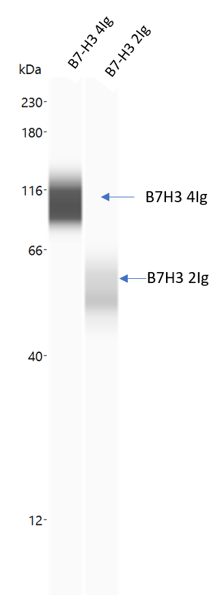
Application: Simple WesternSample Tested: b7h3 4ig protein and Purified proteinSpecies: HumanVerified Customer | Posted 11/02/2018

-
Application: Immunohistochemistry-FrozenSample Tested: See PMID 22080438Species: HumanVerified Customer | Posted 01/05/2015
There are no reviews that match your criteria.
Protocols
Find general support by application which include: protocols, troubleshooting, illustrated assays, videos and webinars.
- 7-Amino Actinomycin D (7-AAD) Cell Viability Flow Cytometry Protocol
- Antigen Retrieval Protocol (PIER)
- Antigen Retrieval for Frozen Sections Protocol
- Appropriate Fixation of IHC/ICC Samples
- Cellular Response to Hypoxia Protocols
- Chromogenic IHC Staining of Formalin-Fixed Paraffin-Embedded (FFPE) Tissue Protocol
- Chromogenic Immunohistochemistry Staining of Frozen Tissue
- ClariTSA™ Fluorophore Kits
- Detection & Visualization of Antibody Binding
- Extracellular Membrane Flow Cytometry Protocol
- Flow Cytometry Protocol for Cell Surface Markers
- Flow Cytometry Protocol for Staining Membrane Associated Proteins
- Flow Cytometry Staining Protocols
- Flow Cytometry Troubleshooting Guide
- Fluorescent IHC Staining of Frozen Tissue Protocol
- Graphic Protocol for Heat-induced Epitope Retrieval
- Graphic Protocol for the Preparation and Fluorescent IHC Staining of Frozen Tissue Sections
- Graphic Protocol for the Preparation and Fluorescent IHC Staining of Paraffin-embedded Tissue Sections
- Graphic Protocol for the Preparation of Gelatin-coated Slides for Histological Tissue Sections
- IHC Sample Preparation (Frozen sections vs Paraffin)
- Immunofluorescent IHC Staining of Formalin-Fixed Paraffin-Embedded (FFPE) Tissue Protocol
- Immunohistochemistry (IHC) and Immunocytochemistry (ICC) Protocols
- Immunohistochemistry Frozen Troubleshooting
- Immunohistochemistry Paraffin Troubleshooting
- Intracellular Flow Cytometry Protocol Using Alcohol (Methanol)
- Intracellular Flow Cytometry Protocol Using Detergents
- Intracellular Nuclear Staining Flow Cytometry Protocol Using Detergents
- Intracellular Staining Flow Cytometry Protocol Using Alcohol Permeabilization
- Intracellular Staining Flow Cytometry Protocol Using Detergents to Permeabilize Cells
- Preparing Samples for IHC/ICC Experiments
- Preventing Non-Specific Staining (Non-Specific Binding)
- Primary Antibody Selection & Optimization
- Propidium Iodide Cell Viability Flow Cytometry Protocol
- Protocol for Heat-Induced Epitope Retrieval (HIER)
- Protocol for Liperfluo
- Protocol for Making a 4% Formaldehyde Solution in PBS
- Protocol for VisUCyte™ HRP Polymer Detection Reagent
- Protocol for the Characterization of Human Th22 Cells
- Protocol for the Characterization of Human Th9 Cells
- Protocol for the Preparation & Fixation of Cells on Coverslips
- Protocol for the Preparation and Chromogenic IHC Staining of Frozen Tissue Sections
- Protocol for the Preparation and Chromogenic IHC Staining of Frozen Tissue Sections - Graphic
- Protocol for the Preparation and Chromogenic IHC Staining of Paraffin-embedded Tissue Sections
- Protocol for the Preparation and Chromogenic IHC Staining of Paraffin-embedded Tissue Sections - Graphic
- Protocol for the Preparation and Fluorescent IHC Staining of Frozen Tissue Sections
- Protocol for the Preparation and Fluorescent IHC Staining of Paraffin-embedded Tissue Sections
- Protocol for the Preparation of Gelatin-coated Slides for Histological Tissue Sections
- Protocol: Annexin V and PI Staining by Flow Cytometry
- Protocol: Annexin V and PI Staining for Apoptosis by Flow Cytometry
- R&D Systems Quality Control Western Blot Protocol
- TUNEL and Active Caspase-3 Detection by IHC/ICC Protocol
- The Importance of IHC/ICC Controls
- Troubleshooting Guide: Fluorokine Flow Cytometry Kits
- Troubleshooting Guide: Immunohistochemistry
- Troubleshooting Guide: Western Blot Figures
- Western Blot Conditions
- Western Blot Protocol
- Western Blot Protocol for Cell Lysates
- Western Blot Troubleshooting
- Western Blot Troubleshooting Guide
- View all Protocols, Troubleshooting, Illustrated assays and Webinars
Loading...