Human Caspase-9 Antibody (LAP6)
R&D Systems | Catalog # MAB8301

Key Product Details
Species Reactivity
Validated:
Human
Cited:
Human
Applications
Validated:
Immunohistochemistry, Western Blot, Simple Western
Cited:
Western Blot
Label
Unconjugated
Antibody Source
Monoclonal Mouse IgG1 Clone # LAP6
Loading...
Product Specifications
Immunogen
E. coli-derived recombinant human Caspase-9
aa 1-134
aa 1-134
Specificity
Detects human Caspase-9 in Western blots and captures Caspase-9 complexed with APAF-1.
Clonality
Monoclonal
Host
Mouse
Isotype
IgG1
Scientific Data Images for Human Caspase-9 Antibody (LAP6)
Capture of Human Caspase-9 and Human Caspase-9 complexed with APAF-1 detected by Western Blot.
Western blot shows Jurkat human acute T cell leukemia cell line lysates untreated (-) or treated (+) with 50 mM dATP and 1 mg/mL rat cytochrome c for 60 minutes, then captured on a 6-well dish coated at 10 µg/mL with Mouse Anti-Human Caspase-9 Monoclonal Antibody (Catalog # MAB8301). PVDF membrane was probed with 1 µg/mL of Mouse Anti-Human Caspase-9 Monoclonal Antibody (Catalog # MAB8301, left side) or Mouse Anti-Human APAF-1 Monoclonal Antibody (MAB868, right side) followed by HRP-conjugated Anti-Mouse IgG Secondary Antibody (HAF007). Specific bands were detected for Caspase-9 Precursor at approximately 46 kDa and the Caspase-9 p37 subunit at approximately 37 kDa (as indicated). A specific band was detected for APAF-1, captured as part of Caspase-9 complexed with APAF-1, at approximately 135 kDa (as indicated). This experiment was conducted under reducing conditions and using Immunoblot Buffer Group 4.
Detection of Human Caspase‑9 by Simple WesternTM.
Simple Western lane view shows lysates of Jurkat human acute T cell leukemia cell line untreated (-) or treated (+) with 1 mM Staurosporine (STS) for 3 hours, loaded at 0.2 mg/mL. A specific band was detected for Caspase‑9 at approximately 53 kDa (as indicated) using 20 µg/mL of Mouse Anti-Human Caspase‑9 Monoclonal Antibody (Catalog # MAB8301). This experiment was conducted under reducing conditions and using the 12-230 kDa separation system.Non-specific interaction with the 230 kDa Simple Western standard may be seen with this antibody.

Detection of Human Caspase‑9 by Western Blot.
Western blot shows lysates of HEK293T human embryonic kidney cell line and HepG2 human hepatocellular carcinoma cell line. PVDF membrane was probed with 1 µg/mL of Mouse Anti-Human Caspase‑9 Monoclonal Antibody (Catalog # MAB8301) followed by HRP-conjugated Anti-Mouse IgG Secondary Antibody (Catalog # HAF018). A specific band was detected for Caspase‑9 at approximately 46 kDa (as indicated). This experiment was conducted under reducing conditions and using Western Blot Buffer Group 4.
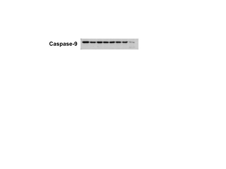
Detection of Human Caspase‑9 by Western Blot.
Western blot shows lysates of Jurkat human acute T cell leukemia cell line untreated (-) or treated (+) with 1 ug/ml Staurosporine (STS) for 2 hours. PVDF membrane was probed with 1 µg/mL of Mouse Anti-Human Caspase‑9 Monoclonal Antibody (Catalog # MAB8301) followed by HRP-conjugated Anti-Mouse IgG Secondary Antibody (Catalog # HAF018). A specific band was detected for Caspase‑9 at approximately 46, 37, 35 kDa (as indicated). This experiment was conducted under reducing conditions and using Western Blot Buffer Group 4.
Detection of Human Caspase-9 by Western Blot
Sorbitol-dependent TDP-43 relocalization is reversible and not dependent on Nup153 cleavage.(A) Western blot of nuclear pore complex protein Nup153, nuclear lamina component lamin B1, procaspases -3 and -9 and their activated forms caspase-3 and caspase-9 in HeLa cells treated with sorbitol after pre-treatment with DMSO control or caspase inhibitor Z-VAD-FMK. alpha -tubulin was used as a loading control. (B) Immunofluorescence staining of nuclear-cytoplasmic shuttling proteins TDP-43 (green) and HuR (red) in sorbitol-stressed HeLa cells after 0 min, 15 min or 60 min rescue in normal medium. Scale bar = 30 μm. (C) Quantification of cytoplasmic TDP-43 and HuR protein in sorbitol-treated HeLa cells after 0 min, 15 min or 60 min rescue (N = 3). Results represent mean percentage of cytoplasmic signal ± SEM;** p < 0.01, *** p < 0.001, **** p < 0.0001. Image collected and cropped by CiteAb from the following publication (https://pubmed.ncbi.nlm.nih.gov/28510586), licensed under a CC-BY license. Not internally tested by R&D Systems.
Detection of Human Human Caspase-9 Antibody by Western Blot
Sorbitol-dependent TDP-43 relocalization is reversible and not dependent on Nup153 cleavage.(A) Western blot of nuclear pore complex protein Nup153, nuclear lamina component lamin B1, procaspases -3 and -9 and their activated forms caspase-3 and caspase-9 in HeLa cells treated with sorbitol after pre-treatment with DMSO control or caspase inhibitor Z-VAD-FMK. alpha -tubulin was used as a loading control. (B) Immunofluorescence staining of nuclear-cytoplasmic shuttling proteins TDP-43 (green) and HuR (red) in sorbitol-stressed HeLa cells after 0 min, 15 min or 60 min rescue in normal medium. Scale bar = 30 μm. (C) Quantification of cytoplasmic TDP-43 and HuR protein in sorbitol-treated HeLa cells after 0 min, 15 min or 60 min rescue (N = 3). Results represent mean percentage of cytoplasmic signal ± SEM;** p < 0.01, *** p < 0.001, **** p < 0.0001. Image collected and cropped by CiteAb from the following publication (https://pubmed.ncbi.nlm.nih.gov/28510586), licensed under a CC-BY license. Not internally tested by R&D Systems.
Detection of Human Caspase-9 by Western Blot
Sorbitol-dependent TDP-43 relocalization is reversible and not dependent on Nup153 cleavage.(A) Western blot of nuclear pore complex protein Nup153, nuclear lamina component lamin B1, procaspases -3 and -9 and their activated forms caspase-3 and caspase-9 in HeLa cells treated with sorbitol after pre-treatment with DMSO control or caspase inhibitor Z-VAD-FMK. alpha -tubulin was used as a loading control. (B) Immunofluorescence staining of nuclear-cytoplasmic shuttling proteins TDP-43 (green) and HuR (red) in sorbitol-stressed HeLa cells after 0 min, 15 min or 60 min rescue in normal medium. Scale bar = 30 μm. (C) Quantification of cytoplasmic TDP-43 and HuR protein in sorbitol-treated HeLa cells after 0 min, 15 min or 60 min rescue (N = 3). Results represent mean percentage of cytoplasmic signal ± SEM;** p < 0.01, *** p < 0.001, **** p < 0.0001. Image collected and cropped by CiteAb from the following open publication (https://pubmed.ncbi.nlm.nih.gov/28510586), licensed under a CC-BY license. Not internally tested by R&D Systems.
Detection of Human Caspase-9 by Western Blot
Sorbitol-dependent TDP-43 relocalization is reversible and not dependent on Nup153 cleavage.(A) Western blot of nuclear pore complex protein Nup153, nuclear lamina component lamin B1, procaspases -3 and -9 and their activated forms caspase-3 and caspase-9 in HeLa cells treated with sorbitol after pre-treatment with DMSO control or caspase inhibitor Z-VAD-FMK. alpha -tubulin was used as a loading control. (B) Immunofluorescence staining of nuclear-cytoplasmic shuttling proteins TDP-43 (green) and HuR (red) in sorbitol-stressed HeLa cells after 0 min, 15 min or 60 min rescue in normal medium. Scale bar = 30 μm. (C) Quantification of cytoplasmic TDP-43 and HuR protein in sorbitol-treated HeLa cells after 0 min, 15 min or 60 min rescue (N = 3). Results represent mean percentage of cytoplasmic signal ± SEM;** p < 0.01, *** p < 0.001, **** p < 0.0001. Image collected and cropped by CiteAb from the following open publication (https://pubmed.ncbi.nlm.nih.gov/28510586), licensed under a CC-BY license. Not internally tested by R&D Systems.Applications for Human Caspase-9 Antibody (LAP6)
Application
Recommended Usage
Immunohistochemistry
8-25 µg/mL
Sample: Immersion fixed paraffin-embedded sections of human colon
Sample: Immersion fixed paraffin-embedded sections of human colon
Simple Western
20 µg/mL
Sample: Jurkat human acute T cell leukemia cell line treated with Staurosporine (STS)
Sample: Jurkat human acute T cell leukemia cell line treated with Staurosporine (STS)
Western Blot
1 µg/mL
Sample: Jurkat cells treated with staurosporine (STS), Jurkat cells treated with dATP and rat cytochrome c, HEK293T cells, HepG2 cells
Sample: Jurkat cells treated with staurosporine (STS), Jurkat cells treated with dATP and rat cytochrome c, HEK293T cells, HepG2 cells
Reviewed Applications
Read 1 review rated 5 using MAB8301 in the following applications:
Formulation, Preparation, and Storage
Purification
Protein A or G purified from hybridoma culture supernatant
Reconstitution
Reconstitute at 0.5 mg/mL in sterile PBS. For liquid material, refer to CoA for concentration.
Loading...
Formulation
Lyophilized from a 0.2 μm filtered solution in PBS with Trehalose. *Small pack size (SP) is supplied either lyophilized or as a 0.2 µm filtered solution in PBS.
Shipping
Lyophilized product is shipped at ambient temperature. Liquid small pack size (-SP) is shipped with polar packs. Upon receipt, store immediately at the temperature recommended below.
Stability & Storage
Use a manual defrost freezer and avoid repeated freeze-thaw cycles.
- 12 months from date of receipt, -20 to -70 °C as supplied.
- 1 month, 2 to 8 °C under sterile conditions after reconstitution.
- 6 months, -20 to -70 °C under sterile conditions after reconstitution.
Calculators
Background: Caspase-9
Additional Caspase-9 Products
Product Documents for Human Caspase-9 Antibody (LAP6)
Certificate of Analysis
To download a Certificate of Analysis, please enter a lot or batch number in the search box below.
Note: Certificate of Analysis not available for kit components.
Product Specific Notices for Human Caspase-9 Antibody (LAP6)
For research use only
Related Research Areas
Citations for Human Caspase-9 Antibody (LAP6)
 Powered by Bioz
Customer Reviews for Human Caspase-9 Antibody (LAP6) (1)
5 out of 5
1 Customer Rating
Have you used Human Caspase-9 Antibody (LAP6)?
Submit a review and receive an Amazon gift card!
$25/€18/£15/$25CAN/¥2500 Yen for a review with an image
$10/€7/£6/$10CAN/¥1110 Yen for a review without an image
Submit a review
Customer Images
Showing
1
-
1 of
1 review
Showing All
Filter By:
-
Application: Western BlotSample Tested: MDA-MB-231 human breast cancer cell lineSpecies: HumanVerified Customer | Posted 01/17/2018

There are no reviews that match your criteria.
Protocols
Find general support by application which include: protocols, troubleshooting, illustrated assays, videos and webinars.
- Antigen Retrieval Protocol (PIER)
- Antigen Retrieval for Frozen Sections Protocol
- Appropriate Fixation of IHC/ICC Samples
- Cellular Response to Hypoxia Protocols
- Chromogenic IHC Staining of Formalin-Fixed Paraffin-Embedded (FFPE) Tissue Protocol
- Chromogenic Immunohistochemistry Staining of Frozen Tissue
- ClariTSA™ Fluorophore Kits
- Detection & Visualization of Antibody Binding
- Fluorescent IHC Staining of Frozen Tissue Protocol
- Graphic Protocol for Heat-induced Epitope Retrieval
- Graphic Protocol for the Preparation and Fluorescent IHC Staining of Frozen Tissue Sections
- Graphic Protocol for the Preparation and Fluorescent IHC Staining of Paraffin-embedded Tissue Sections
- Graphic Protocol for the Preparation of Gelatin-coated Slides for Histological Tissue Sections
- IHC Sample Preparation (Frozen sections vs Paraffin)
- Immunofluorescent IHC Staining of Formalin-Fixed Paraffin-Embedded (FFPE) Tissue Protocol
- Immunohistochemistry (IHC) and Immunocytochemistry (ICC) Protocols
- Immunohistochemistry Frozen Troubleshooting
- Immunohistochemistry Paraffin Troubleshooting
- Preparing Samples for IHC/ICC Experiments
- Preventing Non-Specific Staining (Non-Specific Binding)
- Primary Antibody Selection & Optimization
- Protocol for Heat-Induced Epitope Retrieval (HIER)
- Protocol for Making a 4% Formaldehyde Solution in PBS
- Protocol for VisUCyte™ HRP Polymer Detection Reagent
- Protocol for the Preparation & Fixation of Cells on Coverslips
- Protocol for the Preparation and Chromogenic IHC Staining of Frozen Tissue Sections
- Protocol for the Preparation and Chromogenic IHC Staining of Frozen Tissue Sections - Graphic
- Protocol for the Preparation and Chromogenic IHC Staining of Paraffin-embedded Tissue Sections
- Protocol for the Preparation and Chromogenic IHC Staining of Paraffin-embedded Tissue Sections - Graphic
- Protocol for the Preparation and Fluorescent IHC Staining of Frozen Tissue Sections
- Protocol for the Preparation and Fluorescent IHC Staining of Paraffin-embedded Tissue Sections
- Protocol for the Preparation of Gelatin-coated Slides for Histological Tissue Sections
- R&D Systems Quality Control Western Blot Protocol
- TUNEL and Active Caspase-3 Detection by IHC/ICC Protocol
- The Importance of IHC/ICC Controls
- Troubleshooting Guide: Immunohistochemistry
- Troubleshooting Guide: Western Blot Figures
- Western Blot Conditions
- Western Blot Protocol
- Western Blot Protocol for Cell Lysates
- Western Blot Troubleshooting
- Western Blot Troubleshooting Guide
- View all Protocols, Troubleshooting, Illustrated assays and Webinars
Loading...
Associated Pathways

