CCL2, also known as monocyte chemotactic and activating factor (MCAF), was initially purified independently by two groups based on its ability to chemoattract monocytes. The human CCL2 cDNA encodes a 99 amino acid residue precursor protein with a 23 residue hydrophobic signal peptide that is cleaved to generate the 76 residue mature protein. Natural CCL2 is heterogeneous in size due to the addition of O-linked carbohydrates and sialic acid residues. In addition to fibroblasts; tumor cells, smooth muscle cells, endothelial cells, and mononuclear phagocytes can also produce CCL2 either constitutively or upon stimulation by various stimuli. CCL2 is a member of the beta (C-C) subfamily of chemokines. The existence of MCP-2 and MCP-3 with 62% and 73% amino acid identity respectively, to CCL2 have been reported. Consistent with it being a member of the chemokine beta family, CCL2 has been shown to chemoattract monocytes. In addition, it will also activate monocytes to be cytostatic for some human tumor cell lines; to increase cytosolic free calcium; to generate and release monocyte superoxide anions and to release monocyte lysosomal enzymes in vitro. CCL2 was reported to be capable of regulating adhesion molecule expression and cytokine production in human monocytes as well as chemoattracting, activating, and inducing histamine release from basophils. The biological roles played by CCL2 in a number of inflammatory and non-inflammatory disease states characterized by the accumulation of leukocytes at the site of the lesion, including atherosclerosis, delayed hypersensitivity reactions, etc., are being determined. CCL2 can bind to the C-C chemokine receptor-1 that also binds MIP-1 alpha, RANTES and MIP-1 beta. A specific receptor for CCL2 has also been cloned from THP-1 and MonoMac 6 cells.

Key Product Details
Species Reactivity
Validated:
Human
Cited:
Human, Rat, Primate - Macaca mulatta (Rhesus Macaque), Rabbit
Applications
Validated:
Immunohistochemistry, Western Blot, Neutralization
Cited:
Immunohistochemistry-Paraffin, Immunohistochemistry-Frozen, Western Blot, Neutralization, Immunocytochemistry, Bioassay, ELISA Detection
Label
Unconjugated
Antibody Source
Polyclonal Goat IgG
Loading...
Product Specifications
Immunogen
E. coli-derived recombinant human CCL2/JE/MCP-1
Gln24-Thr99
Accession # P13500
Gln24-Thr99
Accession # P13500
Specificity
Detects human CCL2/JE/MCP-1 in direct ELISAs and Western blots.
Clonality
Polyclonal
Host
Goat
Isotype
IgG
Endotoxin Level
<0.10 EU per 1 μg of the antibody by the LAL method.
Scientific Data Images for Human CCL2/JE/MCP-1 Antibody
Detection of Recombinant Human CCL2/JE/MCP‑1 by Western Blot.
Western blot shows 25 ng of Recombinant Human CCL2/JE/MCP-1 (Catalog # 279-MC), Recombinant Mouse CCL2/JE/MCP-1 (Catalog # 479-JE), Recombinant Rat CCL2/JE/MCP-1 (Catalog # 3144-JE), Recombinant Human CCL7/MCP-3/MARC (Catalog # 282-P3), Recombinant Human CCL11/Eotaxin (Catalog # 320-EO), Recombinant Human CCL8/MCP-2 (Catalog # 281-CP), and Recombinant Human CCL13/MCP-4 (Catalog # 327-P4). PVDF Membrane was probed with 0.1 µg/mL of Goat Anti-Human CCL2/JE/MCP-1 Antigen Affinity-purified Polyclonal Antibody (Catalog # AF-279-NA) followed by HRP-conjugated Anti-Goat IgG Secondary Antibody (Catalog # HAF109). A specific band was detected for CCL2/JE/MCP-1 at approximately 12 kDa (as indicated). This experiment was conducted under reducing conditions and using Immunoblot Buffer Group 3.
CCL2/JE/MCP‑1 in Human Crohn's Disease Intestine.
CCL2/JE/MCP-1 was detected in immersion fixed paraffin-embedded sections of human Crohn's disease intestine using Goat Anti-Human CCL2/JE/MCP-1 Antigen Affinity-purified Polyclonal Antibody (Catalog # AF-279-NA) at 25 µg/mL for overnight at 4° C temperature followed by incubation with the Anti-Goat IgG VisUCyte™ HRP Polymer Antibody (Catalog # VC004). Before incubation with the primary antibody, tissue was subjected to heat-induced epitope retrieval using Antigen Retrieval Reagent-Basic (Catalog # CTS013). Tissue was stained using DAB (brown) and counterstained with hematoxylin (blue). Specific staining was localized to epithelial cells. View our protocol for IHC Staining with VisUCyte HRP Polymer Detection Reagents.
Chemotaxis Induced by CCL2/MCP‑1 and Neutral-ization by Human CCL2/ MCP‑1 Antibody.
Recombinant Human CCL2/ MCP-1 (Catalog # 279-MC) chemoattracts the BaF3 mouse pro-B cell line transfected with human CCR2A in a dose-dependent manner (orange line). The amount of cells that migrated through to the lower chemotaxis chamber was measured by Resazurin (Catalog # AR002). Chemotaxis elicited by Recombinant Human CCL2/MCP-1 (150 ng/mL) is neutralized (green line) by increasing concentrations of Goat Anti-Human CCL2/MCP-1 Antigen Affinity-purified Polyclonal Antibody (Catalog # AF-279-NA). The ND50 is typically 1.5-4.5 µg/mL.
Human CCL2 / JE / MCP-1 ELISA Standard Curve
Recombinant Human CCL2/JE/MCP‑1 (Catalog # 279-MC) was serially diluted and captured by Mouse Anti-Human CCL2/JE/MCP‑1 Monoclonal Antibody (Catalog # MAB679) coated on a Clear Polystyrene Microplate (Catalog # DY990). Goat Anti-Human CCL2/JE/MCP‑1 Antigen Affinity-purified Polyclonal Antibody (Catalog # AF-279-NA) was biotinylated and incubated with the protein captured on the plate. Detection of the standard curve was achieved by incubating Streptavidin-HRP (Catalog # DY998)Applications for Human CCL2/JE/MCP-1 Antibody
Application
Recommended Usage
Immunohistochemistry
10-25 µg/mL
Sample: Immersion fixed paraffin-embedded sections of human Crohn's disease intestine
Sample: Immersion fixed paraffin-embedded sections of human Crohn's disease intestine
Western Blot
0.1 µg/mL
Sample: Recombinant Human CCL2/JE/MCP‑1 (Catalog # 279-MC)
Sample: Recombinant Human CCL2/JE/MCP‑1 (Catalog # 279-MC)
Neutralization
Measured by its ability to neutralize CCL2/JE/MCP‑1-induced chemotaxis in the BaF3 mouse pro‑B cell line transfected with human CCR2A. The Neutralization Dose (ND50) is typically 1.5-4.5 µg/mL in the presence of 150 ng/mL Recombinant Human CCL2/JE/MCP‑1.
Reviewed Applications
Read 4 reviews rated 4.8 using AF-279-NA in the following applications:
Formulation, Preparation, and Storage
Purification
Antigen Affinity-purified
Reconstitution
Reconstitute at 0.2 mg/mL in sterile PBS. For liquid material, refer to CoA for concentration.
Loading...
Formulation
Lyophilized from a 0.2 μm filtered solution in PBS with Trehalose. *Small pack size (SP) is supplied either lyophilized or as a 0.2 µm filtered solution in PBS.
Shipping
Lyophilized product is shipped at ambient temperature. Liquid small pack size (-SP) is shipped with polar packs. Upon receipt, store immediately at the temperature recommended below.
Stability & Storage
Use a manual defrost freezer and avoid repeated freeze-thaw cycles.
- 12 months from date of receipt, -20 to -70 °C as supplied.
- 1 month, 2 to 8 °C under sterile conditions after reconstitution.
- 6 months, -20 to -70 °C under sterile conditions after reconstitution.
Calculators
Background: CCL2/JE/MCP-1
Alternate Names
GDCF-2, HC11, HSMCR30, MCAF, MCP-1, SMC-CF
Gene Symbol
CCL2
UniProt
Additional CCL2/JE/MCP-1 Products
Product Documents for Human CCL2/JE/MCP-1 Antibody
Certificate of Analysis
To download a Certificate of Analysis, please enter a lot or batch number in the search box below.
Note: Certificate of Analysis not available for kit components.
Product Specific Notices for Human CCL2/JE/MCP-1 Antibody
For research use only
Citations for Human CCL2/JE/MCP-1 Antibody
 Powered by Bioz
Customer Reviews for Human CCL2/JE/MCP-1 Antibody (4)
4.8 out of 5
4 Customer Ratings
Have you used Human CCL2/JE/MCP-1 Antibody?
Submit a review and receive an Amazon gift card!
$25/€18/£15/$25CAN/¥2500 Yen for a review with an image
$10/€7/£6/$10CAN/¥1110 Yen for a review without an image
Submit a review
Customer Images
Showing
1
-
4 of
4 reviews
Showing All
Filter By:
-
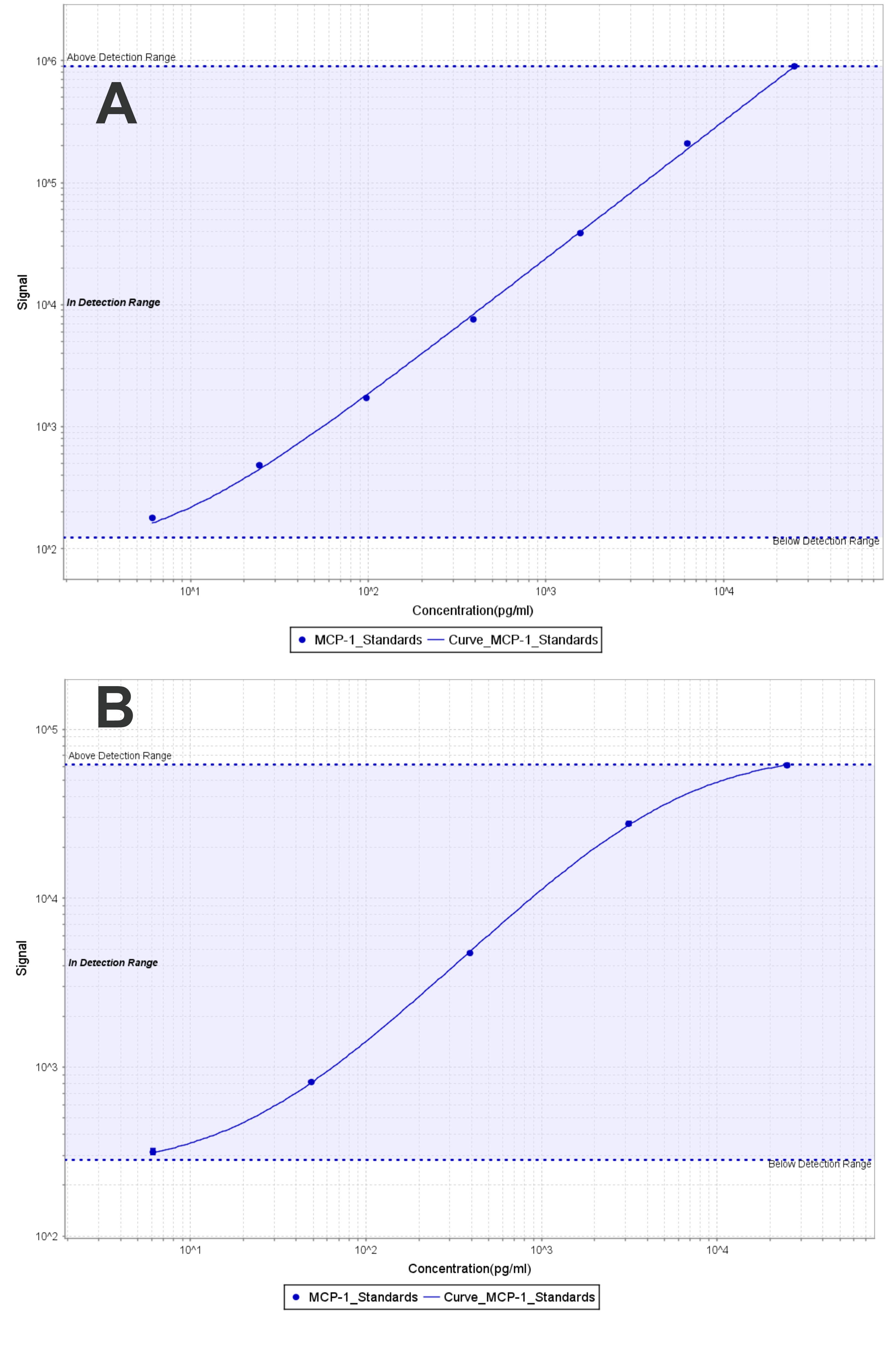
Application: MSD assaySample Tested: Vitreous humorSpecies: Cynomolgus Monkey and PorcineVerified Customer | Posted 11/20/2018After labeling with Sulfo-Tag, used as a detection reagent in MSD assay (Meso Scale Diagnostics LLC). A: Paired with biotinylated anti-human CCL2 antibody (MAB279) as a capture reagent. Calibration curve with Recombinant Human CCL2 (279-MC-010/CF) is shown (dynamic range 6-25,000 pg/ml) B: Paired with biotinylated anti-porcine CCL2 antibody (Abcam, Cat# ab193793). Calibration curve with recombinant swine CCL2 (ImmunoChemistry Technologies, Cat# 6374) is shown (dynamic range 6-25,000 pg/ml).

-
Application: Western BlotSample Tested: CHO Chinese hamster ovary cell lineSpecies: HamsterVerified Customer | Posted 12/13/2017
-
Application: ELISASample Tested: EDTA PlasmaSpecies: HumanVerified Customer | Posted 12/06/2017
-
Application: Block/NeutralizeSample Tested: NHDF human normal dermal fibroblastsSpecies: HumanVerified Customer | Posted 05/10/2017
There are no reviews that match your criteria.
Protocols
Find general support by application which include: protocols, troubleshooting, illustrated assays, videos and webinars.
- Antigen Retrieval Protocol (PIER)
- Antigen Retrieval for Frozen Sections Protocol
- Appropriate Fixation of IHC/ICC Samples
- Cellular Response to Hypoxia Protocols
- Chromogenic IHC Staining of Formalin-Fixed Paraffin-Embedded (FFPE) Tissue Protocol
- Chromogenic Immunohistochemistry Staining of Frozen Tissue
- ClariTSA™ Fluorophore Kits
- Detection & Visualization of Antibody Binding
- Fluorescent IHC Staining of Frozen Tissue Protocol
- Graphic Protocol for Heat-induced Epitope Retrieval
- Graphic Protocol for the Preparation and Fluorescent IHC Staining of Frozen Tissue Sections
- Graphic Protocol for the Preparation and Fluorescent IHC Staining of Paraffin-embedded Tissue Sections
- Graphic Protocol for the Preparation of Gelatin-coated Slides for Histological Tissue Sections
- IHC Sample Preparation (Frozen sections vs Paraffin)
- Immunofluorescent IHC Staining of Formalin-Fixed Paraffin-Embedded (FFPE) Tissue Protocol
- Immunohistochemistry (IHC) and Immunocytochemistry (ICC) Protocols
- Immunohistochemistry Frozen Troubleshooting
- Immunohistochemistry Paraffin Troubleshooting
- Preparing Samples for IHC/ICC Experiments
- Preventing Non-Specific Staining (Non-Specific Binding)
- Primary Antibody Selection & Optimization
- Protocol for Heat-Induced Epitope Retrieval (HIER)
- Protocol for Making a 4% Formaldehyde Solution in PBS
- Protocol for VisUCyte™ HRP Polymer Detection Reagent
- Protocol for the Preparation & Fixation of Cells on Coverslips
- Protocol for the Preparation and Chromogenic IHC Staining of Frozen Tissue Sections
- Protocol for the Preparation and Chromogenic IHC Staining of Frozen Tissue Sections - Graphic
- Protocol for the Preparation and Chromogenic IHC Staining of Paraffin-embedded Tissue Sections
- Protocol for the Preparation and Chromogenic IHC Staining of Paraffin-embedded Tissue Sections - Graphic
- Protocol for the Preparation and Fluorescent IHC Staining of Frozen Tissue Sections
- Protocol for the Preparation and Fluorescent IHC Staining of Paraffin-embedded Tissue Sections
- Protocol for the Preparation of Gelatin-coated Slides for Histological Tissue Sections
- R&D Systems Quality Control Western Blot Protocol
- TUNEL and Active Caspase-3 Detection by IHC/ICC Protocol
- The Importance of IHC/ICC Controls
- Troubleshooting Guide: Immunohistochemistry
- Troubleshooting Guide: Western Blot Figures
- Western Blot Conditions
- Western Blot Protocol
- Western Blot Protocol for Cell Lysates
- Western Blot Troubleshooting
- Western Blot Troubleshooting Guide
- View all Protocols, Troubleshooting, Illustrated assays and Webinars


