CD155 [also known as PVR (poliovirus receptor) and Necl-5 (nectin-like molecule-5)] is a 70 kDa type I transmembrane (TM) glycoprotein that is a member of the nectin-like (Necl) family of nectin-related molecules (1). Like nectins, Necl molecules are Ig superfamily members that contain three Ig-like extracellular domains, a TM segment, and a cytoplasmic tail. Unlike nectins, Necl molecules cannot interact with cytoplasmic afadin (1). While Nectins serve as cell adhesion molecules, the actual functions of most Necls are yet-to-be determined. CD155/PVR was originally isolated based on its ability to mediate polio virus attachment to host cells (2, 3). The full-length (or CD155 alpha isoform) is synthesized as a 417 amino acid (aa) precursor that contains a 20 aa signal sequence, a 323 aa extracellular region, a 24 aa TM segment and a 50 aa cytoplasmic tail. The extracellular region contains one N-terminal V-type and two C2-type Ig-like domains (2, 3). The V-type domain mediates polio virus binding (4). Three other isoforms exist, all of which retain the Ig-like domains. CD155 delta is transmembrane with a shortened cytoplasmic tail of 25 aa. CD155 beta (352 aa) and CD155 gamma (344 aa) are 60‑65 kDa soluble forms that show removal of the TM segment and surrounding amino acids (2, 5). The soluble forms will bind the polio virus (due to the presence of the V-type Ig domain) but afford no protection against polio infection because of low circulating levels (5). CD155 has been demonstrated to bind vitronectin, nectin-3, and DNAM-1 (6‑8). DNAM-1 binding promotes monocyte migration and NK cell killing. CD155 is expressed in all normal tissues and is highly expressed in tumor cells of epithelial and neuronal origin.
Human CD155/PVR Antibody (300907)
R&D Systems | Catalog # MAB25301

Key Product Details
Species Reactivity
Validated:
Human
Cited:
Human
Applications
Validated:
Western Blot, Flow Cytometry, Immunocytochemistry, CyTOF-ready
Cited:
Immunohistochemistry, Western Blot, Flow Cytometry
Label
Unconjugated
Antibody Source
Monoclonal Mouse IgG1 Clone # 300907
Loading...
Product Specifications
Immunogen
Mouse myeloma cell line NS0-derived recombinant human CD155/PVR
Gly27-Asn343
Accession # AAH15542
Gly27-Asn343
Accession # AAH15542
Specificity
Detects human CD155/PVR in direct ELISAs and Western blots.
Clonality
Monoclonal
Host
Mouse
Isotype
IgG1
Scientific Data Images for Human CD155/PVR Antibody (300907)
Detection of CD155/PVR in U937 Human Cell Line by Flow Cytometry.
U937 human histiocytic lymphoma cell line was stained with Mouse Anti-Human CD155/PVR Monoclonal Antibody (Catalog # MAB25301, filled histogram) or isotype control antibody (Catalog # MAB002, open histogram), followed by Phycoerythrin-conjugated Anti-Mouse IgG Secondary Antibody (Catalog # F0102B). View our protocol for Staining Membrane-associated Proteins.
Detection of CD155/PVR in HUVEC Human Cells by Flow Cytometry.
HUVEC human umbilical vein endothelial cells were stained with Mouse Anti-Human CD155/PVR Monoclonal Antibody (Catalog # MAB25301, filled histogram) or isotype control antibody (Catalog # MAB002, open histogram), followed by Allophycocyanin-conjugated Anti-Mouse IgG Secondary Antibody (Catalog # F0101B). View our protocol for Staining Membrane-associated Proteins.
CD155/PVR in Human PBMCs.
CD155/PVR was detected in immersion fixed human peripheral blood mononuclear cells (PBMCs) using Mouse Anti-Human CD155/PVR Monoclonal Antibody (Catalog # MAB25301) at 15 µg/mL for 3 hours at room temperature. Cells were stained using the NorthernLights™ 557-conjugated Anti-Mouse IgG Secondary Antibody (red; Catalog # NL007) and counterstained with DAPI (blue). Specific staining was localized to cytoplasm and plasma membrane. View our protocol for Fluorescent ICC Staining of Non-adherent Cells.Applications for Human CD155/PVR Antibody (300907)
Application
Recommended Usage
CyTOF-ready
Ready to be labeled using established conjugation methods. No BSA or other carrier proteins that could interfere with conjugation.
Flow Cytometry
0.25 µg/106 cells
Sample: U937 human histiocytic lymphoma cell line and HUVEC human umbilical vein endothelial cells
Sample: U937 human histiocytic lymphoma cell line and HUVEC human umbilical vein endothelial cells
Immunocytochemistry
8-25 µg/mL
Sample: Immersion fixed human peripheral blood mononuclear cells (PBMCs)
Sample: Immersion fixed human peripheral blood mononuclear cells (PBMCs)
Western Blot
1 µg/mL
Sample: Recombinant Human CD155/PVR (Catalog # 2530-CD)
Sample: Recombinant Human CD155/PVR (Catalog # 2530-CD)
Reviewed Applications
Read 3 reviews rated 4.7 using MAB25301 in the following applications:
Flow Cytometry Panel Builder
Bio-Techne Knows Flow Cytometry
Save time and reduce costly mistakes by quickly finding compatible reagents using the Panel Builder Tool.
Advanced Features
- Spectra Viewer - Custom analysis of spectra from multiple fluorochromes
- Spillover Popups - Visualize the spectra of individual fluorochromes
- Antigen Density Selector - Match fluorochrome brightness with antigen density

Formulation, Preparation, and Storage
Purification
Protein A or G purified from hybridoma culture supernatant
Reconstitution
Reconstitute at 0.5 mg/mL in sterile PBS. For liquid material, refer to CoA for concentration.
Loading...
Formulation
Lyophilized from a 0.2 μm filtered solution in PBS with Trehalose. *Small pack size (SP) is supplied either lyophilized or as a 0.2 µm filtered solution in PBS.
Shipping
Lyophilized product is shipped at ambient temperature. Liquid small pack size (-SP) is shipped with polar packs. Upon receipt, store immediately at the temperature recommended below.
Stability & Storage
Use a manual defrost freezer and avoid repeated freeze-thaw cycles.
- 12 months from date of receipt, -20 to -70 °C as supplied.
- 1 month, 2 to 8 °C under sterile conditions after reconstitution.
- 6 months, -20 to -70 °C under sterile conditions after reconstitution.
Calculators
Background: CD155/PVR
References
- Takai, Y. et al. (2003) Cancer Sci. 94:655.
- Mendelsohn, C.L. et al. (1989) Cell 56:855.
- Koike, H. et al. (1990) EMBO J. 9:3217.
- Koike, S. et al. (1991) Proc. Natl. Acad. Sci. USA 88:4104.
- Baury, B. et al. (2003) Biochem. Biophys. Res. Commun. 309:175.
- Mueller, S. and E. Wimmer (2003) J. Biol. Chem. 278:31251.
- Reymond, N. et al. (2004) J. Exp. Med. 199:1331.
- Lange, R. et al. (2001) Virology 285:218.
Long Name
Poliovirus Receptor
Alternate Names
CD155, HVED, Necl-5, PVR, PVS
Entrez Gene IDs
Gene Symbol
PVR
UniProt
Additional CD155/PVR Products
Product Documents for Human CD155/PVR Antibody (300907)
Certificate of Analysis
To download a Certificate of Analysis, please enter a lot or batch number in the search box below.
Note: Certificate of Analysis not available for kit components.
Product Specific Notices for Human CD155/PVR Antibody (300907)
For research use only
Citations for Human CD155/PVR Antibody (300907)
 Powered by Bioz
Customer Reviews for Human CD155/PVR Antibody (300907) (3)
4.7 out of 5
3 Customer Ratings
Have you used Human CD155/PVR Antibody (300907)?
Submit a review and receive an Amazon gift card!
$25/€18/£15/$25CAN/¥2500 Yen for a review with an image
$10/€7/£6/$10CAN/¥1110 Yen for a review without an image
Submit a review
Customer Images
Showing
1
-
3 of
3 reviews
Showing All
Filter By:
-
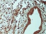
Application: ImmunohistochemistrySample Tested: Pancreatic cancer tissueSpecies: HumanVerified Customer | Posted 10/14/2021

-
Application: ELISASample Tested: Aortic smooth muscle cellsSpecies: HumanVerified Customer | Posted 12/21/2018
-
Application: Western BlotSample Tested: See PMID 23933496Species: HumanVerified Customer | Posted 02/19/2015
There are no reviews that match your criteria.
Protocols
Find general support by application which include: protocols, troubleshooting, illustrated assays, videos and webinars.
- 7-Amino Actinomycin D (7-AAD) Cell Viability Flow Cytometry Protocol
- Appropriate Fixation of IHC/ICC Samples
- Cellular Response to Hypoxia Protocols
- ClariTSA™ Fluorophore Kits
- Detection & Visualization of Antibody Binding
- Extracellular Membrane Flow Cytometry Protocol
- Flow Cytometry Protocol for Cell Surface Markers
- Flow Cytometry Protocol for Staining Membrane Associated Proteins
- Flow Cytometry Staining Protocols
- Flow Cytometry Troubleshooting Guide
- ICC Cell Smear Protocol for Suspension Cells
- ICC Immunocytochemistry Protocol Videos
- ICC for Adherent Cells
- Immunocytochemistry (ICC) Protocol
- Immunocytochemistry Troubleshooting
- Immunofluorescence of Organoids Embedded in Cultrex Basement Membrane Extract
- Immunohistochemistry (IHC) and Immunocytochemistry (ICC) Protocols
- Intracellular Flow Cytometry Protocol Using Alcohol (Methanol)
- Intracellular Flow Cytometry Protocol Using Detergents
- Intracellular Nuclear Staining Flow Cytometry Protocol Using Detergents
- Intracellular Staining Flow Cytometry Protocol Using Alcohol Permeabilization
- Intracellular Staining Flow Cytometry Protocol Using Detergents to Permeabilize Cells
- Preparing Samples for IHC/ICC Experiments
- Preventing Non-Specific Staining (Non-Specific Binding)
- Primary Antibody Selection & Optimization
- Propidium Iodide Cell Viability Flow Cytometry Protocol
- Protocol for Liperfluo
- Protocol for VisUCyte™ HRP Polymer Detection Reagent
- Protocol for the Characterization of Human Th22 Cells
- Protocol for the Characterization of Human Th9 Cells
- Protocol for the Fluorescent ICC Staining of Cell Smears - Graphic
- Protocol for the Fluorescent ICC Staining of Cultured Cells on Coverslips - Graphic
- Protocol for the Preparation and Fluorescent ICC Staining of Cells on Coverslips
- Protocol for the Preparation and Fluorescent ICC Staining of Non-adherent Cells
- Protocol for the Preparation and Fluorescent ICC Staining of Stem Cells on Coverslips
- Protocol for the Preparation of a Cell Smear for Non-adherent Cell ICC - Graphic
- Protocol: Annexin V and PI Staining by Flow Cytometry
- Protocol: Annexin V and PI Staining for Apoptosis by Flow Cytometry
- R&D Systems Quality Control Western Blot Protocol
- TUNEL and Active Caspase-3 Detection by IHC/ICC Protocol
- The Importance of IHC/ICC Controls
- Troubleshooting Guide: Fluorokine Flow Cytometry Kits
- Troubleshooting Guide: Western Blot Figures
- Western Blot Conditions
- Western Blot Protocol
- Western Blot Protocol for Cell Lysates
- Western Blot Troubleshooting
- Western Blot Troubleshooting Guide
- View all Protocols, Troubleshooting, Illustrated assays and Webinars
Loading...