CD4 is an approximately 55 kDa type I membrane glycoprotein that is expressed predominantly on most thymocytes and a subset of mature T lymphocytes. In humans, CD4 is also expressed to a lesser extent on monocytes and macrophage related cells. Human CD4 cDNA encodes a 458 amino acid (aa) precursor protein with a 25 aa signal peptide, a 371 aa extracellular region containing four immunoglobulin homology domains, a 24 aa transmembrane domain and a 38 aa cytoplasmic domain. CD4 is a coreceptor required for T cell recognition of antigens that are presented by class II major histocompatibility complexes. CD4 has been shown to be a coreceptor of HIV entry and specifically binds gp120, the external envelope glycoprotein of HIV.

Key Product Details
Species Reactivity
Validated:
Human
Cited:
Human
Applications
Validated:
Flow Cytometry, Immunocytochemistry, CyTOF-ready
Cited:
Immunohistochemistry, Immunohistochemistry-Paraffin, Immunohistochemistry-Frozen, Western Blot, Flow Cytometry, Immunocytochemistry, Dot Blot, ELISA Capture
Label
Unconjugated
Antibody Source
Monoclonal Mouse IgG1 Clone # 34930
Loading...
Product Specifications
Immunogen
S. frugiperda insect ovarian cell line Sf 21-derived recombinant human CD4
Extracellular domain
Extracellular domain
Specificity
Detects human CD4 in direct ELISAs and Western blots. In direct ELISAs, less than 1% cross-reactivity with recombinant mouse CD4 is observed.
Clonality
Monoclonal
Host
Mouse
Isotype
IgG1
Scientific Data Images for Human CD4 Antibody
CD4 in Human PBMCs.
CD4 was detected in immersion fixed human peripheral blood mononuclear cells (PBMCs) using Mouse Anti-Human CD4 Monoclonal Antibody (Catalog # MAB379) at 10 µg/mL for 3 hours at room temperature. Cells were stained using the NorthernLights™ 493-conjugated Anti-Mouse IgG Secondary Antibody (green; Catalog # NL009) and counterstained with DAPI(blue). Specific staining was localized to the cell surface. View our protocol for Fluorescent ICC Staining of Non-adherent Cells.
Detection of Human CD4 by Immunocytochemistry/Immunofluorescence
CX3CR1 expressions in affected muscle of patients with polymyositis. The muscle tissues of polymyositis (PM) patients were double-stained with CD4, CD8, or CD68, as well as CX3CR1, and were analyzed with fluorescent microscopy: (A) CX3CR1, (B) CD4, (C) merged (A) and (B), (D) CX3CR1, (E) CD8, (F) merged (D) and (E), (G) CX3CR1, (H) CD68, and (I) merged (G) and (H). Arrows indicate double-positive cells. Original magnification, ×400. Image collected and cropped by CiteAb from the following publication (https://arthritis-research.biomedcentral.com/articles/10.1186/ar3761), licensed under a CC-BY license. Not internally tested by R&D Systems.
Detection of Human CD4 by Immunohistochemistry
CX3CR1 expressions in affected muscle of patients with polymyositis. The muscle tissues of polymyositis (PM) patients were double-stained with CD4, CD8, or CD68, as well as CX3CR1, and were analyzed with fluorescent microscopy: (A) CX3CR1, (B) CD4, (C) merged (A) and (B), (D) CX3CR1, (E) CD8, (F) merged (D) and (E), (G) CX3CR1, (H) CD68, and (I) merged (G) and (H). Arrows indicate double-positive cells. Original magnification, ×400. Image collected and cropped by CiteAb from the following open publication (https://pubmed.ncbi.nlm.nih.gov/22394569), licensed under a CC-BY license. Not internally tested by R&D Systems.
Detection of Human CD4 by Immunohistochemistry
CX3CR1 expressions in affected muscle of patients with polymyositis. The muscle tissues of polymyositis (PM) patients were double-stained with CD4, CD8, or CD68, as well as CX3CR1, and were analyzed with fluorescent microscopy: (A) CX3CR1, (B) CD4, (C) merged (A) and (B), (D) CX3CR1, (E) CD8, (F) merged (D) and (E), (G) CX3CR1, (H) CD68, and (I) merged (G) and (H). Arrows indicate double-positive cells. Original magnification, ×400. Image collected and cropped by CiteAb from the following open publication (https://pubmed.ncbi.nlm.nih.gov/22394569), licensed under a CC-BY license. Not internally tested by R&D Systems.Applications for Human CD4 Antibody
Application
Recommended Usage
CyTOF-ready
Ready to be labeled using established conjugation methods. No BSA or other carrier proteins that could interfere with conjugation.
Flow Cytometry
2.5 µg/106 cells
Sample: Human whole blood lymphocytes
Sample: Human whole blood lymphocytes
Immunocytochemistry
8-25 µg/mL
Sample: Immersion fixed human peripheral blood mononuclear cells
Sample: Immersion fixed human peripheral blood mononuclear cells
Reviewed Applications
Read 2 reviews rated 4 using MAB379 in the following applications:
Flow Cytometry Panel Builder
Bio-Techne Knows Flow Cytometry
Save time and reduce costly mistakes by quickly finding compatible reagents using the Panel Builder Tool.
Advanced Features
- Spectra Viewer - Custom analysis of spectra from multiple fluorochromes
- Spillover Popups - Visualize the spectra of individual fluorochromes
- Antigen Density Selector - Match fluorochrome brightness with antigen density

Formulation, Preparation, and Storage
Purification
Protein A or G purified from ascites
Reconstitution
Reconstitute at 0.5 mg/mL in sterile PBS. For liquid material, refer to CoA for concentration.
Loading...
Formulation
Lyophilized from a 0.2 μm filtered solution in PBS with Trehalose. *Small pack size (SP) is supplied either lyophilized or as a 0.2 µm filtered solution in PBS.
Shipping
Lyophilized product is shipped at ambient temperature. Liquid small pack size (-SP) is shipped with polar packs. Upon receipt, store immediately at the temperature recommended below.
Stability & Storage
Use a manual defrost freezer and avoid repeated freeze-thaw cycles.
- 12 months from date of receipt, -20 to -70 °C as supplied.
- 1 month, 2 to 8 °C under sterile conditions after reconstitution.
- 6 months, -20 to -70 °C under sterile conditions after reconstitution.
Calculators
Background: CD4
References
- Capon, D.I. et al. (1991) Annu. Rev. Immunol. 9:649.
Alternate Names
CD4
Entrez Gene IDs
Gene Symbol
CD4
Additional CD4 Products
Product Documents for Human CD4 Antibody
Certificate of Analysis
To download a Certificate of Analysis, please enter a lot or batch number in the search box below.
Note: Certificate of Analysis not available for kit components.
Product Specific Notices for Human CD4 Antibody
For research use only
Citations for Human CD4 Antibody
 Powered by Bioz
Customer Reviews for Human CD4 Antibody (2)
4 out of 5
2 Customer Ratings
Have you used Human CD4 Antibody?
Submit a review and receive an Amazon gift card!
$25/€18/£15/$25CAN/¥2500 Yen for a review with an image
$10/€7/£6/$10CAN/¥1110 Yen for a review without an image
Submit a review
Customer Images
Showing
1
-
2 of
2 reviews
Showing All
Filter By:
-
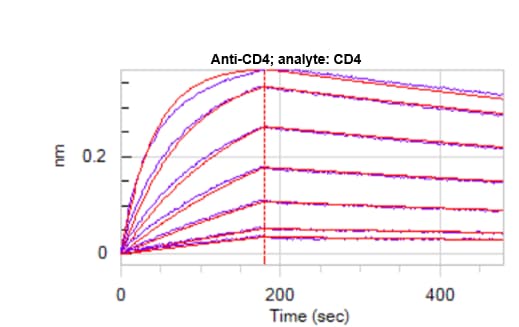
Application: Octet binding assaySample Tested: Recombinant proteinSpecies: HumanVerified Customer | Posted 02/08/2021Immobilize Anti-CD4, flow dose response of CD4 (200-3nM. KD ~5nM

-
Application: Immunocytochemistry/ImmunofluorescenceSample Tested: activated mouse CD8 T cellSpecies: HumanVerified Customer | Posted 01/22/2018
There are no reviews that match your criteria.
Protocols
Find general support by application which include: protocols, troubleshooting, illustrated assays, videos and webinars.
- 7-Amino Actinomycin D (7-AAD) Cell Viability Flow Cytometry Protocol
- Appropriate Fixation of IHC/ICC Samples
- Cellular Response to Hypoxia Protocols
- ClariTSA™ Fluorophore Kits
- Detection & Visualization of Antibody Binding
- Extracellular Membrane Flow Cytometry Protocol
- Flow Cytometry Protocol for Cell Surface Markers
- Flow Cytometry Protocol for Staining Membrane Associated Proteins
- Flow Cytometry Staining Protocols
- Flow Cytometry Troubleshooting Guide
- ICC Cell Smear Protocol for Suspension Cells
- ICC Immunocytochemistry Protocol Videos
- ICC for Adherent Cells
- Immunocytochemistry (ICC) Protocol
- Immunocytochemistry Troubleshooting
- Immunofluorescence of Organoids Embedded in Cultrex Basement Membrane Extract
- Immunohistochemistry (IHC) and Immunocytochemistry (ICC) Protocols
- Intracellular Flow Cytometry Protocol Using Alcohol (Methanol)
- Intracellular Flow Cytometry Protocol Using Detergents
- Intracellular Nuclear Staining Flow Cytometry Protocol Using Detergents
- Intracellular Staining Flow Cytometry Protocol Using Alcohol Permeabilization
- Intracellular Staining Flow Cytometry Protocol Using Detergents to Permeabilize Cells
- Preparing Samples for IHC/ICC Experiments
- Preventing Non-Specific Staining (Non-Specific Binding)
- Primary Antibody Selection & Optimization
- Propidium Iodide Cell Viability Flow Cytometry Protocol
- Protocol for Liperfluo
- Protocol for VisUCyte™ HRP Polymer Detection Reagent
- Protocol for the Characterization of Human Th22 Cells
- Protocol for the Characterization of Human Th9 Cells
- Protocol for the Fluorescent ICC Staining of Cell Smears - Graphic
- Protocol for the Fluorescent ICC Staining of Cultured Cells on Coverslips - Graphic
- Protocol for the Preparation and Fluorescent ICC Staining of Cells on Coverslips
- Protocol for the Preparation and Fluorescent ICC Staining of Non-adherent Cells
- Protocol for the Preparation and Fluorescent ICC Staining of Stem Cells on Coverslips
- Protocol for the Preparation of a Cell Smear for Non-adherent Cell ICC - Graphic
- Protocol: Annexin V and PI Staining by Flow Cytometry
- Protocol: Annexin V and PI Staining for Apoptosis by Flow Cytometry
- TUNEL and Active Caspase-3 Detection by IHC/ICC Protocol
- The Importance of IHC/ICC Controls
- Troubleshooting Guide: Fluorokine Flow Cytometry Kits
- View all Protocols, Troubleshooting, Illustrated assays and Webinars


