CD97 is a 95‑100 kDa member of a protein group known as the LNB-TM7 protein family that evolved from genes of the secretin receptor superfamily (1‑3). Molecules in this family are unique hybrid structures consisting of EGF-like modules coupled to class B G-protein 7-transmembrane (TM) domains by a glycosylated (mucin) stalk. Human CD97 is synthesized as an 835 amino acid (aa) precursor that contains a 20 aa signal sequence, a 532 aa extracellular domain (ECD), a 238 aa “transmembrane” region that includes seven TM segments, and a 45 aa cytoplasmic tail (4). Within the 532 aa ECD, the first 236 aa contains five EGF-like domains, the C-terminal four of which bind calcium, and a juxtamembrane 296 aa RGD-containing mucin stalk (4, 5). The stalk is both glycosylated and proteolytically cleaved (at aa 530) to create a noncovalently linked 65‑70 kDa glycosylated extracellular alpha -subunit and a 28 kDa 7-TM membrane-bound beta -subunit (4). There are two known alternate splice forms in human. Isoform # 1 contains four EGF-like domains (domain # 1, 2, 3 and 5), while isoform # 2 contains three EGF-like domains (domain # 1, 2 and 5) (1, 4, 6). The ECD in isoform 1 is 60 kDa in size, while the ECD in isoform 2 is 55 kDa in size (native molecular weight). The five EGF-like domain region in human is 55% aa identical to that in mouse. Cells known to express CD97 include monocytes, macrophages, T cells, select B cells, dendritic cells and, potentially, vascular and visceral smooth muscle cells (1, 7). There are at least two ligands for CD97. One is chrondroitin sulfate that binds only to the full-length (five domain) form of CD97. Binding is dependent on the presence of EGF-like domain # 4 (3). The second ligand for CD97 is CD55, a GPI-linked cell surface molecule with short consensus repeats that regulates complement activation on cell surfaces (1, 5, 7). CD97 EGF-like domains # 1 and 2 bind CD55 while domain # 5 stabilizes the CD97 molecule. The shortest CD97 isoform shows the strongest binding to CD55 (7, 8).

Key Product Details
Species Reactivity
Validated:
Human
Cited:
Human
Applications
Validated:
Western Blot, Neutralization, Flow Cytometry, CyTOF-ready
Cited:
Western Blot
Label
Unconjugated
Antibody Source
Monoclonal Mouse IgG2A Clone # 380903
Loading...
Product Specifications
Immunogen
Mouse myeloma cell line NS0-derived recombinant human CD97
Gln21-Gln398
Accession # NP_001775.2
Gln21-Gln398
Accession # NP_001775.2
Specificity
Detects human CD97 in direct ELISAs and Western blots. In direct ELISAs and Western blots, no cross‑reactivity with recombinant mouse CD97 is observed.
Clonality
Monoclonal
Host
Mouse
Isotype
IgG2A
Endotoxin Level
<0.10 EU per 1 μg of the antibody by the LAL method.
Scientific Data Images for Human CD97 Antibody (380903)
Cell Adhesion Mediated by CD97 and Neutralization by Human CD97 Antibody.
Recombinant Human CD97 (Catalog # 2529-CD), immobil-ized onto a microplate, supports the adhesion of human red blood cells in a dose-dependent manner (orange line). Adhesion elicited by Recombinant Human CD97 (4 µg/mL) is neutralized (green line) by increasing concentrations of Mouse Anti-Human CD97 Monoclonal Antibody (Catalog # MAB2529). The ND50 is typically 0.5-2.5 µg/mL.
Detection of CD97 in Human Whole Blood by Flow Cytometry.
Human whole blood was stained with Mouse Anti-Human CD97 Monoclonal Antibody (Catalog # MAB2529, filled histogram) or isotype control antibody (Catalog # MAB0031, open histogram), followed by Phy-coerythrin-conjugated Anti-Mouse IgG F(ab')2Secondary Antibody (Catalog # F0102B).
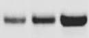
Detection of CD97 by Western Blot
Immunoblot image are shown (A), along with densitometric analysis of (B) PDGFR beta, (C) Cyr61, (D) CD97, (E) Glypican-1, (F) MUC18/CD146 and (G) uPAR. (H) A summary of proteins detected by mass spectrometry and immunoblot. Graphs represent mean ± SEM of n = 3 independent biological experiments, shown as 1,2,3. Unpaired t-test was used as statistical test. *P < 0.05, **P < 0.01, ***P < 0.001 Image collected and cropped by CiteAb from the following open publication (https://pubmed.ncbi.nlm.nih.gov/36171564), licensed under a CC-BY license. Not internally tested by R&D Systems.
Detection of CD97 by Western Blot
Immunoblot image are shown (A), along with densitometric analysis of (B) PDGFR beta, (C) Cyr61, (D) CD97, (E) Glypican-1, (F) MUC18/CD146 and (G) uPAR. (H) A summary of proteins detected by mass spectrometry and immunoblot. Graphs represent mean ± SEM of n = 3 independent biological experiments, shown as 1,2,3. Unpaired t-test was used as statistical test. *P < 0.05, **P < 0.01, ***P < 0.001 Image collected and cropped by CiteAb from the following open publication (https://pubmed.ncbi.nlm.nih.gov/36171564), licensed under a CC-BY license. Not internally tested by R&D Systems.Applications for Human CD97 Antibody (380903)
Application
Recommended Usage
CyTOF-ready
Ready to be labeled using established conjugation methods. No BSA or other carrier proteins that could interfere with conjugation.
Flow Cytometry
2.5 µg/106 cells
Sample: Human whole blood
Sample: Human whole blood
Western Blot
1 µg/mL
Sample: Recombinant Human CD97 (Catalog # 2529-CD) under non-reducing conditions only
Sample: Recombinant Human CD97 (Catalog # 2529-CD) under non-reducing conditions only
Neutralization
Measured by its ability to neutralize CD97-mediated adhesion of human red blood cells. Hamann, J. et al. (1996) J. Exp. Med. 184:1185. The Neutralization Dose (ND50) is typically 0.5-2.5 µg/mL in the presence of 4 µg/mL Recombinant Human CD97.
Reviewed Applications
Read 1 review rated 5 using MAB2529 in the following applications:
Flow Cytometry Panel Builder
Bio-Techne Knows Flow Cytometry
Save time and reduce costly mistakes by quickly finding compatible reagents using the Panel Builder Tool.
Advanced Features
- Spectra Viewer - Custom analysis of spectra from multiple fluorochromes
- Spillover Popups - Visualize the spectra of individual fluorochromes
- Antigen Density Selector - Match fluorochrome brightness with antigen density

Formulation, Preparation, and Storage
Purification
Protein A or G purified from hybridoma culture supernatant
Reconstitution
Reconstitute at 0.5 mg/mL in sterile PBS. For liquid material, refer to CoA for concentration.
Loading...
Formulation
Lyophilized from a 0.2 μm filtered solution in PBS with Trehalose. *Small pack size (SP) is supplied either lyophilized or as a 0.2 µm filtered solution in PBS.
Shipping
Lyophilized product is shipped at ambient temperature. Liquid small pack size (-SP) is shipped with polar packs. Upon receipt, store immediately at the temperature recommended below.
Stability & Storage
Use a manual defrost freezer and avoid repeated freeze-thaw cycles.
- 12 months from date of receipt, -20 to -70 °C as supplied.
- 1 month, 2 to 8 °C under sterile conditions after reconstitution.
- 6 months, -20 to -70 °C under sterile conditions after reconstitution.
Calculators
Background: CD97
References
- McKnight, A.J. and S. Gordon (1998) J. Leukoc. Biol. 63:271.
- Stacey, M. et al. (2000) Trends Biochem. Sci. 25:284.
- Stacey, M. et al. (2003) Blood 102:2916.
- Gray, J.X. et al. (1996) J. Immunol. 157:5438.
- Lin, H-H. et al. (2001) J. Biol. Chem. 276:24160.
- Hamann, J. et al. (1995) J. Immunol. 155:1942.
- Jaspars, L.H. et al. (2001) Tissue Antigens 57:325.
- Hamann, J. et al. (1996) J. Exp. Med. 184:1185.
Alternate Names
CD97
Gene Symbol
ADGRE5
UniProt
Additional CD97 Products
Product Documents for Human CD97 Antibody (380903)
Certificate of Analysis
To download a Certificate of Analysis, please enter a lot or batch number in the search box below.
Note: Certificate of Analysis not available for kit components.
Product Specific Notices for Human CD97 Antibody (380903)
For research use only
Related Research Areas
Citations for Human CD97 Antibody (380903)
 Powered by Bioz
Customer Reviews for Human CD97 Antibody (380903) (1)
5 out of 5
1 Customer Rating
Have you used Human CD97 Antibody (380903)?
Submit a review and receive an Amazon gift card!
$25/€18/£15/$25CAN/¥2500 Yen for a review with an image
$10/€7/£6/$10CAN/¥1110 Yen for a review without an image
Submit a review
Customer Images
Showing
1
-
1 of
1 review
Showing All
Filter By:
-
Application: Western BlotSample Tested: Peripheral blood lymphocytes (PBL)Species: HumanVerified Customer | Posted 05/18/2022

There are no reviews that match your criteria.
Protocols
Find general support by application which include: protocols, troubleshooting, illustrated assays, videos and webinars.
- 7-Amino Actinomycin D (7-AAD) Cell Viability Flow Cytometry Protocol
- Cellular Response to Hypoxia Protocols
- Extracellular Membrane Flow Cytometry Protocol
- Flow Cytometry Protocol for Cell Surface Markers
- Flow Cytometry Protocol for Staining Membrane Associated Proteins
- Flow Cytometry Staining Protocols
- Flow Cytometry Troubleshooting Guide
- Intracellular Flow Cytometry Protocol Using Alcohol (Methanol)
- Intracellular Flow Cytometry Protocol Using Detergents
- Intracellular Nuclear Staining Flow Cytometry Protocol Using Detergents
- Intracellular Staining Flow Cytometry Protocol Using Alcohol Permeabilization
- Intracellular Staining Flow Cytometry Protocol Using Detergents to Permeabilize Cells
- Propidium Iodide Cell Viability Flow Cytometry Protocol
- Protocol for Liperfluo
- Protocol for the Characterization of Human Th22 Cells
- Protocol for the Characterization of Human Th9 Cells
- Protocol: Annexin V and PI Staining by Flow Cytometry
- Protocol: Annexin V and PI Staining for Apoptosis by Flow Cytometry
- R&D Systems Quality Control Western Blot Protocol
- Troubleshooting Guide: Fluorokine Flow Cytometry Kits
- Troubleshooting Guide: Western Blot Figures
- Western Blot Conditions
- Western Blot Protocol
- Western Blot Protocol for Cell Lysates
- Western Blot Troubleshooting
- Western Blot Troubleshooting Guide
- View all Protocols, Troubleshooting, Illustrated assays and Webinars
Loading...