Desmoglein-2 is one of three members of the desmoglein subfamily of calcium-dependent cadherin cell adhesion molecules. Together with desmocollins, another subfamily within the cadherin superfamily, the desmoglein isoforms form the adhesive components of desmosomes, the cell-cell adhesive structures that are found in epithelial cells. Human Desmoglein-2 is a type I transmembrane glycoprotein of 1117 amino acid (aa) residues with a 23 aa signal peptide and a 25 aa propeptide. It differs from other classic cadherins by having four instead of five cadherin repeat domains in its extracellular region, and a much larger cytoplasmic region containing five desmoglein repeat domains which share homology with the cadherin repeats. Instead of having the HAV adhesion motif found in type I cadherins, desmogleins have R/YAL as the adhesion motif on its amino-terminal cadherin repeat. The cytoplasmic tails of desmogleins interact with desmoplakins, plakoglobin and plakophilins. In turn, these proteins link the desmogleins with the intermediate filaments. Desmoglein-2 has been shown to be important in establishing cell-cell adhesion and function in epithelial cells. Desmoglein-2 was originally identified in colon carcinoma and colon, and was named HDGC (human desmoglein colon).
Human Desmoglein-2 Antibody (141409)
R&D Systems | Catalog # MAB947

Key Product Details
Validated by
Knockout/Knockdown
Species Reactivity
Validated:
Human
Cited:
Human, Mouse
Applications
Validated:
Western Blot, Immunocytochemistry, Simple Western
Cited:
Immunohistochemistry, Immunocytochemistry
Label
Unconjugated
Antibody Source
Monoclonal Mouse IgG1 Clone # 141409
Loading...
Product Specifications
Immunogen
Mouse myeloma cell line NS0-derived recombinant human Desmoglein-2
Ala50-Gly608 (predicted)
Accession # CAA81226
Ala50-Gly608 (predicted)
Accession # CAA81226
Specificity
Detects human Desmoglein-2 in direct ELISAs and Western blots. In direct ELISAs and Western blots, 25‑50% cross‑reactivity with recombinant human (rh) Desmoglein-1 is observed and no cross-reactivity with rhDesmoglein-3 is observed.
Clonality
Monoclonal
Host
Mouse
Isotype
IgG1
Scientific Data Images for Human Desmoglein-2 Antibody (141409)
Detection of Human Desmoglein‑2 by Western Blot.
Western blot shows lysates of A431 human epithelial carcinoma cell line and A549 human lung carcinoma cell line. PVDF membrane was probed with 0.5 µg/mL of Mouse Anti-Human Desmoglein-2 Monoclonal Antibody (Catalog # MAB947) followed by HRP-conjugated Anti-Mouse IgG Secondary Antibody (Catalog # HAF018). Specific bands were detected for Desmoglein-2 at approximately 90-160 kDa (as indicated). This experiment was conducted under reducing conditions and using Immunoblot Buffer Group 1.
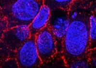
Desmoglein‑2 in NHEK Human Cells.
Desmoglein-2 was detected in immersion fixed NHEK human normal epidermal keratinocytes using Mouse Anti-Human Desmoglein-2 Monoclonal Antibody (Catalog # MAB947) at 10 µg/mL for 3 hours at room temperature. Cells were stained using the NorthernLights™ 557-conjugated Anti-Mouse IgG Secondary Antibody (red; Catalog # NL007) and counterstained with DAPI (blue). Specific staining was localized to cytoplasm and cell junctions. View our protocol for Fluorescent ICC Staining of Cells on Coverslips.
Desmoglein‑2 in A431 Human Cell Line.
Desmoglein-2 was detected in immersion fixed A431 human epithelial carcinoma cell line wildtype and knockout using Mouse Anti-Human Desmoglein-2 Monoclonal Antibody (Catalog # MAB947) at 10 µg/mL for 3 hours at room temperature. Cells were stained using the NorthernLights™ 557-conjugated Anti-Mouse IgG Secondary Antibody (red; Catalog # NL007) and counterstained with DAPI (blue). Specific staining was localized to plasma membrane. View our protocol for Fluorescent ICC Staining of Cells on Coverslips.
Detection of Human Desmoglein‑2 by Simple WesternTM.
Simple Western lane view shows lysates of Jurkat human acute T cell leukemia cell line, loaded at 0.2 mg/mL. A specific band was detected for Desmoglein‑2 at approximately 120 kDa (as indicated) using 20 µg/mL of Mouse Anti-Human Desmoglein‑2 Monoclonal Antibody (Catalog # MAB947). This experiment was conducted under reducing conditions and using the 12-230 kDa separation system.
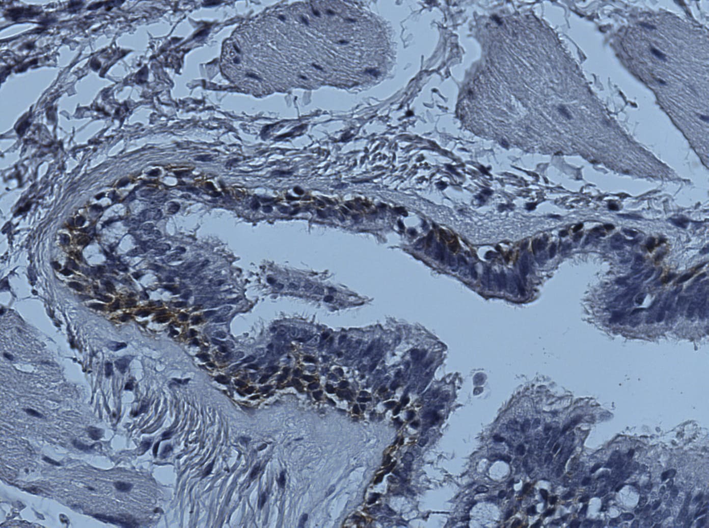
Detection of Desmoglein-2 by Immunohistochemistry
DSG2 promotes the adhesion of MM plasma cells to BM endothelial cells and is co‐regulated with N‐cadherin. (A) BM trephine biopsies from three MM patients were stained for DSG2 by immunohistochemistry; a representative example shows the isotype control (iso ctl) and DSG2‐stained section with a DSG2‐expressing blood vessel (black arrow) and MM PC (red arrows). (B) Expression of DSG2 by the TrHBMEC cell line was assessed by flow cytometry in the parent culture (left); after sorting on DSG2 expression to enrich for BMECDSG2 cells (centre); or after extended passage of the BMECDSG2 cells (right). (C) Adhesion of KMS‐11 cells (± shDSG2) to a monolayer of BMECDSG2 cells for 15 min followed by extensive washing and cell adhesion quantified by imaging the GFP reporter in the KMS‐11 cells. Shown in (C, left image) are representative fluorescent images (scale bar = 100 μm) while (C, right image) shows a summary graph of four independent experiments (Mann–Whitney test *P < 0.05 compared to shNT control). (D) Adhesion of RPMI8226 cells (± siDSG2) to a monolayer of BMECDSG2 cells as above. Representative fluorescent images (scale bar = 100 μm) are shown in (D, left image) while (D, right image) shows a summary graph of three independent experiments (Mann–Whitney test *P < 0.05 compared to siNT control). (E) Gene expression values for DSG2 and CDH2 (N‐cadherin) were extracted from dataset GSE4581. Samples in the MS subgroup are shown in red while others (MS‐negative) are shown in black. Quadrants were set visually to highlight the four distinct subsets defined by individual or co‐expression of DSG2 and CDH2. Image collected and cropped by CiteAb from the following open publication (https://pubmed.ncbi.nlm.nih.gov/34245117), licensed under a CC-BY license. Not internally tested by R&D Systems.Applications for Human Desmoglein-2 Antibody (141409)
Application
Recommended Usage
Immunocytochemistry
5-25 µg/mL
Sample: Immersion fixed NHEK human normal epidermal keratinocytes and A431 human epithelial carcinoma cell line
Sample: Immersion fixed NHEK human normal epidermal keratinocytes and A431 human epithelial carcinoma cell line
Simple Western
20 µg/mL
Sample: Jurkat human acute T cell leukemia cell line
Sample: Jurkat human acute T cell leukemia cell line
Western Blot
2 µg/mL
Sample: Jurkat human acute T cell leukemia cell line under reducing conditions only
Sample: Jurkat human acute T cell leukemia cell line under reducing conditions only
Reviewed Applications
Read 2 reviews rated 5 using MAB947 in the following applications:
Formulation, Preparation, and Storage
Purification
Protein A or G purified from hybridoma culture supernatant
Reconstitution
Reconstitute at 0.5 mg/mL in sterile PBS. For liquid material, refer to CoA for concentration.
Loading...
Formulation
Lyophilized from a 0.2 μm filtered solution in PBS with Trehalose. *Small pack size (SP) is supplied either lyophilized or as a 0.2 µm filtered solution in PBS.
Shipping
Lyophilized product is shipped at ambient temperature. Liquid small pack size (-SP) is shipped with polar packs. Upon receipt, store immediately at the temperature recommended below.
Stability & Storage
Use a manual defrost freezer and avoid repeated freeze-thaw cycles.
- 12 months from date of receipt, -20 to -70 °C as supplied.
- 1 month, 2 to 8 °C under sterile conditions after reconstitution.
- 6 months, -20 to -70 °C under sterile conditions after reconstitution.
Calculators
Background: Desmoglein-2
References
- Nollet, R. et al. (2000) J. Mol. Biol. 299:551.
- Elias, P. et al. (2001) J. Cell Biol. 153:243.
- Arnemann, J. et al. (1992) Genomics 13:484.
Alternate Names
CDHF5, Desmoglein2, DSG2, HDGC
Gene Symbol
DSG2
UniProt
Additional Desmoglein-2 Products
Product Documents for Human Desmoglein-2 Antibody (141409)
Certificate of Analysis
To download a Certificate of Analysis, please enter a lot or batch number in the search box below.
Note: Certificate of Analysis not available for kit components.
Product Specific Notices for Human Desmoglein-2 Antibody (141409)
For research use only
Related Research Areas
Citations for Human Desmoglein-2 Antibody (141409)
 Powered by Bioz
Customer Reviews for Human Desmoglein-2 Antibody (141409) (2)
5 out of 5
2 Customer Ratings
Have you used Human Desmoglein-2 Antibody (141409)?
Submit a review and receive an Amazon gift card!
$25/€18/£15/$25CAN/¥2500 Yen for a review with an image
$10/€7/£6/$10CAN/¥1110 Yen for a review without an image
Submit a review
Customer Images
Showing
1
-
2 of
2 reviews
Showing All
Filter By:
-
Application: Immunocytochemistry/ImmunofluorescenceSample Tested: A431 human epithelial carcinoma cell lineSpecies: HumanVerified Customer | Posted 12/29/2021

-
Application: ImmunohistochemistrySample Tested: Adult lungSpecies: HumanVerified Customer | Posted 02/15/2019
There are no reviews that match your criteria.
Protocols
Find general support by application which include: protocols, troubleshooting, illustrated assays, videos and webinars.
- Appropriate Fixation of IHC/ICC Samples
- Cellular Response to Hypoxia Protocols
- ClariTSA™ Fluorophore Kits
- Detection & Visualization of Antibody Binding
- ICC Cell Smear Protocol for Suspension Cells
- ICC Immunocytochemistry Protocol Videos
- ICC for Adherent Cells
- Immunocytochemistry (ICC) Protocol
- Immunocytochemistry Troubleshooting
- Immunofluorescence of Organoids Embedded in Cultrex Basement Membrane Extract
- Immunohistochemistry (IHC) and Immunocytochemistry (ICC) Protocols
- Preparing Samples for IHC/ICC Experiments
- Preventing Non-Specific Staining (Non-Specific Binding)
- Primary Antibody Selection & Optimization
- Protocol for VisUCyte™ HRP Polymer Detection Reagent
- Protocol for the Fluorescent ICC Staining of Cell Smears - Graphic
- Protocol for the Fluorescent ICC Staining of Cultured Cells on Coverslips - Graphic
- Protocol for the Preparation and Fluorescent ICC Staining of Cells on Coverslips
- Protocol for the Preparation and Fluorescent ICC Staining of Non-adherent Cells
- Protocol for the Preparation and Fluorescent ICC Staining of Stem Cells on Coverslips
- Protocol for the Preparation of a Cell Smear for Non-adherent Cell ICC - Graphic
- R&D Systems Quality Control Western Blot Protocol
- TUNEL and Active Caspase-3 Detection by IHC/ICC Protocol
- The Importance of IHC/ICC Controls
- Troubleshooting Guide: Western Blot Figures
- Western Blot Conditions
- Western Blot Protocol
- Western Blot Protocol for Cell Lysates
- Western Blot Troubleshooting
- Western Blot Troubleshooting Guide
- View all Protocols, Troubleshooting, Illustrated assays and Webinars
Loading...