Human E-Selectin (CD62E)/P-Selectin (CD62P) Antibody (BBIG-E6 (13D5))
R&D Systems | Catalog # BBA1
/P-Selectin (CD62P) in HUVEC Human Cells.")
Key Product Details
Species Reactivity
Validated:
Human
Cited:
Human, Rat, Porcine, Insect - Aedes albopictus (Asian tiger mosquito)
Applications
Validated:
Adhesion Blockade, Flow Cytometry, Immunocytochemistry, Immunoprecipitation, CyTOF-ready
Cited:
Immunohistochemistry, Neutralization, Flow Cytometry, Immunocytochemistry, ELISA Capture
Label
Unconjugated
Antibody Source
Monoclonal Mouse IgG1 Clone # BBIG-E6 (13D5)
Loading...
Product Specifications
Immunogen
Activated HUVEC human umbilical vein endothelial cells
Specificity
Detects human E‑Selectin/P‑Selectin (CD62E/P). Binds to COS cells transfected with human E-Selectin or human P-Selectin. It does not bind to CHO cells transfected with human ICAM-1, L-Selectin, PECAM-1 or VCAM-1.
Clonality
Monoclonal
Host
Mouse
Isotype
IgG1
Scientific Data Images for Human E-Selectin (CD62E)/P-Selectin (CD62P) Antibody (BBIG-E6 (13D5))
E-Selectin (CD62E)/P-Selectin (CD62P) in HUVEC Human Cells.
E-Selectin (CD62E)/P-Selectin (CD62P) was detected in immersion fixed HUVEC human umbilical vein endothelial cells stimulated with TNF-alpha (Catalog # 210-TA-010) using Mouse Anti-Human E-Selectin (CD62E)/P-Selectin (CD62P) Monoclonal Antibody (Catalog # BBA1) at 10 µg/mL for 3 hours at room temperature. Cells were stained using the Northern-Lights™ 557-conjugated Anti-Mouse IgG Secondary Antibody (yellow; Catalog # NL007) and counterstained with DAPI (blue). View our protocol for Fluorescent ICC Staining of Cells on Coverslips.Applications for Human E-Selectin (CD62E)/P-Selectin (CD62P) Antibody (BBIG-E6 (13D5))
Application
Recommended Usage
Adhesion Blockade
The adhesion of U937 human histiocytic lymphoma cells (5 x 10⁴ cells/well) to immobilized Recombinant Human P-Selectin/CD62P (Catalog # ADP3, 10 µg/mL, 100 µL/well) was maximally inhibited (80-100%) by 10 µg/mL of the antibody.
CyTOF-ready
Ready to be labeled using established conjugation methods. No BSA or other carrier proteins that could interfere with conjugation.
Flow Cytometry
2.5 µg/106 cells
Sample: HUVEC human umbilical vein endothelial cells treated with Recombinant Human TNF‑ alpha (Catalog # 210-TA)
Sample: HUVEC human umbilical vein endothelial cells treated with Recombinant Human TNF‑ alpha (Catalog # 210-TA)
Immunocytochemistry
8-25 µg/mL
Sample: Immersion fixed HUVEC human umbilical vein endothelial cells activated with recombinant human TNF-alpha (Catalog # 210-TA-010)
Sample: Immersion fixed HUVEC human umbilical vein endothelial cells activated with recombinant human TNF-alpha (Catalog # 210-TA-010)
Immunoprecipitation
Pigott, R. et al. (1991) J. Immunol. 147:130.
Reviewed Applications
Read 4 reviews rated 3.3 using BBA1 in the following applications:
Flow Cytometry Panel Builder
Bio-Techne Knows Flow Cytometry
Save time and reduce costly mistakes by quickly finding compatible reagents using the Panel Builder Tool.
Advanced Features
- Spectra Viewer - Custom analysis of spectra from multiple fluorochromes
- Spillover Popups - Visualize the spectra of individual fluorochromes
- Antigen Density Selector - Match fluorochrome brightness with antigen density

Formulation, Preparation, and Storage
Purification
Protein A or G purified from hybridoma culture supernatant
Reconstitution
Sterile PBS to a final concentration of 0.5 mg/mL.
Loading...
Formulation
Lyophilized from a 0.2 μm filtered solution in PBS with Trehalose.
Shipping
The product is shipped at ambient temperature. Upon receipt, store it immediately at the temperature recommended below.
Stability & Storage
Use a manual defrost freezer and avoid repeated freeze-thaw cycles.
- 12 months from date of receipt, -20 to -70 °C as supplied.
- 1 month, 2 to 8 °C under sterile conditions after reconstitution.
- 6 months, -20 to -70 °C under sterile conditions after reconstitution.
Calculators
Background: E-Selectin (CD62E)/P-Selectin (CD62P)
Alternate Names
Eselectin, PSelectin
Additional E-Selectin (CD62E)/P-Selectin (CD62P) Products
Product Documents for Human E-Selectin (CD62E)/P-Selectin (CD62P) Antibody (BBIG-E6 (13D5))
Certificate of Analysis
To download a Certificate of Analysis, please enter a lot or batch number in the search box below.
Note: Certificate of Analysis not available for kit components.
Product Specific Notices for Human E-Selectin (CD62E)/P-Selectin (CD62P) Antibody (BBIG-E6 (13D5))
For research use only
Citations for Human E-Selectin (CD62E)/P-Selectin (CD62P) Antibody (BBIG-E6 (13D5))
 Powered by Bioz
Customer Reviews for Human E-Selectin (CD62E)/P-Selectin (CD62P) Antibody (BBIG-E6 (13D5)) (4)
3.3 out of 5
4 Customer Ratings
Have you used Human E-Selectin (CD62E)/P-Selectin (CD62P) Antibody (BBIG-E6 (13D5))?
Submit a review and receive an Amazon gift card!
$25/€18/£15/$25CAN/¥2500 Yen for a review with an image
$10/€7/£6/$10CAN/¥1110 Yen for a review without an image
Submit a review
Customer Images
Showing
1
-
4 of
4 reviews
Showing All
Filter By:
-
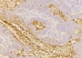
Application: ImmunohistochemistrySample Tested: Сolon adenocarcinoma tissueSpecies: HumanVerified Customer | Posted 10/31/2021

-
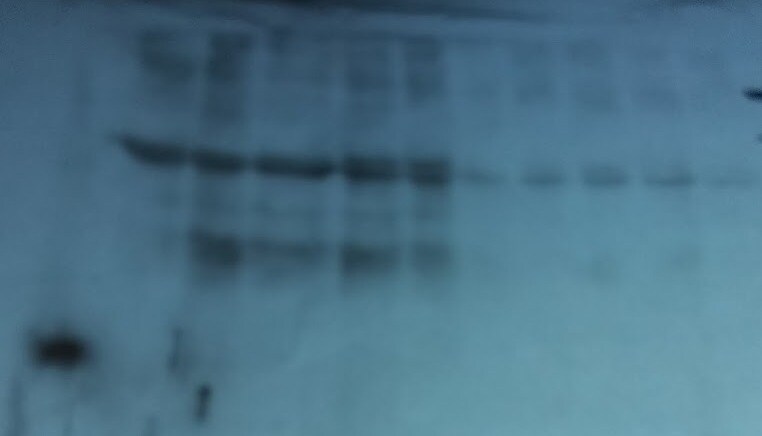
Application: Western BlotSample Tested: HUVEC human umbilical vein endothelial cellsSpecies: HumanVerified Customer | Posted 11/10/2018Western blot using standard conditions on LPS or tumor secretion-stimulated HUVEC cells. No specific bands present.
Bio-Techne ResponseThank you for reviewing our product. We are sorry to hear that this antibody did not perform as expected. We have been in touch with the customer to resolve this issue according to our Product Guarantee and to the customer’s satisfaction.
-
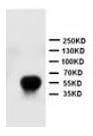
Application: Western BlotSample Tested: Human cellsSpecies: HumanVerified Customer | Posted 07/09/2018
Bio-Techne ResponseThank you for reviewing our product. We are sorry to hear that this product did not perform as expected. We have been in touch with the customer to resolve this issue according to our Product Guarantee and to the customer’s satisfaction.
-
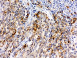
Application: ImmunohistochemistrySample Tested: Human cellsSpecies: HumanVerified Customer | Posted 08/18/2017
There are no reviews that match your criteria.
Protocols
Find general support by application which include: protocols, troubleshooting, illustrated assays, videos and webinars.
- 7-Amino Actinomycin D (7-AAD) Cell Viability Flow Cytometry Protocol
- Appropriate Fixation of IHC/ICC Samples
- Cellular Response to Hypoxia Protocols
- ClariTSA™ Fluorophore Kits
- Detection & Visualization of Antibody Binding
- Extracellular Membrane Flow Cytometry Protocol
- Flow Cytometry Protocol for Cell Surface Markers
- Flow Cytometry Protocol for Staining Membrane Associated Proteins
- Flow Cytometry Staining Protocols
- Flow Cytometry Troubleshooting Guide
- ICC Cell Smear Protocol for Suspension Cells
- ICC Immunocytochemistry Protocol Videos
- ICC for Adherent Cells
- Immunocytochemistry (ICC) Protocol
- Immunocytochemistry Troubleshooting
- Immunofluorescence of Organoids Embedded in Cultrex Basement Membrane Extract
- Immunohistochemistry (IHC) and Immunocytochemistry (ICC) Protocols
- Immunoprecipitation Protocol
- Intracellular Flow Cytometry Protocol Using Alcohol (Methanol)
- Intracellular Flow Cytometry Protocol Using Detergents
- Intracellular Nuclear Staining Flow Cytometry Protocol Using Detergents
- Intracellular Staining Flow Cytometry Protocol Using Alcohol Permeabilization
- Intracellular Staining Flow Cytometry Protocol Using Detergents to Permeabilize Cells
- Preparing Samples for IHC/ICC Experiments
- Preventing Non-Specific Staining (Non-Specific Binding)
- Primary Antibody Selection & Optimization
- Propidium Iodide Cell Viability Flow Cytometry Protocol
- Protocol for Liperfluo
- Protocol for VisUCyte™ HRP Polymer Detection Reagent
- Protocol for the Characterization of Human Th22 Cells
- Protocol for the Characterization of Human Th9 Cells
- Protocol for the Fluorescent ICC Staining of Cell Smears - Graphic
- Protocol for the Fluorescent ICC Staining of Cultured Cells on Coverslips - Graphic
- Protocol for the Preparation and Fluorescent ICC Staining of Cells on Coverslips
- Protocol for the Preparation and Fluorescent ICC Staining of Non-adherent Cells
- Protocol for the Preparation and Fluorescent ICC Staining of Stem Cells on Coverslips
- Protocol for the Preparation of a Cell Smear for Non-adherent Cell ICC - Graphic
- Protocol: Annexin V and PI Staining by Flow Cytometry
- Protocol: Annexin V and PI Staining for Apoptosis by Flow Cytometry
- TUNEL and Active Caspase-3 Detection by IHC/ICC Protocol
- The Importance of IHC/ICC Controls
- Troubleshooting Guide: Fluorokine Flow Cytometry Kits
- View all Protocols, Troubleshooting, Illustrated assays and Webinars
Loading...