
Key Product Details
Species Reactivity
Validated:
Human
Cited:
Human, Hamster
Applications
Validated:
Immunohistochemistry, Western Blot, Flow Cytometry, Simple Western, CyTOF-ready
Cited:
Immunohistochemistry-Paraffin, Western Blot, Bioassay, Mass Spectrometry
Label
Unconjugated
Antibody Source
Polyclonal Goat IgG
Loading...
Product Specifications
Immunogen
E. coli-derived recombinant human EN-RAGE/S100A12
Met1-Glu92
Accession # P80511
Met1-Glu92
Accession # P80511
Specificity
Detects human EN-RAGE in direct ELISAs and Western blots.
Clonality
Polyclonal
Host
Goat
Isotype
IgG
Scientific Data Images for Human EN-RAGE/S100A12 Antibody
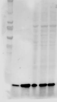
Detection of Human EN‑RAGE/S100A12 by Western Blot.
Western blot shows lysate of human granulocytes. PVDF membrane was probed with 1 µg/mL of Goat Anti-Human EN-RAGE/S100A12 Antigen Affinity-purified Polyclonal Antibody (Catalog # AF1052) followed by HRP-conjugated Anti-Goat IgG Secondary Antibody (HAF017). A specific band was detected for EN-RAGE/S100A12 at approximately 10 kDa (as indicated). This experiment was conducted under reducing conditions and using Immunoblot Buffer Group 1.
EN‑RAGE/S100A12 in Human Tonsil.
EN‑RAGE/S100A12 was detected in immersion fixed paraffin-embedded sections of human tonsil using 15 µg/mL Goat Anti-Human EN‑RAGE/S100A12 Antigen Affinity-purified Polyclonal Antibody (Catalog # AF1052) overnight at 4 °C. Tissue was stained with the Anti-Goat HRP-DAB Cell & Tissue Staining Kit (brown; CTS008) and counterstained with hematoxylin (blue). View our protocol for Chromogenic IHC Staining of Paraffin-embedded Tissue Sections.
Detection of Human EN‑RAGE/S100A12 by Simple WesternTM.
Simple Western lane view shows lysates of human tonsil, loaded at 0.2 mg/mL. A specific band was detected for EN‑RAGE/S100A12 at approximately 11 kDa (as indicated) using 10 µg/mL of Goat Anti-Human EN‑RAGE/S100A12 Antigen Affinity-purified Polyclonal Antibody (Catalog # AF1052). This experiment was conducted under reducing conditions and using the 12-230 kDa separation system.
Detection of EN‑RAGE/S100A12 in Human peripheral blood monocytes by Flow Cytometry
Human peripheral blood monocytes were stained with Goat Anti-Human EN‑RAGE/S100A12 Antigen Affinity-purified Polyclonal Antibody (Catalog # AF1052, filled histogram) or isotype control antibody (Catalog # AB-108-C, open histogram) followed by Phycoerythrin-conjugated Anti-Goat IgG Secondary Antibody (Catalog # F0107). View our protocol for Staining Membrane-associated Proteins.Applications for Human EN-RAGE/S100A12 Antibody
Application
Recommended Usage
CyTOF-ready
Ready to be labeled using established conjugation methods. No BSA or other carrier proteins that could interfere with conjugation.
Flow Cytometry
0.25 µg/106 cells
Sample: Human peripheral blood monocytes
Sample: Human peripheral blood monocytes
Immunohistochemistry
5-15 µg/mL
Sample: Immersion fixed paraffin-embedded sections of human tonsil
Sample: Immersion fixed paraffin-embedded sections of human tonsil
Simple Western
10 µg/mL
Sample: Human tonsil
Sample: Human tonsil
Western Blot
1 µg/mL
Sample: Human granulocytes
Sample: Human granulocytes
Reviewed Applications
Read 4 reviews rated 4.3 using AF1052 in the following applications:
Flow Cytometry Panel Builder
Bio-Techne Knows Flow Cytometry
Save time and reduce costly mistakes by quickly finding compatible reagents using the Panel Builder Tool.
Advanced Features
- Spectra Viewer - Custom analysis of spectra from multiple fluorochromes
- Spillover Popups - Visualize the spectra of individual fluorochromes
- Antigen Density Selector - Match fluorochrome brightness with antigen density

Formulation, Preparation, and Storage
Purification
Antigen Affinity-purified
Reconstitution
Reconstitute at 0.2 mg/mL in sterile PBS. For liquid material, refer to CoA for concentration.
Loading...
Formulation
Lyophilized from a 0.2 μm filtered solution in PBS with Trehalose. See Certificate of Analysis for details.
*Small pack size (-SP) is supplied either lyophilized or as a 0.2 µm filtered solution in PBS.
*Small pack size (-SP) is supplied either lyophilized or as a 0.2 µm filtered solution in PBS.
Shipping
Lyophilized product is shipped at ambient temperature. Liquid small pack size (-SP) is shipped with polar packs. Upon receipt, store immediately at the temperature recommended below.
Stability & Storage
Use a manual defrost freezer and avoid repeated freeze-thaw cycles.
- 12 months from date of receipt, -20 to -70 °C as supplied.
- 1 month, 2 to 8 °C under sterile conditions after reconstitution.
- 6 months, -20 to -70 °C under sterile conditions after reconstitution.
Calculators
Background: EN-RAGE/S100A12
References
- Santamaria-Kisiel, L. et al. (2006) Biochem. J. 396:201.
- Leclerc, E. et al. (2009) Biochim. Biophys. Acta 1793:993.
- Wicki, R. et al. (1996) Cell Calcium 20:459.
- Moroz, O.V. et al. (2009) BMC Biochem. 10:11.
- Miranda, L.P. et al. (2001) FEBS Lett. 488:85.
- Vogl, T. et al. (1999) J. Biol. Chem. 274:25291.
- Xie, J. et al. (2007) J. Biol. Chem. 282:4218.
- Goyette, J. et al. (2009) J. Immunol. 183:593.
- Hatakeyama, T. et al. (2004) Eur. J. Biochem. 271:3765.
- Hofmann, M.A. et al. (1999) Cell 97:889.
- Yang, Z. et al. (2001) J. Leukoc. Biol. 69:986.
- Rouleau, P. et al. (2003) Clin. Immunol. 107:46.
- Yan, W.X. et al. (2008) J. Biol. Chem. 283:13035.
- Yang, Z. et al. (2007) J. Allergy Clin. Immunol. 119:106.
- Mikkelsen, S.E. et al. (2001) J. Neurochem. 79:767.
- Fuellen, G. et al. (2004) OMICS 8:334.
Long Name
Extracellular Newly Identified RAGE-binding Protein
Alternate Names
CAAF1, CAAFI, CAGC, CAGCS100, Calgranulin C, ENRAGE, MRP6, S100A12
Entrez Gene IDs
6283 (Human)
Gene Symbol
S100A12
UniProt
Additional EN-RAGE/S100A12 Products
Product Documents for Human EN-RAGE/S100A12 Antibody
Certificate of Analysis
To download a Certificate of Analysis, please enter a lot or batch number in the search box below.
Note: Certificate of Analysis not available for kit components.
Product Specific Notices for Human EN-RAGE/S100A12 Antibody
For research use only
Related Research Areas
Citations for Human EN-RAGE/S100A12 Antibody
 Powered by Bioz
Customer Reviews for Human EN-RAGE/S100A12 Antibody (4)
4.3 out of 5
4 Customer Ratings
Have you used Human EN-RAGE/S100A12 Antibody?
Submit a review and receive an Amazon gift card!
$25/€18/£15/$25CAN/¥2500 Yen for a review with an image
$10/€7/£6/$10CAN/¥1110 Yen for a review without an image
Submit a review
Customer Images
Showing
1
-
4 of
4 reviews
Showing All
Filter By:
-
Application: Western BlotSample Tested: Recombinant proteinSpecies: HumanVerified Customer | Posted 09/29/2023

-
Application: Western BlotSample Tested: See PMID 19944411Species: HumanVerified Customer | Posted 01/05/2015
-
Application: Immunohistochemistry-ParaffinSample Tested: See PMID 22609404Species: HumanVerified Customer | Posted 01/05/2015
-
Application: Immunohistochemistry-ParaffinSample Tested: See PMID 22609404Species: OtherVerified Customer | Posted 01/05/2015
There are no reviews that match your criteria.
Protocols
Find general support by application which include: protocols, troubleshooting, illustrated assays, videos and webinars.
- 7-Amino Actinomycin D (7-AAD) Cell Viability Flow Cytometry Protocol
- Antigen Retrieval Protocol (PIER)
- Antigen Retrieval for Frozen Sections Protocol
- Appropriate Fixation of IHC/ICC Samples
- Cellular Response to Hypoxia Protocols
- Chromogenic IHC Staining of Formalin-Fixed Paraffin-Embedded (FFPE) Tissue Protocol
- Chromogenic Immunohistochemistry Staining of Frozen Tissue
- ClariTSA™ Fluorophore Kits
- Detection & Visualization of Antibody Binding
- Extracellular Membrane Flow Cytometry Protocol
- Flow Cytometry Protocol for Cell Surface Markers
- Flow Cytometry Protocol for Staining Membrane Associated Proteins
- Flow Cytometry Staining Protocols
- Flow Cytometry Troubleshooting Guide
- Fluorescent IHC Staining of Frozen Tissue Protocol
- Graphic Protocol for Heat-induced Epitope Retrieval
- Graphic Protocol for the Preparation and Fluorescent IHC Staining of Frozen Tissue Sections
- Graphic Protocol for the Preparation and Fluorescent IHC Staining of Paraffin-embedded Tissue Sections
- Graphic Protocol for the Preparation of Gelatin-coated Slides for Histological Tissue Sections
- IHC Sample Preparation (Frozen sections vs Paraffin)
- Immunofluorescent IHC Staining of Formalin-Fixed Paraffin-Embedded (FFPE) Tissue Protocol
- Immunohistochemistry (IHC) and Immunocytochemistry (ICC) Protocols
- Immunohistochemistry Frozen Troubleshooting
- Immunohistochemistry Paraffin Troubleshooting
- Intracellular Flow Cytometry Protocol Using Alcohol (Methanol)
- Intracellular Flow Cytometry Protocol Using Detergents
- Intracellular Nuclear Staining Flow Cytometry Protocol Using Detergents
- Intracellular Staining Flow Cytometry Protocol Using Alcohol Permeabilization
- Intracellular Staining Flow Cytometry Protocol Using Detergents to Permeabilize Cells
- Preparing Samples for IHC/ICC Experiments
- Preventing Non-Specific Staining (Non-Specific Binding)
- Primary Antibody Selection & Optimization
- Propidium Iodide Cell Viability Flow Cytometry Protocol
- Protocol for Heat-Induced Epitope Retrieval (HIER)
- Protocol for Liperfluo
- Protocol for Making a 4% Formaldehyde Solution in PBS
- Protocol for VisUCyte™ HRP Polymer Detection Reagent
- Protocol for the Characterization of Human Th22 Cells
- Protocol for the Characterization of Human Th9 Cells
- Protocol for the Preparation & Fixation of Cells on Coverslips
- Protocol for the Preparation and Chromogenic IHC Staining of Frozen Tissue Sections
- Protocol for the Preparation and Chromogenic IHC Staining of Frozen Tissue Sections - Graphic
- Protocol for the Preparation and Chromogenic IHC Staining of Paraffin-embedded Tissue Sections
- Protocol for the Preparation and Chromogenic IHC Staining of Paraffin-embedded Tissue Sections - Graphic
- Protocol for the Preparation and Fluorescent IHC Staining of Frozen Tissue Sections
- Protocol for the Preparation and Fluorescent IHC Staining of Paraffin-embedded Tissue Sections
- Protocol for the Preparation of Gelatin-coated Slides for Histological Tissue Sections
- Protocol: Annexin V and PI Staining by Flow Cytometry
- Protocol: Annexin V and PI Staining for Apoptosis by Flow Cytometry
- R&D Systems Quality Control Western Blot Protocol
- TUNEL and Active Caspase-3 Detection by IHC/ICC Protocol
- The Importance of IHC/ICC Controls
- Troubleshooting Guide: Fluorokine Flow Cytometry Kits
- Troubleshooting Guide: Immunohistochemistry
- Troubleshooting Guide: Western Blot Figures
- Western Blot Conditions
- Western Blot Protocol
- Western Blot Protocol for Cell Lysates
- Western Blot Troubleshooting
- Western Blot Troubleshooting Guide
- View all Protocols, Troubleshooting, Illustrated assays and Webinars
Loading...
Associated Pathways

