EphB3, also known as Cek10, Tyro6, Sek4, Hek2, and Mdk5, is a 130 kDa member of the transmembrane Eph receptor tyrosine kinase family. The A and B classes of Eph proteins are distinguished by Ephrin ligand binding preference but have a common structural organization. Eph-Ephrin interactions are widely involved in the regulation of cell migration, tissue morphogenesis, and cancer progression (1). The 526 amino acid (aa) extracellular domain (ECD) of mature human EphB3 contains a ligand binding domain followed by a cysteine rich region and two fibronectin type III domains. The 418 aa cytoplasmic domain contains a tyrosine kinase domain, a sterile alpha motif (SAM), and a PDZ binding motif (2). Within the ECD, human EphB3 shares 96% aa sequence identity with mouse and rat EphB3. Binding of EphB3 to its ligands Ephrin-B1, B2, and B3 triggers forward signaling through EphB3 as well as reverse signaling through the Ephrin (1, 3). EphB3 also interacts in cis with the receptor tyrosine kinase Ryk (4). Activation of its kinase is required for some but not all of the effects of EphB3 on cellular adhesion, motility, and morphology (5). EphB3 is widely expressed during development and in the adult; it shows a complementary tissue distribution to the Ephrin-B ligands (6‑9). EphB3 function is important in vascular, nervous system, thymocyte, and palate development (6, 7, 10‑12). It directs embyronic neuronal axon pathfinding, and its upregulation on local macrophages following neuronal injury promotes the growth of regenerating axons (10, 13). EphB3 inhibits colorectal carcinogenesis and invasion by preventing the migration of tumor cells out of the intestinal crypt (9, 14). EphB3 function is supported by the cooperative action of EphB2 in several of these processes (6, 10‑12, 15).

Key Product Details
Species Reactivity
Human
Applications
Western Blot
Label
Unconjugated
Antibody Source
Monoclonal Mouse IgG2B Clone # 647308
Loading...
Product Specifications
Immunogen
Mouse myeloma cell line NS0-derived recombinant human EphB3
Leu38-Ala550
Accession # P54753
Leu38-Ala550
Accession # P54753
Specificity
Detects human EphB3 in direct ELISAs and Western blots. In Western blots, 100% cross-reactivity with recombinant mouse (rm) EphB3, 25% cross‑reactivity with rmEphB2, and no cross-reactivity with recombinant human (rh) EphA1, A2, A5, A6, A10, rmEphA3, A4, A7, B4, B6, or recombinant rat EphB1.
Clonality
Monoclonal
Host
Mouse
Isotype
IgG2B
Scientific Data Images for Human EphB3 Antibody (647308)
Detection of Human EphB3 by Western Blot.
Western blot shows lysates of human cerebellum tissue. PVDF membrane was probed with 2 µg/mL of Mouse Anti-Human EphB3 Monoclonal Antibody (Catalog # MAB5667) followed by HRP-conjugated Anti-Mouse IgG Secondary Antibody (Catalog # HAF018). A specific band was detected for EphB3 at approximately 130 kDa (as indicated). This experiment was conducted under reducing conditions and using Immunoblot Buffer Group 1.Applications for Human EphB3 Antibody (647308)
Application
Recommended Usage
Western Blot
2 µg/mL
Sample: Human cerebellum tissue
Sample: Human cerebellum tissue
Reviewed Applications
Read 1 review rated 4 using MAB5667 in the following applications:
Formulation, Preparation, and Storage
Purification
Protein A or G purified from hybridoma culture supernatant
Reconstitution
Sterile PBS to a final concentration of 0.5 mg/mL. For liquid material, refer to CoA for concentration.
Loading...
Formulation
Lyophilized from a 0.2 μm filtered solution in PBS with Trehalose. *Small pack size (SP) is supplied either lyophilized or as a 0.2 µm filtered solution in PBS.
Shipping
Lyophilized product is shipped at ambient temperature. Liquid small pack size (-SP) is shipped with polar packs. Upon receipt, store immediately at the temperature recommended below.
Stability & Storage
Use a manual defrost freezer and avoid repeated freeze-thaw cycles.
- 12 months from date of receipt, -20 to -70 °C as supplied.
- 1 month, 2 to 8 °C under sterile conditions after reconstitution.
- 6 months, -20 to -70 °C under sterile conditions after reconstitution.
Calculators
Background: EphB3
References
- Pasquale, E.B. (2008) Cell 133:38.
- Bohme, B. et al. (1993) Oncogene 8:2857.
- Pasquale, E.B (2004) Nat. Neurosci. 7:417.
- Trivier, E. and T.S. Ganesan (2002) J. Biol. Chem. 277:23037.
- Miao, H. et al. (2005) J. Biol. Chem. 280:923.
- Adams, R.H. et al. (1999) Genes Dev. 13:295.
- Krull, C.E. et al. (1997) Curr. Biol. 7:571.
- Willson, C.A. et al. (2006) J. Mol. Histol. 37:369.
- Cortina, C. et al. (2007) Nature Genet. 39:1376.
- Birgbauer, E. et al. (2000) Development 127:1231.
- Alfaro, D. et al. (2008) Immunology 125:131.
- Risley, M. et al. (2009) Mech. Dev. 126:230.
- 13. Liu, X. et al. (2006) J. Neurosci. 26:3087.
- 14. Batlle, E. et al. (2005) Nature 435:1126.
- 15. Holmberg, J. et al. (2006) Cell 125:1151.
Long Name
Eph Receptor B3
Alternate Names
Cek10, Hek2, Mdk5, Sek4, Tyro6
Gene Symbol
EPHB3
UniProt
Additional EphB3 Products
Product Documents for Human EphB3 Antibody (647308)
Certificate of Analysis
To download a Certificate of Analysis, please enter a lot or batch number in the search box below.
Note: Certificate of Analysis not available for kit components.
Product Specific Notices for Human EphB3 Antibody (647308)
For research use only
Related Research Areas
Customer Reviews for Human EphB3 Antibody (647308) (1)
4 out of 5
1 Customer Rating
Have you used Human EphB3 Antibody (647308)?
Submit a review and receive an Amazon gift card!
$25/€18/£15/$25CAN/¥2500 Yen for a review with an image
$10/€7/£6/$10CAN/¥1110 Yen for a review without an image
Submit a review
Customer Images
Showing
1
-
1 of
1 review
Showing All
Filter By:
-
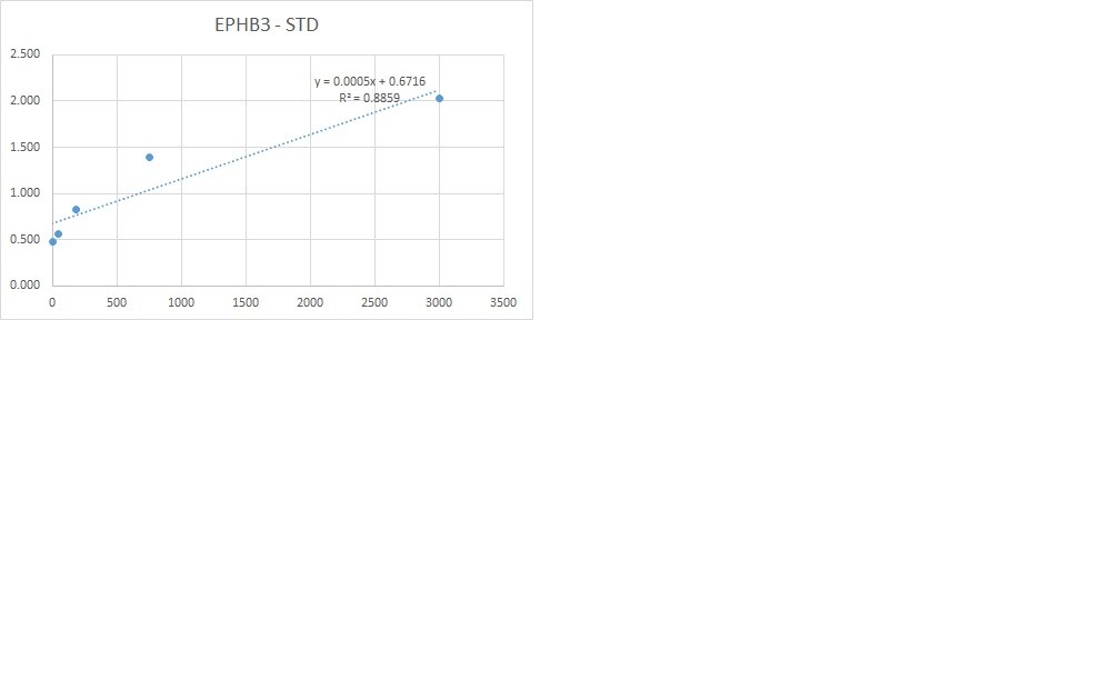
Application: ELISASample Tested: Serum and PlasmaSpecies: HumanVerified Customer | Posted 09/23/2019We used this antibody for developing a sandwich ELISA in combination with pAb (cat. AF5667) and protein (cat. 5667 B3). This combination works well detecting Ephb3 in human serum.

There are no reviews that match your criteria.
Protocols
Find general support by application which include: protocols, troubleshooting, illustrated assays, videos and webinars.
- Cellular Response to Hypoxia Protocols
- R&D Systems Quality Control Western Blot Protocol
- Troubleshooting Guide: Western Blot Figures
- Western Blot Conditions
- Western Blot Protocol
- Western Blot Protocol for Cell Lysates
- Western Blot Troubleshooting
- Western Blot Troubleshooting Guide
- View all Protocols, Troubleshooting, Illustrated assays and Webinars
Loading...