GM-CSF was initially characterized as a factor that can support the in vitro colony formation of granulocyte-macrophage progenitors. It is also a growth factor for erythroid, megakaryocyte, and eosinophil progenitors. GM-CSF is produced by a number of different cell types (including T cells, B cells, macrophages, mast cells, endothelial cells, fibroblasts, and adipocytes) in response to cytokine or inflammatory stimuli. On mature hematopoietic cells, GM-CSF is a survival factor for and activates the effector functions of granulocytes, monocytes/macrophages, and eosinophils (1, 2). GM-CSF promotes a Th1 biased immune response, angiogenesis, allergic inflammation, and the development of autoimmunity (3‑5). It shows clinical effectiveness in ameliorating chemotherapy-induced neutropenia, and GM-CSF transfected tumor cells are utilized as cancer vaccines (6, 7). The 22 kDa glycosylated GM-CSF, similar to IL-3 and IL-5, is a cytokine with a core of four bundled alpha ‑helices (8‑12). Mature human GM-CSF shares 63%‑70% amino acid sequence identity with canine, feline, porcine, and rat GM-CSF and 54% with mouse GM-CSF. GM-CSF exerts its biological effects through a heterodimeric receptor complex composed of GM-CSF R alpha /CD116 and the signal transducing common beta chain (CD131) which is also a component of the high-affinity receptors for IL-3 and IL-5 (13, 14). In addition, GM-CSF binds a naturally occurring soluble form of GM-CSF R alpha (15). Human GM-CSF is active on canine and feline cells but not on murine cells (16‑18).

Key Product Details
Species Reactivity
Validated:
Human
Cited:
Human, Mouse
Applications
Validated:
Western Blot, Neutralization
Cited:
Immunohistochemistry, Western Blot, Neutralization, Immunocytochemistry, ELISA Capture
Label
Unconjugated
Antibody Source
Polyclonal Goat IgG
Loading...
Product Specifications
Immunogen
E. coli-derived recombinant human GM-CSF
Ala18-Glu144
Accession # P04141
Ala18-Glu144
Accession # P04141
Specificity
Detects human GM‑CSF in direct ELISAs and Western blots. In direct ELISAs, less than 1% cross‑reactivity with recombinant mouse GM‑CSF is observed. Neutralizes the biological activity of both recombinant human GM‑CSF and natural human GM‑CSF.
Clonality
Polyclonal
Host
Goat
Isotype
IgG
Endotoxin Level
<0.10 EU per 1 μg of the antibody by the LAL method.
Scientific Data Images for Human GM-CSF Antibody
Cell Proliferation Induced by GM‑CSF and Neutralization by Human GM‑CSF Antibody.
Recombinant Human GM-CSF (Catalog # 215-GM) stimulates proliferation in the TF-1 human erythroleukemic cell line in a dose-dependent manner (orange line). Proliferation elicited by Recombinant Human GM-CSF (0.5 ng/mL) is neutralized (green line) by increasing concentrations of Goat Anti-Human GM-CSF Antigen Affinity-purified Polyclonal Antibody (Catalog # AF-215-NA). The ND50 is typically 0.08-0.16 µg/mL.Applications for Human GM-CSF Antibody
Application
Recommended Usage
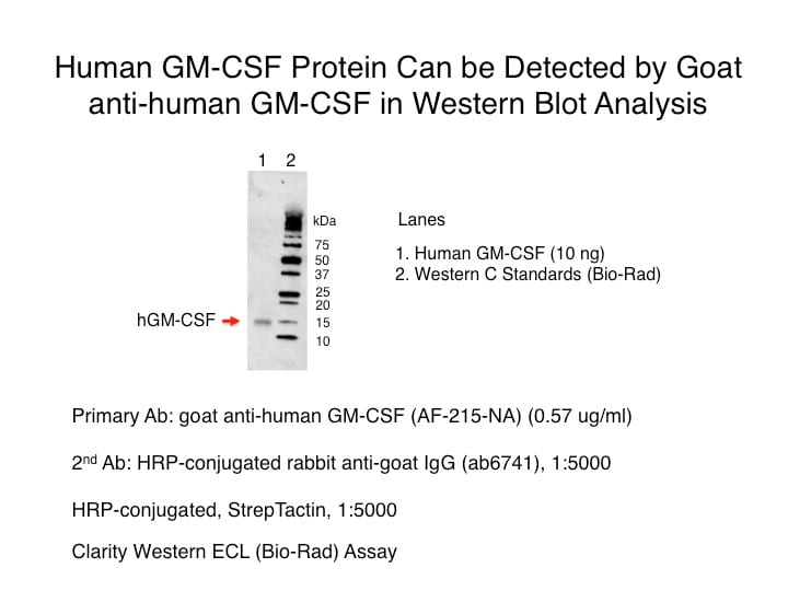
Western Blot
0.1 µg/mL
Sample: Recombinant Human GM-CSF (Catalog # 215-GM)
Sample: Recombinant Human GM-CSF (Catalog # 215-GM)
Neutralization
Measured by its ability to neutralize GM‑CSF-induced proliferation in the TF‑1 human erythroleukemic cell line. Kitamura, T. et al. (1989) J. Cell Physiol. 140:323. The Neutralization Dose (ND50) is typically 0.08-0.16 µg/mL in the presence of 0.5 ng/mL Recombinant Human GM‑CSF.
Reviewed Applications
Read 3 reviews rated 4 using AF-215-NA in the following applications:
Formulation, Preparation, and Storage
Purification
Antigen Affinity-purified
Reconstitution
Reconstitute at 0.2 mg/mL in sterile PBS. For liquid material, refer to CoA for concentration.
Loading...
Formulation
Lyophilized from a 0.2 μm filtered solution in PBS with Trehalose. *Small pack size (SP) is supplied either lyophilized or as a 0.2 µm filtered solution in PBS.
Shipping
Lyophilized product is shipped at ambient temperature. Liquid small pack size (-SP) is shipped with polar packs. Upon receipt, store immediately at the temperature recommended below.
Stability & Storage
Use a manual defrost freezer and avoid repeated freeze-thaw cycles.
- 12 months from date of receipt, -20 to -70 °C as supplied.
- 1 month, 2 to 8 °C under sterile conditions after reconstitution.
- 6 months, -20 to -70 °C under sterile conditions after reconstitution.
Calculators
Background: GM-CSF
References
- Martinez-Moczygemba, M. and D.P. Huston (2003) J. Allergy Clin. Immunol. 112:653.
- Barreda, D.R. et al. (2004) Dev. Comp. Immunol. 28:509.
- Eksioglu, E.A. et al. (2007) Exp. Hematol. 35:1163.
- Cao, Y. (2007) J. Clin. Invest. 117:2362.
- Fleetwood, A.J. et al. (2005) Crit. Rev. Immunol. 25:405.
- Heuser, M. et al. (2007) Semin. Hematol. 44:148.
- Hege, K.M. et al. (2006) Int. Rev. Immunol. 25:321.
- Kaushansky, K. et al. (1992) Biochemistry 31:1881.
- Diederichs, K. et al. (1991) Science 254:1779.
- Cantrell, M.A. et al. (1985) Proc. Natl. Acad. Sci. 82:6250.
- Lee, F. et al. (1985) Proc. Natl. Acad. Sci. 82:4360.
- Wong, G.G. et al. (1985) Science 228:810.
- Onetto-Pothier, N. et al. (1990) Blood 75:59.
- Hayashida, K. et al. (1990) Proc. Natl. Acad. Sci. 87:9655.
- Pelley, J.L. et al. (2007) Exp. Hematol. 35:1483.
- Hogge, G.S. et al. (1990) Cancer Gene Ther. 6:26.
- Sprague, W.S. et al. (2005) J. Comp. Pathol. 133:136.
- Shanafelt, A.B. et al. (1991) J. Biol. Chem. 266:13804.
Long Name
Granulocyte Macrophage Growth Factor
Alternate Names
CSF-2, CSF2, GMCSF, Molgramostim, Sargramostim
Entrez Gene IDs
Gene Symbol
CSF2
UniProt
Additional GM-CSF Products
Product Documents for Human GM-CSF Antibody
Certificate of Analysis
To download a Certificate of Analysis, please enter a lot or batch number in the search box below.
Note: Certificate of Analysis not available for kit components.
Product Specific Notices for Human GM-CSF Antibody
For research use only
Related Research Areas
Citations for Human GM-CSF Antibody
 Powered by Bioz
Customer Reviews for Human GM-CSF Antibody (3)
4 out of 5
3 Customer Ratings
Have you used Human GM-CSF Antibody?
Submit a review and receive an Amazon gift card!
$25/€18/£15/$25CAN/¥2500 Yen for a review with an image
$10/€7/£6/$10CAN/¥1110 Yen for a review without an image
Submit a review
Customer Images
Showing
1
-
3 of
3 reviews
Showing All
Filter By:
-
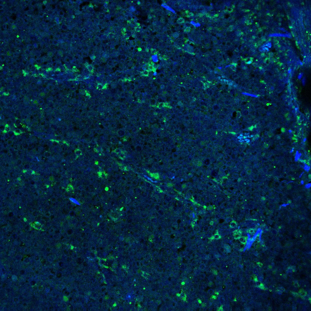
Application: Immunocytochemistry/ImmunofluorescenceSample Tested: Melanoma tissueSpecies: HumanVerified Customer | Posted 10/01/2021

-
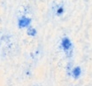
Application: ImmunohistochemistrySample Tested: Adult brainSpecies: HumanVerified Customer | Posted 03/27/2018Published in https://www.ncbi.nlm.nih.gov/pubmed/28169287 Used at 10ug/ml. Briefly, frozen brain sections were fixed in 4% PFA (Fisher Scientific), followed by antigen retrieval using heating in acid citric buffer (Vector, Burlingame, CA, USA). Endogenous avidin-biotin was blocked for 15 min (Vector). Sections were incubated with 10% horse serum in PBS (Biosera, Boussens, France) and Fc Receptor Blocking Solution was added (Human TruStain FcX Biolegend, London, UK). Primary antibodies were added overnight at 4 °C. GM-CSF was detected with donkey anti-goat-biotin (ab6578, Abcam), followed by streptavidin-alkaline phosphatase (SA-5100, Vector) and visualised with the Vector Blue Alkaline Phosphatase Substrate Kit III (Vector).
-
Application: Western BlotSample Tested: Recombinant GM-CSFSpecies: HumanVerified Customer | Posted 07/01/2016
There are no reviews that match your criteria.
Protocols
Find general support by application which include: protocols, troubleshooting, illustrated assays, videos and webinars.
- Cellular Response to Hypoxia Protocols
- R&D Systems Quality Control Western Blot Protocol
- Troubleshooting Guide: Western Blot Figures
- Western Blot Conditions
- Western Blot Protocol
- Western Blot Protocol for Cell Lysates
- Western Blot Troubleshooting
- Western Blot Troubleshooting Guide
- View all Protocols, Troubleshooting, Illustrated assays and Webinars


